Transcription
Darwanto et al. BMC Cancer (2017) 17:358DOI 10.1186/s12885-017-3328-zRESEARCH ARTICLEOpen AccessUse of the QIAGEN GeneReader NGSsystem for detection of KRAS mutations,validated by the QIAGEN Therascreen PCRkit and alternative NGS platformAgus Darwanto1,7 , Anne-Mette Hein2, Sascha Strauss3, Yi Kong4, Andrew Sheridan1, Dan Richards4, Eric Lader5,Monika Ngowe1,8, Timothy Pelletier1, Danielle Adams1,9, Austin Ricker1, Nishit Patel1, Andreas Kühne3,Simon Hughes6, Dan Shiffman4, Dirk Zimmermann3, Kai te Kaat3 and Thomas Rothmann3*AbstractBackground: The detection of somatic mutations in primary tumors is critical for the understanding of cancerevolution and targeting therapy. Multiple technologies have been developed to enable the detection of suchmutations. Next generation sequencing (NGS) is a new platform that is gradually becoming the technology ofchoice for genotyping cancer samples, owing to its ability to simultaneously interrogate many genomic loci atmassively high efficiency and increasingly lower cost. However, multiple barriers still exist for its broader adoptionin clinical research practice, such as fragmented workflow and complex bioinformatics analysis and interpretation.Methods: We performed validation of the QIAGEN GeneReader NGS System using the QIAact Actionable Insights TumorPanel, focusing on clinically meaningful mutations by using DNA extracted from formalin-fixed paraffin-embedded (FFPE)colorectal tissue with known KRAS mutations. The performance of the GeneReader was evaluated and compared to datagenerated from alternative technologies (PCR and pyrosequencing) as well as an alternative NGS platform. The resultswere further confirmed with Sanger sequencing.Results: The data generated from the GeneReader achieved 100% concordance with reference technologies.Furthermore, the GeneReader workflow provides a truly integrated workflow, eliminating artifacts resulting fromroutine sample preparation; and providing up-to-date interpretation of test results.Conclusion: The GeneReader NGS system offers an effective and efficient method to identify somatic (KRAS) cancermutations.Keywords: GeneReader, Kras, Mutation, Cancer, NgsBackgroundSomatic mutations in the KRAS oncogene are common inhuman cancers. They are found in 70-90% of pancreaticcancers [1, 2], 30-50% of colorectal cancers [3–5] and 1030% of Non-Small Cell Lung Cancers (NSCLC) [6–8].Several methods have been developed for the detection ofKRAS mutations, each with specific advantages andlimitations [5, 9, 10].* Correspondence: Thomas.rothmann@qiagen.com3QIAGEN GmbH, QIAGEN Strasse 1, 40724 Hilden, Nordrhein-Westfalen,GermanyFull list of author information is available at the end of the articleSanger sequencing has been the ‘gold standard’ formutation analysis in cancer detection since the 1970s[11]. However, limited by its low sensitivity (10-20%mutant allele frequency (MAF)) and low throughput[10], Sanger sequencing is no longer sufficient for theneeds of today’s cancer molecular diagnostics.The therascreen KRAS RGQ PCR kit is a real-timeqPCR-based assay used to detect the most common KRASmutations including those in codons 12 and 13. It hasgreatly improved sensitivity over Sanger sequencing, andhas been approved by the Food and Drug Administration(FDA) [9] for colorectal cancer patient stratification. The Author(s). 2017 Open Access This article is distributed under the terms of the Creative Commons Attribution 4.0International License (http://creativecommons.org/licenses/by/4.0/), which permits unrestricted use, distribution, andreproduction in any medium, provided you give appropriate credit to the original author(s) and the source, provide a link tothe Creative Commons license, and indicate if changes were made. The Creative Commons Public Domain Dedication o/1.0/) applies to the data made available in this article, unless otherwise stated.
Darwanto et al. BMC Cancer (2017) 17:358Pyrosequencing also offers an attractive alternative toSanger due to its fast turnaround time (TAT) and lowersensitivity threshold, even in tissues with low tumor cellcontent [5].Next-generation sequencing (NGS) differs radicallyfrom the above mentioned methods. Coupled withamplicon-based targeting technology, NGS has the capability to simultaneously sequence in a massively parallelway multiple genetic loci with minimal amounts of nucleic acid input and limited time and expense [12–15].This technology has revolutionized the speed of geneticand genomic discovery, and advanced our understandingof molecular mechanisms of diseases. In recent years,NGS has played an important role in advancing personalized healthcare and precision medicine by enabling theidentification of mutations associated with therapeuticresponse or resistance. As more clinically significantgenetic biomarkers and targeted therapies become available, the profiling of such genetic variations is becomingincreasingly more critical. Several NGS platforms arealready commercially available for sequencing and identification of genetic alterations associated with diseases,such as point mutations, deletions, insertions and copynumber variants [16]. However, QIAGEN’s GeneReaderSystem presented here includes all upstream sampleprocessing steps starting from nucleic acid extraction,together with an integrated downstream bioinformaticssolution that enables a direct access to real-time updatesfrom the rapidly evolving literature, and clinical knowledgeand evidence.To this end, we recently evaluated the QIAGENGeneReader System workflow from DNA extraction andpurification from FFPE tissue samples, to library preparation, sequencing and data analysis and interpretation.Herein we show that the GeneReader presents a unifiedworkflow that provides accurate results and a simplesolution for any laboratory to use in clinical research.MethodsSample and DNA isolationFFPE Tumor material from colorectal cancer tumors(Origene Technologies, MD, USA and Asterand Biosciences, MI, USA) was used to prepare 56 DNA sampleswith known KRAS mutation status, previously determinedusing therascreen assay (Pyrosequencing and PCR) andSanger sequencing according to methods further describedbelow. Tissue sections of 10 μm in thickness, ranging from3 to 20 years of age were used for DNA extraction utilizingeither: i) the QIAamp DNA FFPE Tissue Kit (QIAGEN,Hilden, Germany) or ii) the GeneRead FFPE DNA Kit(QIAGEN, Hilden, Germany) according to manufacturer’sinstructions. DNA concentration was determined usingthe Nanodrop System (Thermo Fisher Scientific, MA,USA) and Qubit dsDNA HS assay (Life Technologies,Page 2 of 8Gaithersburg, USA). The DNA was assessed using theGeneRead DNA QuantiMIZE System (QIAGEN, Hilden,Germany) which utilizes a qPCR-based approach to determine the quality of sample DNA prior to NGS. Furthermore, both NA12878 (Coriell Institute for MedicalResearch) (for which the Genome in the Bottle (GIAB)consortium has published a set of high confident variants [17]) and AcroMetrix (Thermo Fisher Scientific,MA, USA) samples were used as a gold standard set ofvariant calls.GeneReader sample preparation and sequencing runIn total, 40 ng of DNA measured by Qubit (ThermoFisher Scientific, MA, USA) was used as template togenerate libraries for sequencing. Libraries were prepared using the QIAGEN Library Kit v2.0 and theGeneRead QIAact Actionable Insight Tumor Panel(QIAGEN, Hilden, Germany), which amplifies 330amplicons covering 16.7 kb, containing 773 uniquevariant positions in 12 genes (KRAS, NRAS, KIT, BRAF,PDGFRA, ALK, EGFR, ERBB2, PIK3CA, ERBB3, ESR1and RAF1). All steps of library preparation were performed according to the manufacturer’s protocol. Thelibraries were then quantified using a Qubit dsDNA HSAssay Kit (Life Technologies, MA, USA) and QIAxcel(QIAGEN, Hilden, Germany). Ten individual librarieswere pooled prior to emulsion PCR and bead enrichment steps that were carried out using an automatedprotocol on the GeneRead QIAcube (QIAGEN, Hilden,Germany) using the GeneRead Clonal Amp Q Kit(QIAGEN, Hilden, Germany), according to the manufacturer’s protocol. Following bead enrichment, thepooled libraries were sequenced using the GeneReaderplatform (QIAGEN, Hilden, Germany).GeneReader data processingQIAGEN Clinical Insight (QCI ) Analyze software(QIAGEN, Hilden, Germany) was used to QC, align theread data to the hg19 reference genome sequence, callsequence variants, and generate an interactive reportfor visualization of the sequencing results, as well as asummary of the data. QCI Analyze software reports aset of high- and low-confidence variants based on thecoverage of variant positions. Users have an option toanalytically confirm if a variant listed should be valid orinvalid before uploading to QCI Interpret software forthe clinical interpretation. For each sample the reportwas used to assess the quality of the overall sequencingrun and to identify/call the individual variants. Afterreview, variants confirmed as analytically valid wereuploaded to QCI Interpret for creation of a report foreach sample based on detected variants and curatedcontent, with a summary of findings and direct links toevidence sources.
Darwanto et al. BMC Cancer (2017) 17:358Illumina MiSeqThe Actionable Insight Tumor Panel (QIAGEN, Hilden,Germany) was used for a MiSeq (Illumina, CA, USA)sequencing run. The Kapa “with bead” PCR free protocol(KAPABiosystems, MA, USA) was used in further Illuminalibrary preparation steps. Samples were then paired-endsequenced on a MiSeq instrument (Illumina, CA, USA)according to Illumina guidelines. The resulting reads weremapped to the hg19 reference genome sequence usingBWA mem software followed by GATK (best practices) torecalibrate base quality scores. Variants were called usingMuTect. Variants were then filtered using GATK (bestpractice) and annotated using SnpEff. Variants at hotspotpositions were selected using GATK.Pyrosequencing and Sanger analysesThe sample DNA obtained with the QIAamp FFPEDNA Kit (QIAGEN, Hilden, Germany) was subjected toPyrosequencing analysis and Sanger sequencing. ForPyrosequencing the samples were analyzed using thetherascreen RAS Extension Pyro Kit (QIAGEN, Hilden,Germany) which covers mutations in KRAS codons 59,61, 117 and 146 as well as NRAS codons 59, 117 and146. Samples with mutations in KRAS or NRAS codons12 and 13 were further analyzed with the therascreenKRAS or NRAS Pyro Kit (QIAGEN, Hilden, Germany)according to manufacturer’s instructions. In addition,samples that failed the initial PyroMark KRAS analysiswere subjected to a second round of analysis. Sampleswith an initial “check” status, or with an indicated mutation signal of LOD 3% (“Potential low level mutation”)were subjected to a second round of analysis performedin duplicate. Sanger sequencing was performed usingBig Dye Terminator Technology and an ABI 3730xlsequencer (Thermo Fisher Scientific, MA, USA). Mutations were detected by analyzing the sequence trace filesand the quantity of a base at a certain position wascalculated from the area under the curve (AUC) at themutation specific position in the electropherogram.Therascreen qPCRThe therascreen KRAS RGQ PCR Kit (QIAGEN, Hilden,UK) is an allele-specific PCR-based technology with specific primers for the seven most common KRAS codon12 and 13 mutations. The assay screens for the followingmutations: 12 GCT (Ala), 12 GAT (Asp), 12 CGT (Arg),12 TGT (Cys), 12 AGT (Ser), 12 GTT (Val), and 13GAC (Asp). Mutation analysis was performed accordingto manufacturer’s instructions, using the RotorGenereal-time PCR instrument (QIAGEN, Hilden, UK). Analysis of results was performed following the recommendations in the manual, e.g. samples with a control assaywith a cycle threshold (Ct) of 35 or higher were deemedinvalid and excluded from the analysis. Samples werePage 3 of 8called mutation positive based on the delta Ct valuesreported in the handbook. Values over 40 cycles werescored as negative (wild-type).ResultsEvaluation of DNA quality by QuantiMIZEFFPE samples with ages ranging from 3 to 20 years wereused for this study. The quality of the extracted DNA wasmeasured by the GeneRead DNA QuantiMIZE QC assay(QIAGEN, Hilden, UK). Thirteen out of 56 samples failedquality checks and were excluded from further analysis(Additional file 1: Table S1). For the remaining 43 samples, 3 to 9 PCR cycles were added (depending on theQuantiMIZE quality scores) to compensate for differencesin DNA quality during enrichment PCR. The additionalcycles ensured that poor quality (highly fragmented) DNAsamples yielded enough material for downstream librarypreparation. The quality of DNA purified from formalinfixated tissue decreases over the sample storage periodtime [18–20], but also depends on how tissues weretreated, handled and processed before and during samplefixation [19, 21, 22].GeneReader sequencing performanceThe QIAact Actionable Insights Tumor Panel (QIAGEN,Hilden, UK) contains 773 unique variant positions in 12genes (Table 1). An analysis of the reads mapped to thereference showed coverage levels that met the industrystandard 5% sensitivity criteria, even with aged FFPEsamples. A 200 minimum read coverage cutoff wasused for calling a variant at any position in the panel.For the 43 FFPE samples analyzed, an average ampliconcoverage of 97.2% was observed, and an average variantinsight coverage (hotspot coverage) of 99.8% was observed at read depths 200 (Table 1). For NA12878samples, an average amplicon coverage of 98.5% wasobserved and an average variant insight coverage of99.9% was observed at read depths of 200 (Table 1).Table 1 Parameter and sequencing coverage of ActionableInsight Tumor PanelParameterDetailsPanel size12 genes/16.7 kbInsight size773 unique variant positionsAmplicons330Variant allele fraction detection limit5%Frequency cut-off and amplicon coverage 500 : 96.4% (A), 92.0% (B) 200 : 98.5% (A), 97.2% (B)Frequency cut-off and variant insightcoverage 500 : 99.8% (A), 98.6% (B) 200 : 99.9% (A), 99.8% (B)Positive samples included into the study have all been confirmed with Sangersequencing and passed QuantiMIZE ( 0.4). (A) An average of 12 NA12878samples, (B) average of 43 colorectal cancers FFPE samples (ages 3-20 years)
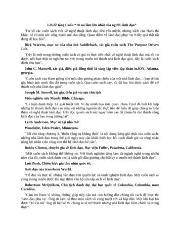
Darwanto et al. BMC Cancer (2017) 17:358Page 4 of 8No false negatives (FN; where an expected variant wasnot detected) were observed.Performance comparison between the QIAamp andGeneRead DNA FFPE kits for DNA purification using theGeneReaderTwo DNA purification kits were used to isolate DNA fromFFPE samples. Table 2 demonstrates the superior performance of the GeneRead DNA FFPE Kit (QIAGEN, Hilden,UK) over the QIAamp DNA FFPE Tissue Kit (QIAGEN,Hilden, UK) in terms of true positives at lower variant calling sensitivity. Fourteen true positive KRAS variants weredetected using an allele fraction cut-off of 5% for DNAisolated by GeneRead DNA FFPE Kit (QIAGEN, Hilden,UK). For the QIAamp DNA FFPE Tissue Kit (QIAGEN,Hilden, UK), 15 KRAS variants were detected using anallele fraction cut-off of 5%. Of the 15 KRAS variantsdetected, 14 were true positive variants and 1 was a falsepositive (Table 2) as confirmed by several independentmethods. Decreasing the allele fraction cutoff to 2.5% resulted in identification of the same 14 KRAS true positivesamples for GeneRead DNA FFPE Kit (QIAGEN, Hilden,UK) extractions. However, for QIAamp DNA FFPE TissueKit (QIAGEN, Hilden, UK) extracted samples at 2.5%allele fraction cut-off, 11 additional false positive KRASmutations (25 variants in total) were detected. Theadditional mutations were mostly C to T transitions. It isknown that FFPE fixation deaminates certain bases, mostprominently cytosine deamination to uracil [23–25]. TheGeneRead DNA FFPE Kit (QIAGEN, Hilden, UK) containsan integrated uracil DNA glycosylase (UDG) step whichremoves uracil from the DNA before the final purificationstep, yielding high-quality DNA with minimal artifacts.Table 3 KRAS agreement study between GeneReader andPyrosequencing and Therascreen PCR Assays 5% KRAS variant allelefrequency cut offPyrosequencing andTherascreen PCR Assays(a)GeneReader NGS 3(a)If KRAS is mutant by Therascreen KRAS RGQ PCR assay or Therascreen RASextension Pyrosequencing assay, the condition is recorded as a mutant (MT)(b)For Actionable Insights Tumor Panel, a 5% allelic frequency cut off was usedto call variants for codon 12, 13, 59, 61, 117 and 146, which are addressed byestablished QIAGENTherascreen PCR assaysThe use of the NA12878 control (Fig. 1, Additionalfile 2: Table S2) and AcroMetrix (Fig. 1, Additionalfile 3: Tables S3) reference standard materials demonstrated the good performance of the GeneReaderplatform on high frequency and low frequency variants,respectively. NA12878 has been used extensively as a reference standard material for verifying NGS platforms [17]and acts as a useful control in establishing backgrounderror. Besides its use as a GeneReader platform performance standard, AcroMetrix has also been used previouslyas a control for variant calls [26].DiscussionA major advantage of NGS over traditional mutationdetection methods is the ability to sequence multiplegenes and variants simultaneously. Other advantagesinclude minimal DNA input, faster turnaround time;Table 4 The concordance study between GeneReader, MiSeq,Pyrosequencingand Therascreen PCR assaysSampleno.KRAS AAchangeKRAS variant allele fraction (%)Therascreen PCR/PyroGeneReaderaMiSeqa1G12D 2172G12D 39123A59T1915144G13D 41155G12D K117 N243439Table 2 The GeneReader FFPE DNA sample preparation kitsuccessfully corrects FFPE artifacts11G13D 471512G12C 3210Type of DNA purification kitAllele frequency cut off13G13D 3910 5% 2.5%14Q61H262123QIAamp FFPE DNA purification Kit1525GeneRead DNA FFPE Kit1414 : Variant identified by Therascreen PCR; allele fraction not availablea: Sample processed from different FFPE section with potentially differenttumor content and variant allele fractionConfirmation of variants by MiSeq, pyrosequencing andtherascreen qPCR assaysThe GeneReader NGS System variant calls demonstrated100% agreement with KRAS mutation status previouslydetermined by either pyrosequencing or therascreenqPCR (Table 3). Of the 43 samples, 14 tested positive forKRAS variants and 29 samples were confirmed as wildtype. The 5% allelic fraction cut-off was used to callKRAS variants for codons 12, 13, 59, 61, 117 and 146.The true positive variants observed by the GeneReaderNGS System share a 100% concordance with MiSeqIllumina (Table 4).
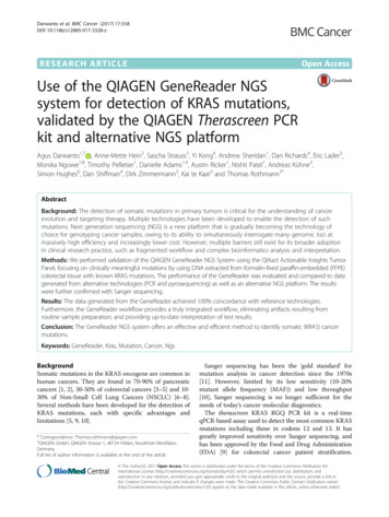
Darwanto et al. BMC Cancer (2017) 17:358Page 5 of 8Fig. 1 Variant calling performances of GeneReader pipeline. Each individual data point was generated from 18 data points (a) NA12878 and (b)AcroMetrix Oncology Hotspotlower overall cost and higher throughput and sensitivitycompared to traditional methods [12, 27–29]. NGS hasrevolutionized the speed of genetic and genomic discovery, and advanced our understanding of the molecularmechanisms of disease and potential treatment options.However, several major hurdles remain and still preventNGS from being broadly adopted in clinical practice.This is especially true for laboratories that are new tothis technology, and may lack the in-house expertiserequired for processing complex bioinformatics data andinterpretation of results. Such expertise is crucial toconstruct a bioinformatics pipeline and to evaluate thesoftware and generate quality reports. The QIAGENGeneReader NGS System allows users to performFig. 2 QCI Analyze report showing the alignment of the reads at the variant positions along with the induced amino acid change
Darwanto et al. BMC Cancer (2017) 17:358experiments from sample to insight, tissue sample todecipherable report based on the interpretation ofsequence variants detected.The QIAGEN GeneReader NGS workflow utilizes ‘QCIAnalyze’ and ‘QCI Interpret’ for bioinformatics analysis andreporting of variants, including read mapping, variant calling and interpretation of results. It provides visualization ofthe alignment of sequencing results (Fig. 2) as well as asummary of the data. Quality assessment is also supported,both at the overall sequencing run level and for the analyticvalidity of individual variants to reduce false positive andnegative results. Using the data visualization tools withinQCI Analyze, it is possible to determine the quality of theresults and assess any variants of interest. Further analysisof variants using QCI Interpret provides access to thecurated information contained within the QIAGEN Knowledge Base enabling a deeper analysis and interpretation ofresults for each sample (Fig. 3). With all relevant information, a report can be created with a summary of findingsand direct links to evidence sources. At the single variantlevel the QCI software is able to identify an individual variant as an actionable cancer mutation, and provides links tocurrent clinical research insights, e.g. the KRAS G12Dsomatic variant it is established to confer resistance to thecolorectal cancer drugs cetuximab and panitumumab,based on evidence curated from their FDA drug labels andclinical practice guidelines. Within QCI-Interpret information on active clinical trials recruiting colorectal cancerpatients with particular mutations are provided with drug,nearest location, and trial phase information.Page 6 of 8The relationship between FFPE DNA quality and sequencing accuracy is a critical point for any sequencinganalysis. The GeneReader workflow starts with the GeneRead FFPE DNA Kit for DNA extraction and is specificallydesigned to reduce artifacts known to commonly occur inFFPE treated samples. As seen in Table 2, by using FFPEsamples aged from 3 to 20 years, the GeneRead FFPEDNA Kit successfully reduced the number of low frequency false positive variants detected. These low frequency false positive variants are likely caused by cytosinedeamination and other fixation associated artifacts. Similarphenomena were observed by Bourgon [23], where pretreatment of FFPE samples with uracil DNA glycosylase(UDG) resulted in a dramatic reduction of false positives,with overall reductions of 77% for C T and 94% forG A changes, respectively. Biochemical removal of deaminated DNA eliminates deamination-associated falsepositive results; however, for samples with very low qualityDNA such as highly fragmented FFPE treated samples,UDG-treated may constitute an issue, as the treatment introduces possible further strand breaks leading to evenhigher fragmentation and lower availability of intact template strands. Therefore, using the QuantiMIZE assay toidentify those samples suitable for sequencing, based onan assessment of original intact and amplifiable templates,before starting an experiment is a critical point for anamplification based NGS technology. Previous reports observed that samples with lower amounts of amplifiableDNA are more likely to give a markedly increased numberof false positive results [30, 31].Fig. 3 QCI Interpret actionable report, showing summary of findings and link to the insights that can be used to guide clinical research
Darwanto et al. BMC Cancer (2017) 17:358ConclusionsIn summary, this study confirms that the GeneReaderNGS System performs consistently and accurately in theidentification of somatic mutations from FFPE samples,with results confirmed by both alternative technologiesas well as an alternative NGS platform. With a full endto-end solution with integrated sample preparation andbioinformatics interpretation, the GeneReader NGSSystem is suitable for any laboratory interested in cancerclinical research.Additional filesAdditional file 1: Table S1. The QC results of the extracted DNAsamples were measured using GeneRead DNA QuantiMIZE. (DOCX 28 kb)Additional file 2: Table S2. List of NA12878 Gold Standard Variantsfrom 18 samples sequenced by GeneReader. (DOCX 27 kb)Additional file 3: Table S3. List of AcroMetrix Oncology HotspotGold Standard Variants from 18 samples sequenced by GeneReader(DOCX 28 kb) (DOCX 27 kb)AbbreviationsCt: Cycle threshold; DNA: Deoxyribonucleic acid; dsDNA: Double-strandedDNA; FDA: Food and Drug Administration; FFPE: Formalin-fixed paraffinembedded; GR: GeneReader; NGS: Next generation sequencing; NSCLC: Nonsmall cell lung cancer; PCR: Polymerase chain reaction; qPCR: QuantitativePCR; QC: Quality control; QCI: QIAGEN clinical insight; TAT: Turn-around time;UDG: Uracil-DNA glycosylaseAcknowledgementsWe greatly appreciate Drs. Scott Steelman, Kathleen Steinmann and RobertLintner from Broad Technology Labs (Broad Institute of MIT and Harvard, 75Ames Street (Rm 8021), Cambridge, MA 02141) for their support in samplesprocessing, GeneReader testing, and manuscript revisions. We thanks to Drs.Vikas Gupta, Naomi Thompson Kiran Divakar and Dietrich Lueerssen fromQIAGEN for the input of the manuscript.FundingThis work was supported by QIAGEN.DisclaimerThe sequencing chemistry used in this manuscript is currently available outsidethe US. The GeneReader NGS System is for research use only. An upgraded anddifferent sequencing chemistry has meanwhile been made available in the USsince April 2017 and will become available worldwide later in 2017. For therelease of the new sequencing chemistry in the US in April, we have shownequivalency to the sequencing chemistry used in the manuscript.Availability of data and materialsThe analyzed data sets generated during the study are available from thecorresponding author on reasonable request.Authors’ contributionsAD, AMH, SS, AS, EL, SH, TR designed the study. AD, AMH, SS, AS, DR, MN, TP,DA, AR, NP, DS, TR performed experiments and analyzed data. AD, YK and TRwrote the manuscript. AK, DS, DZ, KK assisted in preparing the manuscript.All authors read and approved the final manuscript.Competing interestsAt the time of the work was being done the authors where employees ofQIAGEN, and QIAGEN funded the research and the publication costs. Wedeclare that our current or previous employment with QIAGEN did notinfluence our interpretation of data or presentation of information.Consent for publicationNot applicable.Page 7 of 8Ethics approval and consent to participateFFPE Tumor material from colorectal cancer tumors (Origene Technologies,MD, USA and Asterand Biosciences, MI, USA). We refer to Origene’s andAsterand’s quality system as well as ethics and compliance processes withinformed consent for commercial clinical samples. We expect Origene andAsterand to follow industry standard ethics approval and consent processes.Publisher’s NoteSpringer Nature remains neutral with regard to jurisdictional claims inpublished maps and institutional affiliations.Author details1QIAGEN Waltham, 35 Gatehouse Dr, Waltham, MA 02451, USA. 2QIAGENArhus, Silkeborgvej 2, 8000 Aarhus, Denmark. 3QIAGEN GmbH, QIAGENStrasse 1, 40724 Hilden, Nordrhein-Westfalen, Germany. 4QIAGEN RedwoodCity, 1700 Seaport Blvd, Redwood, CA 94063, USA. 5QIAGEN Frederick, 6951Executive Way, Frederick, MD 21703, USA. 6QIAGEN Manchester, SkeltonHouse Lloyd Street North, Manchester M15 6SH, UK. 7Novartis Institutes forBioMedical Research, Cambridge, MA 02139, USA. 8T2 Biosystems, Lexington,MA 02421, USA. 9Macherey-Nigel, Bethlehem, PA 18020, USA.Received: 28 November 2016 Accepted: 5 May 2017References1. Di Marco M, Astolfi A, Grassi E, Vecchiarelli S, Macchini M, Indio V, Casadei R,Ricci C, D’Ambra M, Taffurelli G, Serra C, Ercolani G, Santini D, D’Errico A,Pinna AD, Minni F, Durante S, Martella LR, Biasco G. Characterization ofpancreatic ductal adenocarcinoma using whole transcriptome sequencingand copy number analysis by single-nucleotide polymorphism array. MolMed Rep. 2015;12:7479–84.2. Huang J, Löhr JM, Nilsson M, Segersvärd R, Matsson H, Verbeke C, Heuchel R,Kere J, Iafrate AJ, Zheng Z, Ye W. Variant profiling of candidate genes inpancreatic Ductal Adenocarcinoma. Clin Chem. 2015;61:1408–16.3. Gao J, Wu H, Wang L, Zhang H, Duan H, Lu J, Liang Z. Validation oftargeted next-generation sequencing for RAS mutation detection in FFPEcolorectal cancer tissues: comparison with Sanger sequencing andARMS-scorpion real-time PCR. BMJ Open. 2016;6:e009532.4. Sakai K, Yoneshige A, Ito A, Ueda Y, Kondo S, Nobumasa H, Fujita Y, Togashi Y,Terashima M, De Velasco MA, Tomida S, Nishio K. Performance of a novel KRASmutation assay for formalin-fixed paraffin embedded tissues of colorectalcancer. Spring. 2015;4:7.5. Sundström M, Edlund K, Lindell M, Glimelius B, Birgisson H, Micke P, Botling J.KRAS analysis in colorectal carcinoma: analytical aspects of pyrosequencingand allele-specific PCR in clinical practice. BMC Cancer. 2010;10:660.6. Casadio C, Guarize J, Donghi S, Di Tonno C, Fumagalli C, Vacirca D, Dell’Orto P,De Marinis F, Spaggiari L, Viale G, Barberis M. Molecular testing for targetedtherapy in advanced non-small cell lung cancer: suitability of Endobronchialultrasound Transbronchial needle aspiration. Am J Clin Pathol. 2015;144:629–34.7. Papadopoulou E, Tsoulos N, Tsirigoti A, Apessos A, Agiannitopoulos K,Metaxa-Mariatou V, Zarogoulidis K, Zarogoulidis P, Kasarakis D, Kakolyris S,Dahabreh J, Vlastos F, Zoublios C, Rapti A, Papageorgiou NG, Veldekis D,Gaga M, Aravantinos G, Karavasilis V, Karagiannidis N, Nasioulas G.Determination of EGFR and KRAS mutational status in Greek non-small-celllung cancer patients. Oncol Lett. 2015;10:2176–84.8. Lièvre A, Bachet JB, Boige V, Cayre A, Le Corre D, Buc E, Ychou M, Bouché O,Landi B, Louvet C, André T, Bibeau F, Diebold MD, Rougier P, Ducreux M,Tomasic G, Emile JF, Penault-Llorca F, Laurent-Puig P. KRAS mutations as anindependent prognostic factor in patients with advanced colorectal cancertreated with cetuximab. J Clin Oncol. 2008;26:374–9.9. Rodriguez R. Biomarker testing for treatment of metastatic colorectal cancer:role of the pathologist in community practice. J Community Support Oncol.2014;12:27–32.10. Martinez DA, Ne
variant positions in 12 genes (KRAS, NRAS, KIT, BRAF, PDGFRA, ALK, EGFR, ERBB2, PIK3CA, ERBB3, ESR1 and RAF1). All steps of library preparation were per-formed according to the manufacturer's protocol. The libraries were then quantified using a Qubit dsDNA HS Assay Kit (Life Technologies, MA, USA) and QIAxcel (QIAGEN, Hilden, Germany).