Transcription
REVIEWSThe sympathetic control ofblood pressurePatrice G.GuyenetAbstract Hypertension — the chronic elevation of blood pressure — is a major human healthproblem. In most cases, the root cause of the disease remains unknown, but there is mountingevidence that many forms of hypertension are initiated and maintained by an elevatedsympathetic tone. This review examines how the sympathetic tone to cardiovascular organsis generated, and discusses how elevated sympathetic tone can contribute to hypertension.PreganglionicAutonomic neurons that havetheir cell bodies in thebrainstem or spinal cord andsynapse onto visceral motorneurons (sympathetic orparasympathetic) in peripheralganglia.Department ofPharmacology, HealthSciences Center, University ofVirginia, 1300 Jefferson ParkAvenue, Charlottesville,Virginia 22908-0735, USA.e-mail: pgg@virginia.edudoi:10.1038/nrn1902The autonomic nervous system is a collection of afferent and efferent neurons that link the CNS with visceraleffectors1,2. The two efferent arms of the autonomicnervous system — the sympathetic and parasympatheticarms — consist of parallel and differentially regulatedpathways made up of cholinergic neurons (preganglionicneurons) located within the CNS that innervate ganglia(for example, para- or pre-vertebral sympathetic ganglia), glands (adrenal glands) or neural networks of varying complexity (enteric or cardiac ganglionic networks)located outside the CNS1,2. These peripheral ganglia andnetworks contain the motor neurons (ganglionic neurons) that control smooth muscles and other visceraltargets. The sympathetic ganglionic neurons that controlcardiovascular targets are primarily noradrenergic2.Blood pressure (BP) fluctuates substantially withbehaviour, but the 24-h average BP is tightly regulated.Hypertension is, by definition, a chronic elevation of the24-h average BP, and the disease is known as neurogenicif the probable cause is an abnormality of the autonomicnervous system rather than a primary vascular or renaldefect. This abnormality can originate in the afferentarm of the system (for example, baroreceptors, chemoreceptors and renal afferents) or in the central circuitry.The neural control of the circulation operates viaparasympathetic neurons that innervate the heart andvia three main classes of sympathetic efferent — barosensitive, thermosensitive and glucosensitive cardiovascular — that innervate blood vessels, the heart, thekidneys and the adrenal medulla. The barosensitivesympathetic efferents are under the control of arterial baroreceptors. This large group of efferents has adominant role in both short-term and long-term BPregulation. Their level of activity at rest is presumed tobe the most crucial parameter for long-term BP control.This background activity is set by a core network ofneurons that reside in the rostral ventrolateral medullaNATURE REVIEWS NEUROSCIENCE(RVLM), the spinal cord, the hypothalamus and thenucleus of the solitary tract (NTS). These structures arethe primary focus of this review (FIG. 1). Limbic, corticaland midbrain structures (not discussed in this review)are responsible for the rapid changes in sympathetictone that relate to behaviour. It is generally assumedthat these changes are not pertinent to the long-termregulation of BP, except perhaps in the context of stressrelated hypertension.I begin by discussing the determinants of BP andthe cardiovascular sympathetic efferents that controlit. The three central control regions — the RVLM,NTS and hypothalamus — that regulate the barosensitive sympathetic efferents, and consequently BP, aredescribed, together with their potential contributionto various forms of neurogenic hypertension.Determinants and neural control of BPBP is a function of vascular resistance and cardiac output, two variables that are controlled by the autonomicnervous system. In turn, cardiac output is dependent onthree regulated variables: end-diastolic volume; myocardial contractility; and heart rate. End-diastolic volume isthe volume reached by the ventricular chamber beforecontraction and is determined by venous pressure, whichis related to blood volume and venous smooth muscletone, both of which are under sympathetic control.Myocardial contractility and heart rate are regulated byboth the sympathetic and parasympathetic divisions ofthe autonomic nervous system.On a short timescale (seconds to hours), the autonomic nervous system adjusts the circulation in keepingwith behaviour (for example, feeding and exercise), theenvironment (for example, thermoregulation) and emotions (for example, fright)1. These circulatory changesare components of more global autonomic responsepatterns that are elaborated in large portions of theVOLUME 7 MAY 2006 335
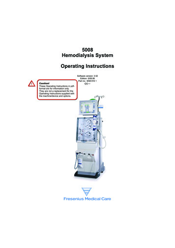
REVIEWSAng IIAng IIRegions of brainrenin–angiotensinsystem activityAldosteroneNa SFO,OVLTAP?CardiopulmonarymechanoreceptorsCO2, O2, ionsand cytokinesTissuemetabolitesNTSNa s,O2, pHSGNTo heart, arteriolesand kidneysAng IIFigure 1 CNS network that regulates the basal sympathetic tone. The backgroundlevel of sympathetic tone present at rest is presumably crucial for long-term bloodpressure (BP) control. The network that sets this background level is located in therostral ventrolateral medulla (RVLM), the spinal cord, the hypothalamus and the nucleusof the solitary tract (NTS). Limbic, cortical and midbrain structures (not representedhere) are responsible for rapid behaviour-related adjustments of sympathetic tone butare probably not involved in the long-term regulation of BP, except perhaps in thecontext of stress-related hypertension. The core sympathetic network is regulated bymany classes of sensory afferent that project either to the NTS (for example,baroreceptors and other mechanoreceptors from the cardiopulmonary region) or to thespinal cord (somatic and sympathetic afferents that detect a range of chemical orphysical parameters from muscle stretch to tissue hypoxia and metabolites). The centralportion of the network is also regulated at multiple levels by circulating hormones andblood-borne factors. Peptide hormones (for example, angiotensin II (ang II)) andcytokines (for example, interleukin-1) influence this network via circumventricularorgans (subfornical organ (SFO), organum vasculosum lamina terminalis (OVLT) and areapostrema (AP)) or through endothelial receptors that trigger the release of mediatorsthat subsequently cross the blood–brain barrier (for example, nitric oxide andprostaglandins41,75). These transendothelial mechanisms operate in the hypothalamus,the RVLM and the NTS. Freely diffusible hormones (for example, ouabain-likesubstance115 and aldosterone) also act on this network, but their sites of action in thebrain are not conclusively known104,113. The central network also responds to changes insodium and osmolality that are detected at multiple hypothalamic sites, to carbondioxide (CO2) via brainstem chemoreceptors, and could detect hypoxia directly in thebrainstem. Moreover, virtually every component of the central network is influenced bythe brain renin–angiotensin system through increased production of radical oxygenspecies and, possibly, other mechanisms8,119. Finally, the sympathetic ganglia are alsoinfluenced by hormones, such as angiotensin II, and transmitter release by sympatheticganglionic neurons (SGNs) is regulated presynaptically by angiotensin II andcatecholamines. SPGN, sympathetic preganglionic neuron. Black arrows indicateexternal effect; green arrows show interactions within the network.BaroreflexReflex decrease in sympatheticnerve activity that is initiatedby the activation of stretchsensitive afferents located inthe arterial wall.336 MAY 2006 VOLUME 7midbrain, limbic forebrain and cortex3–5. They occurvia rapid changes in cardiac output and regional arteriolar resistance, and can be associated with substantialBP increases that are, in most instances, physiologicallyadaptive, thereby facilitating gas and nutrient exchangein metabolically active tissues (for example, muscles during exercise). Behaviour-dependent rises in BP are bothenabled and moderated by the baroreflex.Numerous brain manipulations (including lesions,overexpression of nitric oxide synthase and brain-specificexpression of various components of the renin–angiotensinsystem) produce long-term changes in mean BP6–8, therebydemonstrating that the CNS normally contributes to thelong-term regulation of BP. The fact that renal denervationor specific brain lesions attenuate or delay the developmentof hypertension9,10 also indicates that the CNS contributesto the hypertensive process. However, the exact role ofthe CNS in long-term BP control is not well understood.From a neurophysiological perspective, the most fundamental and still unanswered question is whether the brainis a controller of BP in the strict engineering sense (thatis, has the capacity to detect changes in BP and to initiateappropriate responses)11,12. How a set-point for BP mightbe encoded by the CNS and the nature of the error signalshave yet to be established. The only well identified neuralsensors that encode BP are the baroreceptors, but theircontribution to the long-term regulation of BP has beenrepeatedly questioned (discussed later)11,12. Numeroushumoral factors (for example, sodium, angiotensin IIand mineralocorticoids) alter the activity of the centralautonomic network via neural mechanisms that arebeing described in ever greater detail. However, evidencethat these substances provide error signals for a CNS BPcontroller is tenuous12. Indeed, it could be argued that theneural control of the circulation is primarily designed toregulate blood volume and blood flow (cardiac output andits apportionment) at the expense of BP.Any discussion of neurogenic hypertension must consider the role of the kidneys. The influential model developed by Guyton postulates that the relationship betweenrenal sodium excretion and BP (the pressure–natriuresisrelationship) defines the BP homeostatic set-point13.According to this model, any increase in sodium retention produces an initial blood volume expansion, causing BP to increase via a rise in cardiac output. Eventually,tissue over-perfusion leads to an increase in peripheralresistance (whole-body autoregulation) that returns resting cardiac output towards normal13. According to thiswidely held theory, a resetting of the pressure–natriuresisrelationship inevitably leads to hypertension, regardlessof the cause of the resetting, whether it be humoral, neural, degenerative or genetic.Although evidence that the brain regulates the 24-haverage BP and contributes to the hypertensive process isvery persuasive, the mechanisms are not well understood.Elevated sympathetic nerve activity (SNA) is present inmost forms of human hypertension14 (FIG. 2) and a causalrelationship is suggested by the well-documented antihypertensive efficacy of sympatholytic drugs (for example,α1- or β-adrenergic receptor antagonists)15. However,elevated SNA might not be the sole mechanism involvedin neurogenic hypertension, and how an increase in SNAraises the 24-h mean BP has not been established. Themost commonly invoked mechanism is resetting of therenal BP–natriuresis relationship to higher levels of BPby either a rise in sympathetic tone to the kidney or byhormones whose production is partly controlled by theautonomic nervous system (for example, angiotensin II).However, abnormalities in the neural control of the heartand blood vessels are not ruled out9,13,16,17.Sympathetic efferents that regulate BPCardiovascular sympathetic efferents can be broadlyclassified into three groups according to their dominantcharacteristic: thermosensitivity; glucosensitivity; orbarosensitivity18–20. This section describes the generalcharacteristics of each group, with a focus on thephysiological properties of the barosensitive efferents.www.nature.com/reviews/neuro
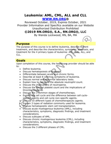
REVIEWSabDorsalrootganglionNormotensive Hypertensive(107/59 mm Hg) (148/102 mm Hg)Spinal cordPeripheralnerveEKGMSNAMSNA150BP(mm Hg)Ventral chainSkeletalmuscleSensory afferentBloodvesselSympatheticganglionic neuronMotor neuronSympatheticpreganglionic neuron50c100p 0.01MSNA (bursts/100 heart beats)Skinbranch806040200NTEHFigure 2 Sympathetic tone and hypertension. a Sympathetic nerve activity (SNA) can be measured directly in awakehumans by the insertion of a metal electrode into a somatic nerve under conditions in which sensory and skeletomotornerve activity are negligible21. MSNA, muscle SNA. b,c Show examples of multifibre recordings from the peroneal nerve ofnormotensive (NT) and hypertensive participants, representing the resting level of activity of sympathetic postganglionicneurons that innervate muscle resistance arterioles14. MSNA represents the activity of a fairly homogeneous functionalclass of sympathetic efferent that is subject to a powerful feedback from arterial baroreceptors and has a central role inblood pressure (BP) homeostasis. Barosensitive sympathetic efferents innervate the kidneys, the heart, resistancearterioles and capacitance veins throughout the body (except in the skin). Their discharge occurs in bursts, because thepulsatile nature of arterial baroreceptor activity is transmitted polysynaptically through the entire brainstem baroreflexcircuitry1. MSNA also fluctuates with respiration owing to feedback from other cardiopulmonary afferents and the factthat the autonomic circuits in the brainstem receive inputs from the central respiratory network19,23. The intensity andfrequency of MSNA bursts is elevated in human essential hypertension (EH), several other forms of hypertension14 (such asthose in obesity or obstructive sleep apnoea) and in many other pathological conditions (for example, heart failure,haemorrhage and dehydration). The increased burst frequency is clearly of CNS origin. The increased intensity of thebursts is probably also of central origin but could conceivably be due, in part, to hormone-induced changes in ganglionictransmission (FIG. 1). Panels b and c adapted, with permission, from REF. 14 (2004) American Heart Association. EKG,electrocardiogram.Renin–angiotensin systemThis is a regulated biochemicalpathway with paracrinefunction that leads to theproduction of angiotensin IIand related bioactive peptidesin the brain. This system isactive in most brain regionsthat regulate the sympatheticoutflow and is activated invarious forms of hypertensionand heart failure, although thecauses of its activation are stillnot clear.NatriuresisSodium excretion by thekidney.SympatholyticA drug that reduces SNA by aCNS or peripheral action orreduces transmission betweensympathetic ganglionicneurons and their peripheraltargets.Classes of cardiovascular efferents. The thermosensitivegroup of cardiovascular efferents consists primarily ofcutaneous vasoconstrictors that are activated by hypothermia, emotional stimuli and hyperventilation19,21. Theglucosensitive group controls adrenaline release from theadrenal medulla and is activated by hypoglycaemia andphysical exercise22. These two types of cardiovascularefferent are only weakly, if at all, regulated by arterialbaroreceptors, and presumably have a secondary role inshort- and long-term BP stability.The third class, which is by far the largest group ofcardiovascular sympathetic efferents, is the barosensitivegroup. Regardless of the organ or tissue that they innervate,these neurons show ongoing activity at rest (sympathetictone) and they discharge in bursts that are highly synchronized with the arterial pulse and respiration19,21,23 (FIG. 2b).Barosensitive sympathetic efferents control the heart andthe kidneys, the release of noradrenaline from a subset ofadrenal chromaffin cells, and constrict resistance arterioles, with the exception of those in the skin19. Barosensitiveefferents are responsible for short-term BP fluctuations1,19.They are also likely to be a key determinant of theNATURE REVIEWS NEUROSCIENCElong-term neural control of BP, in part because renin secretion, renal tubular sodium reabsorption and renal bloodflow are apparently all under the control of this type ofsympathetic efferent9.Properties of barosensitive efferents. The physiological properties of barosensitive sympathetic efferentsare fairly uniform and have been thoroughly characterized from recordings in anaesthetized or awakeanimals and from numerous recordings of ganglionicneurons in awake humans19,21,23 (FIG. 2). Barosensitiveefferents are subject to numerous reflex regulationsthat operate as either feedback or feedforward mechanisms19,24. For example, the activation of stretch-sensitive afferents by ventilation (lung afferents) and arterialpressure (carotid and aortic receptors) inhibits SNA.By contrast, muscle receptors (group III and IV) thatare activated by stretch and metabolites (for example,ATP, lactate and pH) raise the discharge of barosensitive sympathetic fibres during exercise25. The activation of visceral nociceptors (for example, by angina)or cutaneous nociceptors elevates the activity ofVOLUME 7 MAY 2006 337
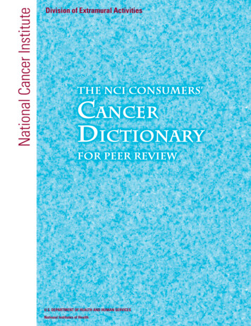
REVIEWSbarosensitive sympathetic efferents, as does the activationof peripheral (by hypoxia or hypercapnia) and central(by hypercapnia) chemoreceptors19,26. Barosensitive sympathetic fibres are activated by mental stress and in manydisease states1,19,21. On the basis of recordings made whenanimals were anaesthetized and awake, the response MCVLMCPARVLMA5To heart, arteriolesand kidneysSpinal cordcBaroreceptorsNTSPonsVRC1 mmRVLMInhibitory inputs(for example,containing GABAand glycine)Excitatory inputs(for example,containingglutamate)Mixed or ympatheticneuronTo heartPostganglionicparasympathetic neuronTo heart,arterioles andkidneysFigure 3 The rostral ventrolateral medulla and barosensitive sympatheticefferents. a All sympathetic preganglionic neurons (SPGNs), regardless of theirfunction, receive monosynaptic inputs from overlapping subsets of neurons located ineach of the regions indicated30,36. The extent to which each of these regions contributesto the activity of the barosensitive system of sympathetic efferents probably depends onthe physiological state and the type of sympathetic efferents. The rostral ventrolateralmedulla (RVLM) is the dominant source of excitatory drive to the barosensitive class ofsympathetic efferent under anaesthesia. Its role is assumed, but not proved, to be equallydominant in the awake state. The RVLM input originates from a neurochemicallyheterogeneous collection of glutamatergic neurons, a large subset (70%) of which alsosynthesize adrenaline. These are called C1 neurons30,33,36. Spinal interneurons areconsidered unimportant in regulating barosensitive efferents in intact mammals, butbecome dominant after spinal cord damage. b RVLM barosensitive neurons receiveinputs from multiple areas of the brain and spinal cord. Only a few of the inputs from themedulla oblongata are represented. These inputs presumably mediate some of the manycardiovascular reflexes that are integrated by the RVLM neurons. c Anatomically correctlocation of the RVLM and caudal ventrolateral medulla (CVLM): the parasagittal sectionof the rat medulla oblongata 1.8 mm lateral to the midline. RVLM barosensitive neuronsinnervate numerous pontomedullary regions in addition to SPGNs. This fact issymbolized by a collateral to the dorsal pons. The RVLM and CVLM are both coextensivewith the ventral respiratory column (VRC; outlined in blue). The cholinergicparasympathetic neurons that control the heart are also located in the same region.Parasympathetic neurons and the barosensitive RVLM neurons receive inputs fromunidentified VRC neurons that coordinate respiration and circulation. A5, noradrenergiccluster located at the pontomedullary junction; CPA, caudal pressor area; Lat. hyp.,lateral hypothalamus; LTF, lateral tegmental field; NTS, nucleus of the solitary tract; PVH,paraventricular nucleus of the hypothalamus; RVMM, rostral ventromedial medulla;GABA, γ-aminobutyric acid.338 MAY 2006 VOLUME 7barosensitive efferents to the above-mentioned list ofstimuli or physiological conditions is typically in thesame direction but variable in intensity depending onthe organ targeted by these neurons. An importantexception is the selective inhibition of renal SNA byatrial stretch or volume expansion, a reflex that is crucial for the regulation of blood volume27,28. Contrary toprevious assumptions, a decrease in barosensitive muscleSNA does not contribute to muscle vasodilation duringexercise. Reflexly, and through central command, musclesympathetic tone actually increases monotonically withthe level of exercise, possibly to curb the hypotensionthat might otherwise result from excessive vasodilationdue to local metabolites21,25.In summary, barosensitive sympathetic efferentsare regulated in parallel under most circumstances,but target-specific differences in their level of activityshow that these efferents are, to some extent, differentially regulated. The selective control of renal SNA byvolume receptors could be the most important of thesedifferential regulations.The rostral ventrolateral medullaAlthough anatomical experiments suggest that everysympathetic preganglionic neuron (SPGN) receivessome synaptic input from the same general areas of thespinal cord, medulla oblongata and hypothalamus29–31(FIG. 3a), physiological evidence indicates that these CNSregions contribute unequally to the various sympatheticoutflows. Barosensitive sympathetic efferents appear tobe regulated primarily through the RVLM24, whereasthe cutaneous circulation is regulated predominantlythrough the rostral ventromedial medulla (RVMM) andmedullary raphe19,20,24. The central control of adrenalinesecretion is less well understood. Although not underbaroreceptor control, it is regulated, at least in part, bythe RVLM22,32. The next sections focus on the anatomyof the RVLM, its role in regulating the activity of thebarosensitive sympathetic efferents and its potential rolein neurogenic hypertension.C1 and other RVLM BP-regulating neurons. The C1neurons (FIG. 3) are, by definition, one of only three clusters of adrenaline-synthesizing cells in the CNS33. In theearly 1980s, the RVLM — the portion of the ventrolateralmedulla that is coextensive with C1 neurons (FIG. 3b,c)— was definitively identified as a key BP regulatory centre1,24,34. The RVLM neurons that are most directly linkedto BP control are cells that innervate SPGNs monosynaptically (FIG. 3). These neurons have a discharge pattern that is similar to that of barosensitive sympatheticefferents and they are a nodal point for most, if not all,sympathetic reflexes that involve cardiovascular targets, with the exception of cutaneous arterioles1,20,35–37.All these RVLM neurons probably release glutamate,but they also synthesize various additional combinations of transmitters, including adrenaline. Those thatsynthesize adrenaline ( 70%) belong, by definition, tothe C1 group34,38,39. However, not all C1 cells are underbaroreceptor control; the best-documented exampleof non-barosensitive C1 cells is those that controlwww.nature.com/reviews/neuro
REVIEWSadrenaline-releasing chromaffin cells22,32. Furthermore,neither RVLM barosensitive neurons nor the C1 cellsshould be viewed strictly as ‘central sympathetic neurons’ because these cells, as well as innervating SPGNs,also innervate many regions of the medulla, pons andmidbrain36.The RVLM also contains C1 cells that innervate thehypothalamus. These neurons are different from thosethat innervate the spinal cord, but they have a range ofneurochemical and electrophysiological properties thatare similar to those of their bulbospinal counterparts40.Some of these cells presumably contribute a baroreceptor-modulated excitatory drive to the hypothalamiccentres (paraventricular and median preoptic nuclei)that regulate aspects of circulation, including sodiumand water balance. Other C1 cells are probably not underbaroreceptor control40 and mediate, or at least enable,the activation of the hypothalamic–pituitary axis duringa range of physical stresses that is clearly not limited tocardiovascular challenges32,41.BulbospinalNeurons located in thebrainstem and innervatingneurons in the spinal cord,such as sympatheticpreganglionic neurons.Sympathoexcitatory reflexAny reflex that causes anincrease in SNA (the oppositeis a sympathoinhibitory reflex).Vigilance-regulatingnetworkNetwork of neurons thatregulate the sleep–wake cycle.This network includes thesuprachiasmatic and otherhypothalamic nuclei andvarious brainstem aminergiccell groups.RVLM and sympathetic vasomotor tone. A backgroundlevel of SNA that can be either withdrawn or enhancedis required for the short- and long-term stabilization ofBP. As this background level is largely determined bythe level of activity of RVLM barosensitive neurons, theintrinsic properties and inputs of these cells are centralto understanding sympathetic tone and its pathological abnormalities. Under most anaesthetic conditions,ionotropic glutamate transmission is a minor source ofdrive for barosensitive neurons36,42. However, glutamatetransmission makes a much greater contribution to theactivity of these neurons in animals that are dehydratedor have abnormal blood gases (that is, high CO2 and lowO2), or when any of a large number of sympathoexcitatoryreflexes are elicited36,37,43,44. In short, the activity of RVLMneurons appears to depend on ionotropic drives andmetabotropic transmission (for example, neuropeptides;discussed below) in proportions that vary according tothe physiological circumstances.In brain slices, C1 neurons have beating propertiesthat rely to some extent on a persistent sodium current45.Dissociated C1 neurons are not spontaneously active,which suggests that their autoactivity in slices relies inpart on dendritic properties or requires unidentifiedextracellular signals46. So, whether autoactivity contributes to the discharge of the barosensitive neurons,and therefore to basal vasomotor tone in vivo, has yetto be determined42. Besides GABA (γ-aminobutyricacid) and glutamate, the list of transmitters that regulatethe barosensitive neurons is extensive. Acetylcholine,serotonin, corticotropin-releasing factor (CRF), oxytocin, substance P, vasopressin and orexin have all beenidentified in nerve terminals that synapse onto identified or presumed BP-regulating neurons (usually C1cells)36. Some of these inputs (for example, acetylcholine,serotonin and orexin) probably originate from vigilanceregulating networks and could contribute to the circadianrhythm of SNA and BP16. Other inputs originate from thehypothalamus (for example, vasopressin, oxytocin, CRFand angiotensin II) and have a role in the cardiovascularNATURE REVIEWS NEUROSCIENCEresponse to internal (for example, infection, dehydration, haemorrhage and heart failure) and external (forexample, social) stresses36,43,47–50.RVLM neurons also receive inputs from numerous sources in the medulla oblongata and pons. Fewof these inputs are thoroughly characterized, with theexception of a GABA-mediated input from the caudalventrolateral medulla (CVLM) that is crucial to thebaroreflex1,51 (FIG. 3b,c). The remaining sources of inputhave been identified primarily as sites at which electrical or chemical stimulation elicits changes in BP: that is,the caudal pressor area; midline depressor area; varioussubnuclei of the NTS; and the gigantocellular depressor area1 (FIG. 3b). These brainstem regions are probablerelays for the various somatic and visceral sympatheticreflexes (exercise pressor reflex, nociceptive reflexes andcardiopulmonary reflexes) that are mediated, at least inpart, through the RVLM52–54. Other pontomedullaryareas probably serve as an interface between the central respiratory network and the sympathetic outflow,and are responsible for the stimulatory effect of centraland peripheral chemoreceptor activation on barosensitive SNA44 (FIG. 3c). The RVLM could also containinterneurons that regulate the barosensitive neurons,given the differential sensitivity of various sympatheticreflexes to the microinjection of pharmacological agentsinto the RVLM (for an example, see REF. 55).The organotopy hypothesis. The ‘organotopy’ theorystates that separate groups of RVLM barosensitive neurons preferentially control, for example, skeletal musclearteries, splanchnic arteries, the heart and the kidneys56–58.Anatomical studies have yet to provide convincing evidence in support of this hypothesis29,31,59,60, but there isphysiological evidence for some input–output diversityamong RVLM barosensitive neurons. The best evidencefor output diversity comes from RVLM microstimulation, which produces different activation of various sympathetic nerves, depending on the site of stimulation56–58.Input diversity is supported by unit recordings that showcell-specific responses to the intravenous injection ofcholecystokinin and the activation of central and peripheral chemoreceptors61,62, but these cells have a uniformresponse to many other stimuli. In any event, the targetspecific responses of barosensitive sympathetic efferentsare unlikely to be entirely due to differential recruitmentof RVLM barosensitive neurons. For example, directprojections from the paraventricular nucleus of thehypothalamus (PVH) to SPGNs probably contribute tothe selective control of renal SNA by volume receptors27.The scheme proposed in FIG. 4 is an attempt to reconcilethe contradictory evidence regarding the RVLM.RVLM and long-term BP control. Adenovirus-mediatedoverexpression of endothelial nitric oxide synthase(eNOS) in the RVLM leads to reductions in BP 5–10 daysafter injection of the viral vector, presumably by e
Sciences Center, University of Virginia, 1300 Jefferson Park Avenue, Charlottesville, Virginia 22908-0735, USA. e-mail: pgg@virginia.edu doi:10.1038/nrn1902 Preganglionic Autonomic neurons that have their cell bodies in the brainstem or spinal cord and synapse onto visceral motor neurons (sympathetic or parasympathetic) in peripheral ganglia.