Transcription
Kawasaki Medical Journal 47:69 75,2021doi:10.11482/KMJ-E20214706969〈Case Report〉A Case of Takotsubo syndrome after Surgeryfor Papillary Thyroid CancerYutaka YAMAMOTO1), Junichi KUREBAYASHI1), Hiroshi OKAMOTO2)Yuka MIKAMI3), Takuya MORIYA3), Yuna FUKUMA1), Tsuyoshi MIKAMI1)Shiori KAWANO1), Emi KISHINO1), Ryohei OGATA1), Wataru SAITO1)Yoshikazu KOIKE1), Mai SOHDA1), Tsunehisa NOMURA1), Katsuhiro TANAKA1)1) Department of Breast and Thyroid Surgery,2) Department of Cardiology,3) Department of Pathology, Kawasaki Medical SchoolABSTRACTThe patient was an 82-year-old woman undergoing treatment for bronchialasthma. In December 200X, she visited her former doctor because of a left cervical mass andpain, and was referred to our hospital for detailed examination. A 19-mm mass was found inthe left lobe of the thyroid gland and there were many swollen lymph nodes on both sides ofher neck. Fine-needle aspiration cytology revealed malignant and papillary cancer in both theleft lobe and left cervical lymph node, but no lung metastasis was found in chest CT, and itwas judged to be T1bN1bM0 Stage IVa. Total thyroidectomy bilateral cervival lymphnodedissection was performed. The left lobe mass of the thyroid gland invaded the sterno-thyroidmuscle, but no infiltration to other organs, including the lymph nodes on both sides, wasobserved. On the night of the operation, wheezing, dyspnea, lower mandibular breathing,and impaired consciousness suddenly developed, and the oxygen saturation of peripheralartery (SpO2) decreased to 60%. No postoperative bleeding was observed. Chest CTdemonstrated no signs of heart failure, but based on thickening of the bronchial wall, the causeof hypoxemia was considered to be bronchial asthma. Steroids and oxygen (high-dose, 15 L/min) were administered, but respiratory acidosis developed and non-invasive positive pressureventilation (NIPPV) was started. The respiratory condition gradually improved thereafter. Acutecoronary syndrome (ACS) was suspected based on symptoms, increased troponin T, and STelevation on ECG. Echocardiography revealed akinesis and left ventricular apical and basalhypercontractility. No coronary arterial stenosis was noted on coronary angiography and leftventriculography demonstrated Takotsubo-like wall movement (hypercontraction of the baseand contraction failure of the apex), leading to a diagnosis of Takotsubo syndrome (TS). TheCorresponding authorYutaka YamamotoDepartment of Breast and Thyroid Surgery, KawasakiMedical School, 577 Matsushima, Kurashiki, 701-0192,JapanPhone : 81 86 462 1111Fax : 81 86 462 1199E-mail: yamayou@med.kawasaki-m.ac.jp
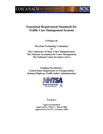
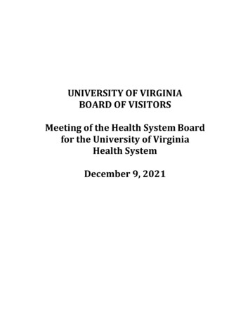
70Kawasaki Medical Journalsubsequent course was good and she was discharged on the 10th postoperative day. doi:10.11482/KMJ-E202147069(Accepted on April 20, 2021)Key words:Papillary thyroid cancer, Takotsubo syndromeINTRODUCTIONTa k o t s u b o s y n d r o m e ( T S ) i s g e n e r a l l ywith papillary thyroid cancer and referred to ourdepartment for consultation.[Past history] Bronchial asthma and postoperativecharacterized by transient and reversible ventricularwall dyskinesias caused by physical or mental stresscataract1,2). TS causes symptoms and ECG findings similar[Family history] Not contributoryto acute coronary syndrome (ACS); therefore, an[Physical findings] A tumor of approximately 20accurate diagnosis is essential 3,4). As there aremm was observed in the left lobe of the thyroidfew reports of TS associated with thyroid cancer,gland and the 15-mm left cervical lymph node waswe report a case of TS after postoperative papillarypalpable.thyroid cancer.[Image findings]Ultrasonography:A 19 18 18-mm hypoechoic mass was foundCASE REPORT[Patient] 82-year-old womanin the middle left lobe of the thyroid gland. The[Chief complaint] Left cervical massmass had a rough boundary and heterogeneous[Present illness] In 201X, she noted a left cervicalinternal structure (Fig. 1a), and the internal bloodmass and visited a neighborhood hospital. Basedflow was abundant (Fig. 1d). Left deep cervicalon detailed examination, she was diagnosedlymph node: Lymphadenopathy was observed andFigure 1acbdFig. 1. Ultrasonographic findings: Lt. lobe mass (a). Lt. cervical lymph node (b). Rt. cervical lymph node (c).Color Doppler (d).
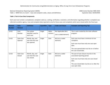
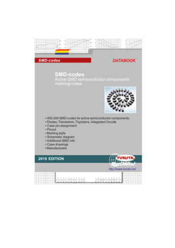
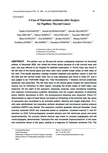
Figure 2Yamamoto Y, et al. : A Case of Takotsubo Syndrome After Surgery for Papillary Thyroid Cancer71abFigure3 MRI (b) showing a lt. lobe mass (white arrow) and a lt. cervical lymph node (gray arrow).Fig. 2. CT(a) andabcdFig. 3. Histopathological examination of the resected specimen: Papillary cell proliferation wasobserved (a), and ground glass-like nuclei (arrowhead), nuclear grooves (white arrow), andintranuclear cytoplasmic inclusion bodies (gray arrow) were noted (b, c). Lymph node metastases (d)were widespread.the maximum diameter was 25 mm, suggestingmetastasis (Fig. 1b). It was in contact with theinternal jugular vein and infiltration was suspected.Right deep cervical lymph node: At least two sitesof lymphadenopathy were found, also suggestingmetastasis (Fig. 1c).internal jugular vein. No lung metastasis was found.[Fine-needle aspiration cytology]Malignant and papillary cancer in both the leftlobe mass and left cervical lymph node[Blood test at first visit]Thyroid function was normal. The thyroglobulinCT (Fig. 2a), MRI (Fig. 2b):level was high at 313 ng/mL, but anti-thyroglobulinNo notable tracheal infiltration of the tumor wasantibody was negative. There were no otherobserved. The tumor may have invaded the leftabnormal findings.
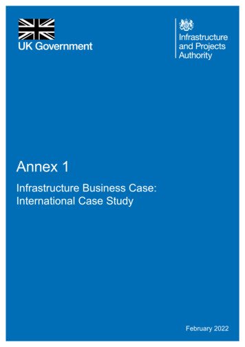
Figure 472Kawasaki Medical JournalFig. 4. Chest CT: Thickening of the tracheal wall was observed (white arrow).[Operation]performed to search for the cause of hypoxemia.Total thyroidectomy bilateral cervical lymphnodeThere was no evidence of heart failure, but baseddissection was performed. The left lobe mass of theon thickening of the bronchial wall (Fig. 4), thethyroid gland invaded the sterno-thyroid muscle,cause of hypoxemia was considered to be bronchialmany sites of bilateral cervical lymphadenopathyasthma.were noted, but there was no infiltration into theinternal jugular vein.[Histopathological examination]Although steroids and oxygen (high-dose, 15 L/min) were administered, respiratory acidosis (pH6.978, pCO2 106.1 mmHg, pO2 138.5 mmHg, BEPapillary cell proliferation was observed (Fig.-10.0 mEq/L) was confirmed on blood gas analysis3a), and ground glass-like nuclei, nuclear grooves,and non-invasive positive pressure ventilationand intranuclear cytoplasmic inclusion bodies(NIPPV) was started. The respiratory conditionwere noted (Fig. 3b, 3c), leading to a diagnosis ofgradually improved thereafter.papillary thyroid cancer (PTC). PTC of the thyroidBlood tests demonstrated an increased troponingland infiltrated the surrounding muscles andT level (0.232 ng/ml). ST elevation in leads V1-adipose tissue, but the surgical margin was negative.V3 and aVR was noted on ECG (Fig. 5b, Fig. 5aLymph node metastases (Fig. 3d) were widespreadshows ECG on admission). Echocardiography(maximum diameter of metastases 27 mm, a total ofdemonstrated akinesis of left ventricular apex, and12 metastatic lymphnodes).basal hypercontractility. Therefore, the differential[Postoperative course]diagnosis included TS and ACS. Because theOn the night of the operation, the patientregional wall motion abnormalities extend beyondsuddenly developed wheezing, respiratory distress,a single epicardial vascular distribution, TS wasmandibular breathing, and impaired consciousness,strongly suspected.and the oxygen saturation of peripheral artery16 hours later after onset, the patient underwent( S p O 2 ) d e c r e a s e d t o 6 0 % a t r o o m a i r. N ocoronary angiography to differentiate acute coronarypostoperative bleeding was observed. Chest CT wassyndrome (ACS) from stress cardiomyopathy.
Figure 5Yamamoto Y, et al. : A Case of Takotsubo Syndrome After Surgery for Papillary Thyroid Cancer73abFigureFig. 5. 6ECG: On admission (a). At sudden change (b).ST elevation was noted in leads V1-V3and aVR (white arrow).abFig. 6. Left ventriculography: Diastolic phase (a). Systolic phase (b).No coronary arterial stenosis was present and leftechocardiography, she was discharged on her ownventriculography (Fig. 6) revealed Takotsubo-on the 10th postoperative day.like wall movement (hypercontraction of the baseand contraction failure of the apex), leading to adiagnosis of TS.DISCUSSIONTakotsubo Cardiomyopathy was first describedThe subsequent course was good. Although shein Japan in 1990 by Dote and colleagues5). Thehad reduced left ventricular systolic function onname“Takotsubo”was used to describe the
74Kawasaki Medical Journalcardiomyopathy due to the characteristic apicaldischarged on the 10th postoperative day.ballooning on left ventriculography, which wasCancer is a chronic condition that inducessimilar in shape to a Japanese octopus trap. Itsignificant emotional and physical stress, increasingis also referred to as“broken heart”,“apicalthe risk of stress cardiomyopathy. The potentialballooning syndrome”,“myocardial stunning”, ortriggers of TS in cancer patients include the6), however“Takotsubo“stress cardiomyopathy”emotional turmoil of the diagnosis, the inflammatorysyndrome”is common internationally that causesstate of cancer, and the physical stress of cancercontraction disorders in the heart associated withtreatments, including chemotherapy10,11).various pathological conditions.In a recent International Takotsubo RegistryTS is a reversible disease commonly affecting(InterTAK) 12), malignancy was found in 16.6%postmenopausal women, characterized by transientof 1,604 TS patients. The most frequent type ofventricular wall motion abnormalities mediated bymalignancy was breast cancer, followed by tumors1,2). The well-acceptedof the gastrointestinal system, respiratory tract, andcatecholamine hypothesis suggests that stress-internal sex organs. Long-term mortality was higherinduced release of epinephrine and norepinephrinein TS patients with malignancy13).physical or mental stressadversely affects cardiac and microvasculaturefunctions, leading to myocardial stunning7).In this case, the cause of TS may have been relatedto postoperative bronchial asthma attack in additionto the stress caused by thyroid cancer surgery.The symptoms of TS mimics those of ACS, aspatients often present with chest pain, dyspnea,4)Only two cases of TS associated with thyroidcancer have been reported, one that developedduring lenvatinib treatment 14) and one thatdeveloped postoperatively15).Hayashi et al. 15) reported a case of TS thatdeveloped after surgery for thyroid cancer.A 72-year-old woman developed sudden dyspneaand hypotensive shock , in addition to ECGand arrhythmia on the 3rd day after surgery, and amanifestations akin to those of myocardialnegative T wave was observed on ECG. However,infarction 5). In this case, ACS was suspectedshe did not exhibit significant stenosis on coronarybased on symptoms, increased troponin T level,angiography and there was no contraction of theand ECG findings. However, Mayo Clinic criteriaapex of the heart on left ventricular angiography, butfor TS described“Transient hypokinesis, akinesis,she was diagnosed with TS. She had a good courseor dyskinesis of the left ventricular midsegmentsand was discharged 11 days after surgery15).with or without apical involvement; the regionalIf cardiovascular complications are observedwall motion abnormalities extend beyond a singleduring the perioperative period for thyroid glandepicardial vascular distribution”8). In this case,surgery, TS should be kept in mind.Echocardiography showed similar findings, soTS was strongly suspected. After that, we wereable to make a definitive diagnosis by coronaryangiography and left ventriculography.CONCLUSIONSWe reported a case of TS after surgery forpapillary thyroid cancerTS has a favorable long-term prognosis becausesupportive treatment leads to spontaneous rapid9)CONFLICTS OF INTERESTrecovery in nearly all patients . In this case, theJK received advisory/consultation fees andpatient was treated with supportive treatment andresearch funding from Takeda Pharmaceutical Co.her general condition recovered. And then she wasJK also received research funding from Eisai Co.
Yamamoto Y, et al. : A Case of Takotsubo Syndrome After Surgery for Papillary Thyroid CancerThe other authors declare that they have no conflictsof interest.REFERENCES1)Hachamovitch R, Chang JD, Kuntz RE, PapageorgiouP, Levin MS, Goldberger AL: Recurrent reversiblecardiogenic shock triggered by emotional distress withno obstructive coronary disease. Am Heart J. 1995; 129:1026-1028. doi: 10.1016/0002-8703(95)90127-2.2)Vriz O, Brosolo G, Martina S, Pertoldi F, Citro R, MosL, Ferrara F, Bossone E: In-hospital and long-termmortality in Takotsubo cardiomyopathy: a communityhospital experience. J Community Hosp Intern MedPerspect. 2016; 6: 31082. doi: 10.3402/jchimp.v6.31082.3)Templin C, Ghadri JR, Diekmann J, et al.: ClinicalF e a t u r e s a n d O u t c o m e s o f Ta k o t s u b o ( S t r e s s )Cardiomyopathy. N Engl J Med. 2015; 373: 929-938.doi: 10.1056/NEJMoa1406761.4)Gopalakrishnan P, Zaidi R, Sardar MR: Takotsubocardiomyopathy: pathophysiology and role of cardiacbiomarkers in differential diagnosis. World J Cardiol.2017; 9: 723-730. doi: 10.4330/wjc.v9.i9.723.5)Dote K, Sato H, Tateishi H, Uchida T, Ishihara M:Myocardial stunning due to simultaneous multivesselcoronary spasms: a review of 5 cases. J Cardiol. 1991;21: 203-214. (Article in Japanese).6)Desai A, Noor A, Joshi S, Kim AS: Takotsubocardiomyopathy in cancer patients. Cardiooncology.2019; 5: 7. doi: 10.1186/s40959-019-0042-9.7)K o m a m u r a K , F u k u i M , I w a s a k u T, H i r o t a n iS , M a s u y a m a T: Ta k o t s u b o c a r d i o m y o p a t h y :Pathophysiology, diagnosis and treatment. World JCardiol. 2014; 6: 602-609. doi: 10.4330/wjc.v6.i7.602.8)Prasad A, Lerman A, Rihal CS : Apical ballooningsyndrome (Tako-Tsubo or stress cardiomyopathy): amimic of acute myocardial infarction. Am Heart J. 2008;75155 : 408-417. doi: 10.1016/j.ahj.2007.11.008.9)Sheppard MN: Takotsubo Syndrome-Stress-inducedHeart Failure Syndrome. Eur Cardiol. 2015; 10: 83-88.doi: 10.15420/ecr.2015.10.2.83.10)Madias JE: What is/are the trigger(s) of takotsubosyndrome in cancer patients receiving chemotherapy?.Int J Cardiol. 2016; 222: 253. doi: 10.1016/j.ijcard.2016.07.246.11)De Pasquale MD, Mastronuzzi A, De Sio L, Serra A,Grimaldi C, Chinali M, Giordano U: Transient globalventricular dysfunction in an adolescent affected bypancreatic adenocarcinoma. BMC Pediatr. 2016; 16: 99.doi: 10.1186/s12887-016-0628-4.12)Cammann VL, Sarcon A, Ding KJ, et al.: ClinicalFeatures and Outcomes of Patients With Malignancyand Takotsubo Syndrome: Observations From theInternational Takotsubo Registry. J Am Heart Assoc.2019; 8: e010881.doi: 10.1161/JAHA.118.010881.13)Stępień K, Nowak K, Pasieka P, Warmuz K, StępieńA, Nessler J, Zalewski J: Typical variant of takotsubocardiomyopathy in oncological patients. Two casereports and review of the literature. Folia Med Cracov.2020; 60: 45-54. doi: 10.24425/fmc.2020.133485.14)Chae YK, Chiec L, Adney SK, Waitzman J, Costa R,Carneiro B, Matsangou M, Agulnik M, Kopp P, GilesF: Posterior reversible encephalopathy syndrome andtakotsubo cardiomyopathy associated with lenvatinibtherapy for thyroid cancer: a case report and review.Oncotarget. 2018; 9: 28281-28289. doi: 10.18632/oncotarget.25606.15)Masatoshi Hayashi, Kouya Tochii, Masao Niwa, KeiTakahashi, Kouzo Kawamura, Kentarou Kokubo: A caseof takotsubo cardomyopathy after surgery for thyroidcancer. J JAES JSTS. 2017; 34: 191-194. doi: 10.11226/jaesjsts.34.3 191. (Article in Japanese).
[Postoperative course] On the night of the operation, the patient suddenly developed wheezing, respiratory distress, mandibular breathing, and impaired consciousness, and the oxygen saturation of peripheral artery (SpO2) decreased to 60% at room air. No postoperative bleeding was observed. Chest CT was performed to search for the cause of .