Transcription
Hemmat et al. Molecular Cytogenetics 2014, /1/35CASE REPORTOpen AccessSubmicroscopic deletion of 5q involving tumorsuppressor genes (CTNNA1, HSPA9) and copyneutral loss of heterozygosity associated withTET2 and EZH2 mutations in a case of MDS withnormal chromosome and FISH resultsMorteza Hemmat1*, Weina Chen2, Arturo Anguiano1, Mohammed El Naggar1, Frederick K Racke1, Dan Jones3,Yongbao Wang3, Charles M Strom1, Karl Chang1 and Fatih Z Boyar1AbstractAdvances in genome-wide molecular cytogenetics allow identification of novel submicroscopic DNA copy numberalterations (aCNAs) and copy-neutral loss of heterozygosity (cnLOH) resulting in homozygosity for known genemutations in myeloid neoplasms. We describe the use of an oligo-SNP array for genomic profiling of aCNA andcnLOH, together with sequence analysis of recurrently mutated genes, in a patient with myelodysplastic syndrome(MDS) presenting with normal karyotype and FISH results. Oligo-SNP array analysis revealed a hemizygous deletionof 896 kb at chromosome 5q31.2, representing the smallest 5q deletion reported to date. The deletion involvedmultiple genes, including two tumor suppressor candidate genes (CTNNA1 and HSPA9) that are associated withMDS/AML. The SNP-array study also detected 3 segments of somatic cnLOH: one involved the entire long arm ofchromosome 4; the second involved the distal half of the long arm of chromosome 7, and the third encompassedthe entire chromosome 22 (UPD 22). Sequence analysis revealed mutations in TET2 (4q), EZH2 (7q), ASXL1(20q11.21), and RUNX1 (21q22.3). Coincidently, TET2 and EZH2 were located at segments of cnLOH resulting in theirhomozygosity. Loss of heterozygosity affecting these two chromosomes and mutations in TET2 and EZH2 are indicativeof a myelodysplastic syndrome with a poor prognosis. Deletion of the tumor suppressor genes CTNNA1 and HSPA9 isalso likely to contribute to a poor prognosis. Furthermore, the original cnLOHs in multiple chromosomes and additionalcnLOH 14q in the follow-up study suggest genetic evolution of the disease and poor prognosis. This study attests tothe fact that some patients with a myelodysplastic syndrome who exhibit a normal karyotype may have underlyinggenetic abnormalities detectable by chromosomal microarray and/or targeted mutation analyses.Keywords: Copy neutral loss of heterozygosity (cnLOH), Uniparental disomy (UPD), MDS, TET2, EZH2, RUNX1, EZH2,ASXL1, CTNNA1, HSPA9BackgroundRecent advances in genome-wide molecular cytogeneticsallow the identification of novel molecular abnormalities[1-8]. Emerging data demonstrate that myelodysplasticsyndrome (MDS) exhibits abundant CNAs and cnLOH,often in the setting of a normal karyotype [9-11]. Loss of* Correspondence: morteza.x.hemmat@questdiagnostics.com1Cytogenetics Department, Quest Diagnostics Nichols Institute, 33608 OrtegaHwy, 92675 San Juan Capistrano, CA, USAFull list of author information is available at the end of the articleheterozygosity (LOH) is an indicator of neoplastic evolution and disease progression [12,13]. Copy-neutralLOH (cnLOH) arises either via a hemizygous deletion inone homolog and duplication of the other, or uniparentaldisomy (UPD). Both types of somatic LOH have been observed in studies of various cancer types and may explainsome of the mechanisms by which tumor suppressorgenes (TSGs) are inactivated or activating mutations inoncogenes are duplicated. Acquired UPD (aUPD) is nowunderstood to be common in oncogenesis and appears to 2014 Hemmat et al.; licensee BioMed Central Ltd. This is an Open Access article distributed under the terms of the CreativeCommons Attribution License (http://creativecommons.org/licenses/by/4.0), which permits unrestricted use, distribution, andreproduction in any medium, provided the original work is properly credited. The Creative Commons Public DomainDedication waiver ) applies to the data made available in this article,unless otherwise stated.
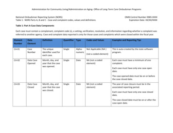
Hemmat et al. Molecular Cytogenetics 2014, /1/35be a mechanism to increase the allelic burden of themutated genes [14-24]. Thus, it is important to know theregions of cnLOH to determine new regions containingpotential mutational targets affecting disease pathogenesisand treatment outcome [25,26].Application of SNP-array technology has led to the identification of recurrent regions of cnLOH in a majority ofthe chromosomes [4,13,25,27-30] and recurrent pathogenicmutations. These findings have greatly advanced our understanding of the molecular mechanisms of cancer evolutionand have led to the development of therapeutics and diagnostic tests. In this respect, mutation analysis helps furtherstratify neoplasms and their treatment outcome [31-34].In this study, we used SNP array-based genomicprofiling to detect acquired copy number alterations(aCNA) and cnLOHs, together with sequence analysisof genes commonly mutated in MDS, in a patient withnormal chromosome and MDS FISH (fluorescence insitu hybridization) panel results.Clinical presentationMorphologic and immunophenotypic findingsAn 88-year-old woman presented with leukocytosis(16.2 109/L) including minimal monocytosis (1.0 109/L),mild anemia (HGB 105 g/L), and moderate thrombocytopenia(79 109/L). Bone marrow morphologic and flow cytometricexaminations revealed hypercellular bone marrow withgranulocytic predominance with left-shifted and unusualPage 2 of 9maturation, and occasional atypical granulocytes andmegakaryocytes, but no significant increase in monocytes or myeloblasts (Figure 1). A myeloid neoplasmpositive for CD13 and CD33 was diagnosed, best classified as MDS, refractory cytopenia with multilineagedysplasia. Data on therapeutic interventions were notavailable for this study. At the 5-month follow-up, theoverall morphologic and immunophenotypic findingswere similar to those at initial presentation.Chromosome and FISH studyBone marrow cells were cultivated for 24 and 48 hoursin RPMI 1640 or Marrow Max Medium with 10% fetal calfserum (Life Technologies, Grand Island, NY 14072, USA).In total, 20 mitoses were analyzed according to theISCN (International System for Human CytogeneticNomenclature), with a resolution of 300 bands perhaploid karyotype. FISH analyses using a panel of MDSprobes ( 5/5q-, 7/7q-, 8 and 20q-; Vysis, DownersGrove, IL, USA) and BlueGnome probes RP11-114B12(Illumina, San Diego, CA, USA) for the deleted region5q31.2 were performed on interphase cells accordingto the manufacturer’s protocol. Subsequently, 200 cellswere examined carefully.Oligo SNP arrayMicrodeletion/microduplication screening was performedusing an SNP-array platform (CytoScan HD SNP array;Figure 1 Morphologic and immunophenotypic findings. A) At presentation, there was granulocytic predominance with progressivematuration and no significant increase in blasts or monocytes. The core biopsy shows hypercellular bone marrow with left-shifted granulocyticpredominance. Flow cytometry reveals granulocytic predominance (in green) but no significant increase in monocytes (in blue) or myeloblasts(in red). B) At follow-up, the morphologic and immunophenotypic findings are similar to those at presentation.
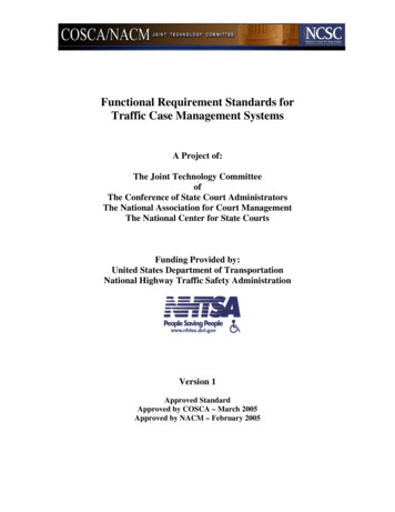
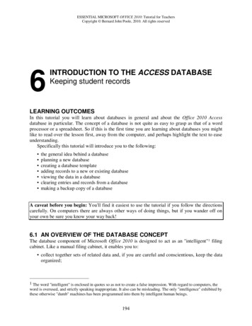
Hemmat et al. Molecular Cytogenetics 2014, /1/35Affymetrix, Santa Clara, CA), following the manufacturer’sinstructions. The CytoScan HD array has 2.67 millionprobes, including 1.9 million copy number probes and0.75 million SNP probes. Array data were analyzedusing the Chromosome Analysis Suite (ChAS) softwarev 2.0 (Affymetrix).Mutation analysisGenomic DNA was also tested for mutations in 19 genesthat are recurrently mutated in myeloid neoplasms,including ASXL1, EZH2, RUNX1, IDH1, IDH2, KRAS,NRAS and TET2. Sequencing was performed using aTruSeq custom amplicon assay on the MiSeq sequencingplatform (Illumina, Hayward, CA). Analysis was performedusing SeqPilot software (JSI Medical Systems, Costa Mesa,CA). The assay had sufficient read depth to provide a minimum sensitivity of 5% to 10% for mutation detection.Page 3 of 9Results and discussionDuring the initial evaluation of the patient, cytogeneticanalysis revealed a normal karyotype and FISH studies werenegative for aCNAs commonly seen in MDS (MDS panel)(Figure 2A-D). Microarray analysis revealed a microdeletionof approximately 896 kb at the 5q31.2 chromosomal regionand three segments of somatic cnLOH for the entirelong arm of chromosome 4 (136 Mb), the distal half ofthe long arm of chromosome 7 (50 Mb), and the entirechromosome 22 (31 Mb). The microdeletion at 5q31.2extended from 137,821,899 to 138,718,504 bp (UCSC genome Browser; http://genome.ucsc.edu/; hg19 release) andincluded the ETF1, HSPA9, SNORD63, CTNNA1, LRRTM2,SIL1, SNHG4, MATR3, SNORA74A, PAIP2, and SLC23A1genes (Figure 3).Of the genes deleted due to this microdeletion, twoare tumor suppressor candidate genes associated withFigure 2 Routine cytogenetic and fluorescence insitu hybridization (FISH) results. A) G-banded chromosome analysis shows a normalfemale karyotype. B-D) FISH analyses show normal hybridization with an MDS panel using probes for EGR1 (5q31, red signal) and its controlprobe D5S23 (5p15.2, green signal) in B; D7S486 (7q31, red signal) and its control probe D7Z1 (7centromere, green signal) in C; CEP8(green signal) for chromosome 8 centromere and D20S108 (20q12, red signal) in D.
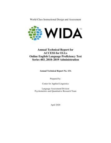
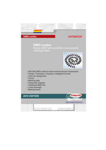
Hemmat et al. Molecular Cytogenetics 2014, /1/35Page 4 of 9ABCFigure 3 5q31.2 deletion. A) Chromosome 5 with deletion at q31.2. B) SNP-array results, including the weighted log2 ratio, copy number state,and allele peaks at the deleted region. C) Database of genomic variants showing an 896-kb deletion in the short arm of chromosome 5 withinband q31.2 (position 137,821,899 to 138,718,504), including the CTNNA1 and HSPA9 genes.MDS/AML: CTNNA1, which encodes alpha-1 catenin,and HSPA9, which encodes heat-shock 70-KD protein9 (mortalin) [35]. Deletion of CTNNA1 was confirmedby applying BlueGnome FISH probes (RP11-114B12)(Figure 4). Alpha-catenins such as that encoded byCTNNA1 are essential for the regulation of cell-cell andcell-matrix interactions in tissues [36]. Loss of expressionof the CTNNA1 tumor suppressor gene in hematopoieticstem cells may provide a growth advantage that contributesto human MDS/AML with 5q deletion [37]. Furthermore,loss of the CTNNA1 expression has been associated withleukemia progression or transformation of MDS to AML[38]. The HSPA9 gene is also located at the 5q31.2 regionfrequently deleted in MDS/AML, making it a candidatetumor suppressor gene; this is consistent with the biological function of its murine homologue. Human mortalin(encoded by HSPA9) was originally identified by its closehomology to murine mortalins, which play importantroles in cellular senescence [39]. The HSPA9 gene is anovel negative regulator of Raf/MEK/ERK pathway thatmay be a potential therapeutic target [40].The combined size of cnLOHs spanning at least 10 Mbacross the genome was approximately 217.6 Mb (Figure 5).These were detected at the long arm of chromosomes4, 7, and 22. The cnLOH might result from mitotic recombination or nondisjunction which leads to segmental
Hemmat et al. Molecular Cytogenetics 2014, /1/35Figure 4 FISH inverted DAPI image showing deletion of theCTNNA1 gene using the BlueGnome FISH probe RP11-114B12(5q31.2, red signal). The deleted chromosome 5 is indicated by an arrow.Page 5 of 9or whole chromosomal UPD, respectively [13]. AcquiredUPD (cnLOH) at diagnosis in our case is indicative ofneoplastic evolution [12,13]. An additional UPD was identified at 14q in the follow-up study 5 months later, confirming the genetic progression of disease (Figure 6).Recent investigations have indicated that cnLOH canbe responsible for homozygosity of mutations in criticalgenes within the region. Reduction to homozygosity as aconsequence of cnLOH was initially thought to be amechanism for only inactivation of tumor suppressorgenes [14,41,42]; however, identification of cnLOH inleukemia has shown that oncogeneic mutations arealso targeted [13,16,17].Mutation analysis of 19 MDS-associated genes revealed an ASXL1 frameshift/stop mutation (Y591*, 41%of reads), an EZH2 point mutation (R690H, 85%), twoRUNX1 frameshift/stop mutations (c.474dupT, 19% andc.424 425ins11bp, 7%), and two TET2 frameshift/stopmutations (c.1510 1513delAAAA, 77% and R1465*, 10%).These mutated genes are located on chromosomes 20, 7,21, and 4, respectively. Recent studies have shown thatTET2 mutations are present in up to 30% of MDS/MPNFigure 5 SNP-array results at diagnosis showing loss of heterozygosity (LOH) at the long arm of chromosomes 4, 7, and 22 as purplebars next to the corresponding chromosomes.
Hemmat et al. Molecular Cytogenetics 2014, /1/35Page 6 of 9Figure 6 SNP-array results at the follow-up study showing loss of heterozygosity (LOH) of chromosome 14, in addition to the LOHchromosomes 4, 7, and 22 found at initial diagnosis. LOH is shown as purple bar next to the corresponding chromosome.cases, with mutations in ASXL1, EZH2, and RUNX1 alsocommonly reported [43]. The finding of mutations inall four of these myeloid regulatory genes suggests thatthey play a critical role in the pathogenesis of this case anddemonstrate that mutation analysis is useful in cytogenetically normal myeloid disorders [43].The cnLOH affecting chromosome 7q and homozygousEZH2 mutation have been reported in 10% of AMLand MDS cases. They have been associated with a poorprognosis [9,26,44] and clonal evolution [45,46], supportingthe possible role of EZH2 as a tumor suppressor genefor myeloid malignancies.In contrast to the mutated TET2 and EZH2 genes,no LOH was found for the other two mutated genes(RUNX1 and ASXL1). RUNX1 mutations have beenproposed as clinically useful biomarkers to follow diseaseprogression from MDS to AML as well as to monitorminimal residual disease (MRD) [47]. Moreover, RUNX1mutations were demonstrated to be frequent in de novoAML with non-complex karyotypes and conferred anunfavorable prognosis [48] explained by an associationwith resistance to chemotherapy [49].Mutations in ASXL1 have been identified in MDS,AML, chronic myeloid leukemia, chronic myelomonocyticleukemia (CMML), and juvenile myelomonocytic leukemia[50-54], and act as a tumor suppressor in myeloid malignancies [50]. Mutations in ASXL1, TET2, and EZH2 havebeen found in 41% of MDS cases in a Chinese population[44], similar to the data reported in patients of Europeandecent with MDS [21,23,24,55,56].ConclusionIn conclusion, our study identified four large cnLOH anda microdeletion at 5q31 harboring two tumor suppressor genes (CTNNA1 and HSPA9) in an MDS case withan apparently normal karyotype. The regions of cnLOH atchromosomes 4, 7, 14, and 22 confirm and extend previousstudies, supporting that cnLOHs in myeloid disorders arecommon and nonrandom. Moreover, the presence of anadditional aUPD 14q at follow up, along with persistence of
Hemmat et al. Molecular Cytogenetics 2014, /1/35the 5q31 microdeletion and all cnLOHs detected at presentation, indicates genetic progression of the disease. Thedeletion of tumor suppressor genes CTNNA1 and HSPA9,along with mutation of candidate myeloid regulatory genesTET2 and EZH2, supports the diagnosis of MDS and likelyportends a poor prognosis.This study attests to the fact that some patients with amyelodysplastic syndrome exhibiting a normal karyotypemay have underlying genetic abnormalities detectableby chromosomal microarray and/or targeted mutationanalyses. Further genomic and molecular studies on aseries of patients with MDS may yield information onhow to stratify this category of disease to seek furthermolecular definition.Ethical approval and consentThese studies were performed on anonymized samplesreceived in the clinical laboratory and thus were exemptedfrom the requirement for consent by an opinion for theWestern Institutional Review Board.Page 7 of 95.6.7.8.9.10.AbbreviationsaCNA: Acquired copy number alteration; cnLOH: Loss of heterozygosity;aUPD: Acquired uniparental disomy.11.Competing interestsThe authors declare that they have no competing interests.Authors’ contributionsMH, First author; performed chromosome, FISH, and microarray analysis,interpretation of the results, drafting and finalization of the manuscript. WCperformed the morphology and immunophenotypic analysis. DJ and YWperformed the genomic sequencing. FR performed the comprehensivereview of the clinical and laboratory findings. AA reviewed and reported theSNP-array analyses. MEN reviewed the manuscript. FZB reviewed and finalizedthe manuscript. All authors read and approved the final manuscript.12.13.14.AcknowledgmentsThe authors would like to express their thanks to Maryam Talai and RobertMcgough (Quest Diagnostics) for their technical support.Author details1Cytogenetics Department, Quest Diagnostics Nichols Institute, 33608 OrtegaHwy, 92675 San Juan Capistrano, CA, USA. 2University of Texas southwesternMedical Center, 5323 Harry Hines Blvd, 75235 Dallas, TX, USA. 3QuestDiagnostics Nichols Institute, 14225 Newbrook Drive, 20151 Chantilly, VA, USA.15.16.Received: 30 March 2014 Accepted: 19 May 2014Published: 27 May 201417.References1. Mullighan CG, Goorha S, Radtke I, Miller CB, Coustan-Smith E, Dalton JD,Girtman K, Mathew S, Ma J, Pounds SB, Su X, Pui CH, Relling MV, Evans WE,Shurtleff SA, Downing JR: Genome-wide analysis of genetic alterations inacute lymphoblastic leukemia. Nature 2007, 446:758–764.2. Suela J, Alvarez S, Cifuentes F, Largo C, Ferreira BI, Blesa D, Ardanaz M,Garcia R, Marquez JA, Odero MD, Calasanz MJ, Cigudosa JC: DNA profilinganalysis of 100 consecutive de novo acute myeloid leukemia casesreveals patterns of genomic instability that affect all cytogenetic riskgroups. Leukemia 2007, 21:1224–1231.3. Tyyba kinoja A, Elonen E, Piippo K, Porkka K, Knuutila S: Oligonucleotidearray-CGH reveals cryptic gene copy number alterations in karyotypicallynormal acute myeloid leukemia. Leukemia 2007, 21:571–574.4. Gupta M, Raghavan M, Gale RE, Chelala C, Allen C, Molloy G, Chaplin T,Linch DC, Cazier JB, Young BD: Novel regions of acquired uniparental18.19.20.disomy discovered in acute myeloid leukemia. Genes ChromosomesCancer 2008, 47:729–739.Kawamata N, Ogawa S, Zimmermann M, Kato M, Sanada M, Hemminki K,Yamatomo G, Nannya Y, Koehler R, Flohr T, Miller CW, Harbott J, LudwigWD, Stanulla M, Schrappe M, Bartram CR, Koeffler HP: Molecularallelokaryotyping of pediatric acute lymphoblastic leukemias byhigh-resolution single nucleotide polymorphism oligonucleotidegenomic microarray. Blood 2008, 111(2):776–84.Akagi T, Ogawa S, Dugas M, Kawamata N, Yamamoto G, Nannya Y, SanadaMiller CW, Yung A, Schnittger S, Haferlach T, Haferlach C, Koeffler HP: Frequentgenomic abnormalities in acute myeloid leukemia/myelodysplasticsyndrome with normal karyotype. Haematologica 2009, 94:213–223.Maciejewski JP, Tiu RV, O’Keefe C: Application of array based wholegenome scanning technologies as a cytogenetic tool in haematologicalmalignancies. Br J Hematol 2009, 146:479–488.Tiu RV, Gondek LP, O’Keefe CL, Huh J, Sekeres MA, Elson P, McDevitt MA,Wang XF, Levis MJ, Karp JE, Advani AS, Maciejewski JP: New lesionsdetected by single nucleotide polymorphism array-based chromosomalanalysis have important clinical impact in acute myeloid leukemia. J ClinOncol 2009, 27:5219–5226.Heinrichs S, Kulkarni RV, Bueso-Ramos CE, Levine RL, Loh ML, Li C, NeubergD, Kornblau SM, Issa JP, Gilliland DG, Garcia-Manero G, Kantarjian HM, Estey EH,Look AT: Accurate detection of uniparental disomy and microdeletions bySNP array analysis in myelodysplastic syndromes with normal cytogenetics.Leukemia 2009, 23(9):1605–13.Thiel A1, Beier M, Ingenhag D, Servan K, Hein M, Moeller V, Betz B,Hildebrandt B, Evers C, Germing U, Royer-Pokora B: Comprehensive arrayCGH of normal karyotype myelodysplastic syndromes reveals hiddenrecurrent and individual genomic copy number alterations withprognostic relevance. Leukemia 2011, 25:387–399.Tiu RV1, Gondek LP, O’Keefe CL, Elson P, Huh J, Mohamedali A, KulasekararajA, Advani AS, Paquette R, List AF, Sekeres MA, McDevitt MA, Mufti GJ,Maciejewski JP: Prognostic impact of SNP array karyotyping inmyelodysplastic syndromes and related myeloid malignancies.Blood 2011, 117(17):4552–60.Andersen CL, Wiuf C, Kruhøffer M, Korsgaard M, Laurberg S, Ørntoft TF:Frequent occurrence of uniparental disomy in colorectal cancer.Carcinogenesis 2007, 28(1):38–48.Raghavan M, Smith LL, Lillington DM, Chaplin T, Kakkas I, Molloy G, ChelalaC, Cazier JB, Cavenagh JD, Fitzgibbon J, Lister TA, Young BD: Segmentaluniparental disomy is a commonly acquired genetic event in relapsedacute myeloid leukemia. Blood 2008, 112(3):814–21.Flotho C, Steinemann D, Mullighan CG, Neale G, Mayer K, Kratz CP,Schlegelberger B, Downing JR, Niemeyer CM: Genome-wide single-nucleotidepolymorphism analysis in juvenile myelomonocytic leukemia identifiesuniparental disomy surrounding the NF1 locus in cases associated withneurofibromatosis but not in cases with mutant RAS or PTPN11.Oncogene 2007, 26(39):5816–21.Fitzgibbon J, Smith LL, Raghavan M, Smith ML, Debernardi S, Skoulakis S,Lillington D, Lister TA, Young BD: Association between acquireduniparental disomy and homozygous gene mutation in acute myeloidleukemias. Cancer Res 2005, 65:9152–9154.Kralovics R, Passamonti F, Buser AS, Teo SS, Tiedt R, Passweg JR, TichelliA, Cazzola M, Skoda RC: A gain-of-function mutation of JAK2 inmyeloproliferative disorders. N Engl J Med 2005, 352(17):1779–90.Kralovics R, Guan Y, Prchal JT: Acquired uniparental disomy of chromosome9p is a frequent stem cell defect in polycythemia vera. Exp Hematol 2002,30(3):229–36.Grand FH, Hidalgo-Curtis CE, Ernst T, Zoi K, Zoi C, McGuire C, Kreil S, JonesA, Score J, Metzgeroth G, Oscier D, Hall A, Brandts C, Serve H, Reiter A,Chase AJ, Cross NC: Frequent CBL mutations associated with 11qacquired uniparental disomy in myeloproliferative neoplasms.Blood 2009, 113(24):6182–92.Sanada M, Suzuki T, Shih LY, Otsu M, Kato M, Yamazaki S, Tamura A, HondaH, Sakata-Yanagimoto M, Kumano K, Oda H, Yamagata T, Takita J, Gotoh N,Nakazaki K, Kawamata N, Onodera M, Nobuyoshi M, Hayashi Y, Harada H,Kurokawa M, Chiba S, Mori H, Ozawa K, Omine M, Hirai H, Nakauchi H,Koeffler HP, Ogawa S: Gain-of-function of mutated C-CBL tumoursuppressor in myeloid neoplasms. Nature 2009, 460(7257):904–8.Dunbar AJ, Gondek LP, O’Keefe CL, Makishima H, Rataul MS, Szpurka H,Sekeres MA, Wang XF, McDevitt MA, Maciejewski JP: 250 K single
Hemmat et al. Molecular Cytogenetics 2014, nucleotide polymorphism array karyotyping identifies acquireduniparental disomy and homozygous mutations, including novelmissense substitutions of c-Cbl, in myeloid malignancies. Cancer Res2008, 68(24):10349–57.Ernst T, Chase AJ, Score J, Hidalgo-Curtis CE, Bryant C, Jones AV, Waghorn K,Zoi K, Ross FM, Reiter A, Hochhaus A, Drexler HG, Duncombe A, CervantesF, Oscier D, Boultwood J, Grand FH, Cross NC: Inactivating mutations ofthe histone methyltransferase gene EZH2 in myeloid disorders. Nat Genet2010, 42(8):722–6.Langemeijer SM, Kuiper RP, Berends M, Knops R, Aslanyan MG, Massop M,Stevens-Linders E, van Hoogen P, van Kessel AG, Raymakers RA, Kamping EJ,Verhoef GE, Verburgh E, Hagemeijer A, Vandenberghe P, de Witte T, van derReijden BA, Jansen JH: Acquired mutations in TET2 are common inmyelodysplastic syndromes. Nat Genet 2009, 41(7):838–42.Nikoloski G, Langemeijer SM, Kuiper RP, Knops R, Massop M, Tönnissen ER,van der Heijden A, Scheele TN, Vandenberghe P, de Witte T, van der ReijdenBA, Jansen JH: Somatic mutations of the histone methyltransferase geneEZH2 in myelodysplastic syndromes. Nat Genet 2010, 42(8):665–7.Delhommeau F, Dupont S, Della Valle V, James C, Trannoy S, Massé A,Kosmider O, Le Couedic JP, Robert F, Alberdi A, Lécluse Y, Plo I, Dreyfus FJ,Marzac C, Casadevall N, Lacombe C, Romana SP, Dessen P, Soulier J, ViguiéF, Fontenay M, Vainchenker W, Bernard OA: Mutation in TET2 in myeloidcancers. N Engl J Med 2009, 360(22):2289–301.Mohamedali A, Gäken J, Twine NA, Ingram W, Westwood N, Lea NC,Hayden J, Donaldson N, Aul C, Gattermann N, Giagounidis A, Germing U,List AF, Mufti GJ: Prevalence and prognostic significance of allelicimbalance by single-nucleotide polymorphism analysis in low-riskmyelodysplastic syndromes. Blood 2007, 110(9):3365–73.Gondek LP, Tiu R, O'Keefe CL, Sekeres MA, Thiel KS, Maciejewski JP:Chromosomal lesions and uniparental disomy detected by SNP arrays inMDS. Blood 2008, 111:1534–42.Gorletta TA, Gasparini P, D’Elios MM, Trubia M, Pelicci PG, Di Fiore PP:Frequent loss of heterozygosity without loss of genetic material inacute myeloid leukemia with a normal karyotype. Genes ChromosomesCancer 2005, 44:334–337.Raghavan M, Lillington DM, Skoulakis S, Debernardi S, Chaplin T, Foot NJ,Lister TA, Young BD: Genome-wide single nucleotide polymorphismanalysis reveals frequent partial uniparental disomy due to somaticrecombination in acute myeloid leukemias. Cancer Res 2005, 65:375–378.Fitzgibbon J, Smith LL, Raghavan M, Smith ML, Debernardi S, Skoulakis S,Lillington D, Lister TA, Young BD: Association between acquireduniparental disomy and homozygous gene mutation in acute myeloidleukemias. Cancer Res 2005, 65:9152–9154.Gondek LP, Dunbar AJ, Szpurka H, McDevitt MA, Maciejewski JP: SNP arraykaryotyping allows for the detection of uniparental disomy and crypticchromosomal abnormalities in MDS/MPD-U and MPD. PLoS One 2007,2(11):e122.Kottaridis PD, Gale RE, Frew ME, Harrison G, Langabeer SE, Belton AA, WalkerH, Wheatley K, Bowen DT, Burnett AK, Goldstone AH, Linch DC: Thepresence of a FLT3 internal tandem duplication in patients with acutemyeloid leukemia (AML) adds important prognostic information tocytogenetic risk group and response to the first cycle of chemotherapy:Analysis of 854 patients from the United Kingdom medical researchcouncil. AML 10 and 12 trials. Blood 2001, 98:1752–1759.Falini B, Mecucci C, Tiacci E, Alcalay M, Rosati R, Pasqualucci L, La Starza R,Diverio D, Colombo E, Santucci A, Bigerna B, Pacini R, Pucciarini A, Liso A,Vignetti M, Fazi P, Meani N, Pettirossi V, Saglio G, Mandelli F, Lo-Coco F,Pelicci PG, Martelli MF: Cytoplasmic nucleophosmin in acutemyelogenous leukemia with a normal karyotype. N Engl J Med 2005,352:254–266.Baldus CD, Mrozek K, Marcucci G, Bloomfield CD: Clinical outcome of denovo acute myeloid leukaemia patients with normal cytogenetics isaffected by molecular genetic alterations: a concise review. Br JHaematol 2007, 137:387–400.Mead AJ, Linch DC, Hills RK, Wheatley K, Burnett AK, Gale RE: FLT3tyrosine kinase domain mutations are biologically distinct from andhave a significantly more favorable prognosis than FLT3 internaltandem duplications in patients with acute myeloid leukemia.Blood 2007, 110:1262–1270.Horrigan SK, Arbieva ZH, Xie HY, Kravarusic J, Fulton NC, Naik H, Le TT,Westbrook CA: Delineation of a minimal interval and identification of 9Page 8 of andidates for a tumor suppressor gene in malignant myeloid disorderson 5q31. Blood 2000, 95(7):2372–7.Kask M1, Pruunsild P, Timmusk T: Bidirectional transcription from humanLRRTM2/CTNNA1 and LRRTM1/CTNNA2 gene loci leads to expression ofN-terminally truncated CTNNA1 and CTNNA2 isoforms. Biochem BiophysRes Commun 2011, 411(1):56–61.Liu TX, Becker MW, Jelinek J, Wu WS, Deng M, Mikhalkevich N, Hsu K,Bloomfield CD, Stone RM, DeAngelo DJ, Galinsky IA, Issa JP, Clarke MF, LookAT: Chromosome 5q deletion and epigenetic suppression of the geneencoding alpha-catenin (CTNNA1) in myeloid cell transformation.Nat Med 2007, 13(1):78–83.Ye Y, McDevitt MA, Guo M, Zhang W, Galm O, Gore SD, Karp JE, MaciejewskiJP, Kowalski J, Tsai HL, Gondek LP, Tsai HC, Wang X, Hooker C, Smith BD,Carraway HE, Herman JG: Progressive chromatin repression and promotermethylation of CTNNA1 associated with advanced myeloidmalignancies. Cancer Res 2009, 69(21):8482–90.Xie H, Hu Z, Chyna B, Horrigan SK, Westbrook CA: Human mortalin(HSPA9): a candidate for the myeloid leukemia tumor suppressor geneon 5q31. Leukemia 2000, 14(12):2128–34.Wu PK, Hong SK, Veeranki S, Karkhanis M, Starenki D, Plaza JA, Park JI: Amortalin/HSPA9-mediated switch in tumor-suppressive signaling ofRaf/MEK/extracellular signal-regulated kinase. Mol Cell Biol 2013,33(20):4051–67.Cavenee WK, Dryja TP, Phillips RA, Benedict WF, Godbout R, Gallie BL, MurphreeAL, Strong LC, White RL: Expression of recessive alleles by chromosomalmechanisms in retinoblastoma. Nature 1983, 305(5937):779–84.Fearon ER, Vogelstein B, Feinberg AP: Somatic deletion and duplication ofgenes on chromosome 11 in Wilms’ tumours. Nature 1984, 309:176–8.Jankowska AM, Szpurka H, Tiu RV, Makishima H, Afable M, Huh J, O’Keefe CL,Ganetzky R, McDevitt MA, Maciejewski JP: Loss of heterozygosity 4q24 andTET2 mutations associated with myelodysplastic/myeloproliferativeneoplasms. Blood 2009, 113(25):6403–10.Wang J, Ai X, Gale RP, Xu Z, Qin T, Fang L, Zhang H, Pan L, Hu N, Zhang Y,Xiao Z: TET2, ASXL1 and EZH2 mutations in Chinese withmyelodysplastic syndromes. Leuk Res 2013, 37(3):305–11.Jerez A1, Sugimoto Y, Makishima H, Verma A, Jankowska AM, PrzychodzenB, Visconte V, Tiu RV, O’Keefe CL, Mohamedali AM, Kulasekararaj AG,Pellagatti A, McGraw K, Muramatsu H, Moliterno AR, Sekeres MA, McDevittMA, Kojima S, List A, Boultwood J, Mufti GJ, Maciejewski JP: Loss ofheterozygosity in 7q myeloid disorders: clinical associations andgenomic pathogenesis. Blood 2012, 119(25):6109–17.Khan SN, Jankowska AM, Mahfouz R, Dunbar AJ, Sugimoto Y, Hosono N, HuZ
TruSeq custom amplicon assay on the MiSeq sequencing platform (Illumina, Hayward, CA). Analysis was performed using SeqPilot software (JSI Medical Systems, Costa Mesa, CA). The assay had sufficient read depth to provide a mini-mum sensitivity of 5% to 10% for mutation detection. Results and discussion During the initial evaluation of the patient, cytogenetic analysis revealed a normal .