Transcription
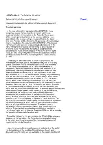
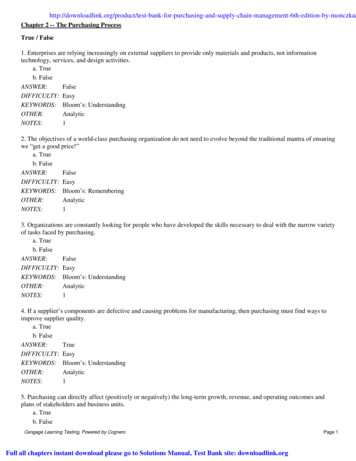
C CHHAAP PT TE ER R28Paul S. Masters Stanley PerlmanCoronaviridaeHistoryClassificationVirion StructureVirus and NucleocapsidVirion Structural ProteinsGenome Structure and OrganizationBasic and Accessory GenesCoronavirus GeneticsCoronavirus ReplicationVirion Attachment to Host CellsViral Entry and UncoatingExpression of the Replicase-Transcriptase ComplexViral RNA SynthesisAssembly and Release of VirionsPathogenesis and Pathology of Coronavirus InfectionsGeneral PrinciplesAnimal Coronavirus InfectionsHuman Coronavirus InfectionsImmune Response and Viral Evasion of the Immune ResponseEpidemiologyHuman Coronaviruses Other Than Severe Acute Respiratory Syndrome CoronavirusSevere Acute Respiratory SyndromeGenetic Diversity of CoronavirusesClinical FeaturesHuman Coronaviruses Other Than Severe Acute Respiratory Syndrome CoronavirusSevere Acute Respiratory Syndrome Coronavirus HISTORYCoronaviruses are enveloped RNA viruses that are broadly distributed among humans, other mammals, and birds, causingacute and persistent infections. Members of this family wereisolated as early as the 1930s as the causative agents of infectiousbronchitis in chickens,25 transmissible gastroenteritis in pigs,142and severe hepatitis and neurologic diseases in mice.75,186 It wasnot until the 1960s, however, that these viruses,27,32 as well ascertain human respiratory viruses,8,391 were recognized to sharecharacteristics that merited their being grouped together. Theirmost notable common feature, revealed by electron microscopy,was a fringe of widely spaced, club shaped spikes that projectedfrom the virion surface; these spikes were morphologicallydistinct from the surface projections of ortho and paramyxo viruses. The halo of spikes was described as giving the viral par ticle the appearance of the solar corona, which prompted thename that was adopted for this new virus group.7Over the next 40 years, coronaviruses were studied mainlybecause they cause economically signiicant respiratory andgastrointestinal diseases in domestic animals and because theyprovide unique models for viral pathogenesis. In humans, twocoronaviruses were known to be responsible for a substantialfraction of common colds, particularly those that circulate inwinter months. This situation changed dramatically with theemergence in 2002 of a devastating new human disease, severeacute respiratory syndrome (SARS), which was caused by a previously unknown coronavirus.143,288,440 Research stimulated bythe SARS outbreak has led to great strides in our understandingof coronaviruses; by 2005, two additional, widespread humanrespiratory coronaviruses had been discovered.573,615 Moreover,the search for animal virus reservoirs has nearly tripled the totalnumber of identiied coronaviruses,255,394,616 although most of therecently discovered species are known only as genomic sequencesand have yet to be isolated or propagated experimentally.CLASSIFICATIONThe coronaviruses are the largest group within the Nidovirales(Fig. 28.1), an order that comprises the families Coronaviridae,Arteriviridae,524 and Roniviridae.102 The arteriviruses, a smallgroup of mammalian pathogens, are discussed in Chapter 29.The roniviruses, which infect shrimp, and a very recently isolated mosquito-borne virus,416,663 which is not yet classiied, arecurrently the only members of the order having invertebratehosts. Nidoviruses are membrane-enveloped, nonsegmentedpositive-strand RNA viruses that are set apart from other RNAviruses by certain distinctive characteristics.194 Their most signiicant common features are (a) an invariant general genomicorganization, with a very large replicase gene upstream of thestructural protein genes; (b) the expression of the replicasetranscriptase polyprotein by means of ribosomal frame shifting; (c) a collection of unique enzymatic activities containedwithin the replicase-transcriptase protein products; and (d) theexpression of downstream genes via transcription of multiple825
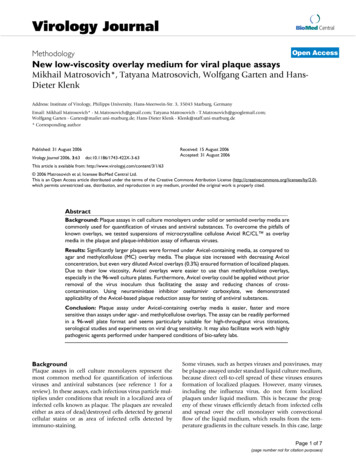
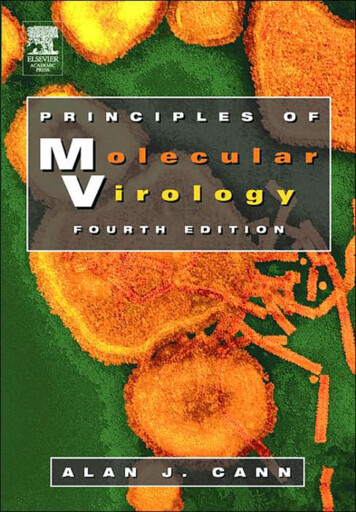
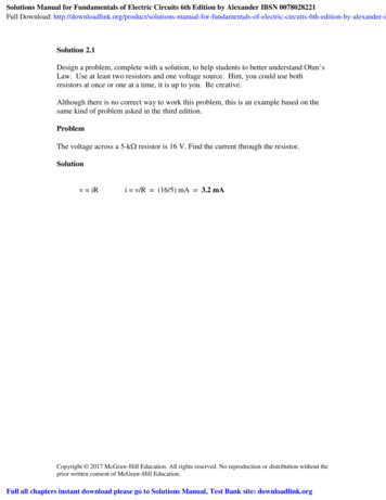
826SECTION II SPECIFIC VIRUS E 28.1. Taxonomy of the order Nidovirales.3′ nested subgenomic messenger RNAs (mRNAs). This lastproperty has provided the name for the order, which comesfrom the Latin nido, for “nest”.157 It should be noted that thereplicative similarities among the three nidovirus families areoffset by marked differences in the numbers, types, and sizesof their structural proteins and great variation among themorphologies of their virions and nucleocapsids.Coronaviruses are now classiied as one of two subfamilies(Coronavirinae) in the family Coronaviridae (see Fig. 28.1).The other subfamily, Torovirinae, includes the toroviruses,which are pathogens of cattle, horses, and swine,523 and thebainiviruses, whose sole member is the only nidovirus currently known to infect ish.505 This chapter will concentratealmost exclusively on the Coronavirinae.Coronaviruses have long been sorted into three groups, originally on the basis of serologic relationships and, subsequently,on the basis of phylogenetic clustering.193,195 Following proposals that were recently ratiied by the International Committeeon Taxonomy of Viruses (ICTV),57 these groups—the alpha-,beta-, and gammacoronaviruses—have now been accorded thetaxonomic status of genera (see Fig. 28.1). The ICTV classiications have also established rigorous criteria for coronavirusspecies deinitions, in a manner consistent with those used forother viral families. As a consequence, some viruses previouslyconsidered to be separate species are currently recognized as asingle species—for example, the viruses now grouped withinalphacoronavirus 1 or betacoronavirus 1 (Table 28.1). Additionally, the new classiication criteria resolve any previousuncertainty about the taxonomic assignment of the virus thatcaused SARS (severe acute respiratory syndrome coronavirus[SARS-CoV]) as a ost all alpha- and betacoronaviruses have mammalian hosts. In contrast, the gammacoronaviruses, with a singleexception, have been isolated from avian hosts. Several of theviruses listed in Table 28.1 have been studied for decades, speciically those included in the species alphacoronavirus 1, betacoronavirus 1, murine coronavirus, and avian coronavirus. Thefocus on these viruses came about largely because they wereamenable to isolation and growth in tissue culture. However,since 2004, molecular surveillance and genomics efforts initiated in the wake of the SARS epidemic have led to the discovery of a multitude of previously unknown coronaviruses thatnow constitute most members of this subfamily.616 Notably,most of the newly recognized species were identiied in bats,which constitute one of the largest orders within the mammals.Diverse coronaviruses have been described from bats, principally in Asia but also in Africa, Europe, and North and SouthAmerica. These viruses include likely predecessors of SARSCoV308,332 but also four unique species of alphacoronavirusesand three species of betacoronaviruses. Birds have also provento be a rich source of new viruses. Novel avian coronaviruseshave been found to infect geese, pigeons, and ducks,255 andhighly divergent coronaviruses recently identiied in bulbuls,thrushes, and munias617 have the potential to deine a fourthgenus in the Coronavirinae. It has been proposed that bats andbirds are ideally suited as reservoirs for the incubation and evolution of coronaviruses, owing to their common ability to lyand their propensity to roost and lock.616Five of the viruses in Table 28.1 are associated with humandisease. The most categorically harmful of these, SARS-CoV,which is discussed at length later in this chapter, does not currently infect the human population. The remaining four humancoronaviruses (HCoVs), the alphacoronaviruses HCoV-229Eand HCoV-NL63, and the betacoronaviruses HCoV-OC43and HCoV-HKU1, typically cause common colds. Remarkably, HCoV-NL63 and HCoV-HKU1 were only discoveredrecently, in the post-SARS era,573,615 despite the fact that eachhas a worldwide prevalence and has been in circulation fora long time.461,618 Although generally associated with upperrespiratory tract infections, the extant HCoVs can also causelower respiratory tract infections and have more serious consequences in the young, the elderly, and immunocompromisedindividuals. In particular, HCoV-NL63 is strongly associatedwith childhood croup,574 and the most severe HCoV-HKU1,-OC43, and -229E infections are manifest in patients withother underlying illnesses.460VIRION STRUCTUREVirus and NucleocapsidVirions of coronaviruses are roughly spherical and exhibit amoderate degree of pleomorphism. In the earlier literature,viral particles were reported to have average diameters of 80 to120 nm but were far from uniform, with extreme sizes from50 to 200 nm.389 The spikes of coronaviruses, typically describedas club-like or petal-shaped, emerge from the virion surface asstalks with bulb-like distal termini. Some of the variation in particle size and shape was likely attributable to stresses exerted byvirion puriication or distortions introduced by negative staining of samples for electron microscopy. More recent studies,employing cryo-electron microscopy and cryo-electron tomography,21,30,413,415 have produced images (e.g., Fig. 28.2A) inwhich virion size and shape are far more regular, although still
CHAPTER 28 CORONAVIRIDAETABLE 28.1827Classification of CoronavirusesSpeciesaGenus AlphacoronavirusAlphacoronavirus 1Human coronavirus 229E (HCoV-229E)Human coronavirus NL63 (HCoV-NL63)Porcine epidemic diarrhea virus (PEDV)Rhinolophus bat coronavirus HKU2 (Rh-BatCoV HKU2)Scotophilus bat coronavirus 512 (Sc-BatCoV 512)Miniopterus bat coronavirus 1 (Mi-BatCoV 1)Miniopterus bat coronavirus HKU8 (Mi-BatCoV HKU8)Genus BetacoronavirusBetacoronavirus 1cMurine coronavirusdHuman coronavirus HKU1 (HCoV-HKU1)Severe acute respiratory syndrome–related coronavirus(SARSr-CoV)GenBank 11AY278741DQ022305DQ071615Tylonycteris bat coronavirus HKU4 (Ty-BatCoV HKU4)Pipistrellus bat coronavirus HKU5 (Pi-BatCoV HKU5)Rousettus bat coronavirus HKU9 (Ro-BatCoV HKU9)Genus GammacoronavirusAvian coronaviruseBeluga whale coronavirus SW1Previous names for viruses included innewly defined speciesFeline coronavirus type I (FeCoV I)Feline coronavirus type II (FeCoV II), Felineinfectious peritonitis virus (FIPV)Canine coronavirus (CCoV)Transmissible gastroenteritis virus (TGEV)Bovine coronavirus (BCoV)Equine coronavirus (EqCoV)Human coronavirus OC43 (HCoV-OC43)Porcine hemagglutinating encephalomyelitisvirus (PHEV)Mouse hepatitis virus (MHV)Rat coronavirus (RCoV)Human severe acute respiratory syndromecoronavirus (SARS-CoV)Severe acute respiratory syndrome–relatedRhinolophus bat coronavirus HKU3(SARSr-Rh-BatCoV HKU3)Severe acute respiratory syndrome–relatedRhinolophus bat coronavirus Rp3 (SARSr-RhBatCoV 42Infectious bronchitis virus (IBV)Turkey coronavirus (TuCoV)Listed viruses are those for which complete genome sequences are available. Novel viruses that have not yet been formally classified include Bulbul coronavirus HKU11,617Thrush coronavirus HKU12,617 Munia coronavirus HKU13,617 Asian leopard cat coronavirus,139 and Mink coronavirus.592bRepresentative GenBank accession numbers are given for viruses in each species; in many cases, multiple genomic sequences for a given virus are available.cOther viruses included in the species Betacoronavirus 1 are Human enteric coronavirus (HECoV) and Canine respiratory coronavirus (CRCoV), for which only partial genomicsequences are available.dOther viruses included in the species Murine coronavirus are Puffinosis virus (PCoV) and Sialodacryoadenitis virus (SDAV), for which only partial genomic sequences areavailable.eOther viruses included in the species Avian coronavirus are Pheasant coronavirus (PhCoV), Goose coronavirus (GCoV), Pigeon coronavirus (PCoV), and Duck coronavirus(DCoV), for which only partial genomic sequences are available.255apleomorphic. These studies, which examined a number of alpha and betacoronaviruses, converge on mean particle diameters of118 to 136 nm, including the contributions of the spikes, whichproject some 16 to 21 nm from the virion envelope.Enclosed within the virion envelope is the nucleocapsid—a ribonucleoprotein that contains the viral genome. The struc ture of this component is relatively obscure in images of wholevirions; however, its makeup has been partially displayed byelectron micrographs of spontaneously disrupted virions orof virions solubilized with nonionic detergents.59,109,183,269,366Such studies revealed another distinguishing characteristic ofcoronaviruses: They have helically symmetric nucleocapsids.
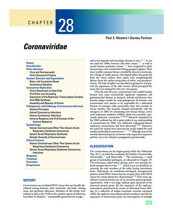
828SECTION II SPECIFIC VIRUS FAMILIESABESMN gRNAlipid bilayer envelopeFIGURE 28.2. Coronavirus structure. A: Cryo-electron tomographic image of purified virions of mouse hepatitis virus (MHV), reconstructed as described in reference 415. (Courtesy of Benjamin Neuman, David Bhella, and Stanley Sawicki.) B: Schematic showing themajor structural proteins of the coronavirus virion: S, spike protein; M, membrane protein; E, envelope protein; and N, nucleocapsid protein.Helical symmetry is common for negative strand RNA virusnucleocapsids, although it is highly unusual for positive strandRNA animal viruses, almost all of which have icosahedral cap sids. The best resolved images of the coronavirus nucleocapsid,which were obtained with HCoV 229E, showed ilamentousstructures 9 to 13 nm in diameter, with 3- to 4-nm-widecentral canals59; these ilaments were th
826 SECTION II SPECIFIC VIRUS FAMILIES 3′ nested subgenomic messenger RNAs (mRNAs). This last property has provided the name for the order, which comes from the Latin nido, for “nest”.157 It should be noted that the replicative similarities among the three nidovirus families are