Transcription
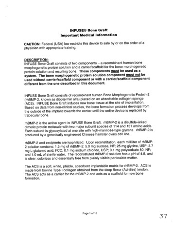
INFUSE Bone GraftImportant Medical InformationCAUTION: Federal (USA) law restricts this device to sale by or on the order of aphysician with appropriate training.DESCRIPTION:INFUSE Bone Graft consists of two components - a recombinant human bonemorphogenetic protein solution and a carrier/scaffold for the bone morphogeneticprotein solution and resulting bone. These components must be used as asystem. The bone morphogenetic protein solution component must not beused without carrier/scaffold component or with a carrier/scaffold componentdifferent from the one described in this document.INFUSE Bone Graft consists of recombinant human Bone Morphogenetic Protein-2(rhBMP-2, known as dibotermin alfa) placed on an absorbable collagen sponge(ACS). INFUSE Bone Graft induces new bone tissue at the site of implantation.Based on data from non-clinical studies, the bone formation process develops fromthe outside of the implant towards the center until the entire device is replaced bytrabecular bone.rhBMP-2 is the active agent in INFUSE Bone Graft. rhBMP-2 is a disulfide-linkeddimeric protein molecule with two major subunit species of 114 and 131 amino acids.Each subunit is glycosylated at one site with high-mannose-type glycans. rhBMP-2 isproduced by a genetically engineered Chinese hamster ovary cell line.rhBMP-2 and excipients are lyophilized. Upon reconstitution, each milliliter of rhBMP2 solution contains: 1.5 mg of rhBMP-2; 5.0 mg sucrose, NF; 25 mg glycine, USP; 3.7mg L-glutamic acid, FCC; 0.1 mg sodium chloride, USP; 0.1 mg polysorbate 80, NF;and 1.0 mL of sterile water. The reconstituted rhBMP-2 solution has a pH of 4.5, andis clear, colorless and essentially free from plainly visible particulate matter.The ACS is a soft, white, pliable, absorbent implantable matrix for rhBMP-2. ACS ismade from bovine Type I collagen obtained from the deep flexor (Achilles) tendon.The ACS acts as a carrier for the rhBMP-2 and acts as a scaffold for new boneformation.Page 1 of 1537
Each kit contains all the components necessary to prepare INFUSE Bone Graft: therhBMP-2 which must be reconstituted, sterile water, absorbable collagen sponges,syringes with needles, this package insert and instructions for preparation.The rhBMP-2 is provided as a lyophilized powder in vials delivering 12 mg of protein.After appropriate reconstitution, the concentration of rhBMP-2 is 1.5 mg/mL. Thesolution is then applied to the provided absorbable collagen sponge. INFUSE BoneGraft is prepared at the time of surgery and allowed a prescribed amount of time (noless than 15 minutes) before placement at the fracture site. The Instructions forPreparation contain complete details on preparation of INFUSE Bone GraftINDICATIONS:INFUSE Bone Graft is indicated for treating acute, open tibial shaft fractures that havebeen stabilized with IM nail fixation after appropriate wound management. INFUSEBone Graft must be applied within 14 days after the initial fracture. Prospectivepatients should be skeletally mature.CONTRAINDICATIONS*INFUSE Bone Graft is contraindicated for patients with a known hypersensitivity torecombinant human Bone Morphogenetic Protein-2, bovine Type I collagen or toother components of the formulation.*INFUSE Bone Graft should not be used in the vicinity of a resected or extanttumor, in patients with any active malignancy or patients undergoing treatment fora malignancy.*INFUSE Bone Graft should not be used in patients who are skeletally immature( 118 years of age or no radiographic evidence of epiphyseal closure).*INFUSE Bone Graft should not be used in patients with an inadequateineurovascular status, e.g., high risk of amputation.*INFUSE Bone Graft should not be used in patients with compartment syndrome of-the affected limb.*INFUSE Bone Graft should not be used in pregnant women. The potential effectsof rhBMP-2 on the human fetus have not been evaluated.*INFUSE Bone Graft should not be implanted in patients with an active infection atthe operative site.Page 2 of 153
WARNINGS:*Women of childbearing potential should be advised that antibody formation torhBMP-2 or its influence on fetal development have not been assessed. In theclinical trial supporting the safety and effectiveness of INFUSE Bone Graft in tibialfracture, 9/149 (6.0%) patients treated with INFUSE Bone Graft and 1/150 (0.7%)patients treated without exposure to rhBMP-2 developed antibodies to rhBMP-2.The effect of maternal antibodies to rhBMP-2, as might be present for severalmonths following device implantation, on the unborn fetus is unknown.Additionally, it is unknown whether fetal expression of BMP-2 could re-exposemothers who were previously antibody positive, thereby eliciting a more powerfulimmune response to BMP-2 with adverse consequences for the fetus. Studies ingenetically altered mice indicate that BMP-2 is critical to fetal development andthat lack of BMP-2 activity, as might be induced by antibody formation, may causeneonatal death or birth defects.*'The safety and effectiveness of INFUSE Bone Graft in nursing mothers has notbeen established. It is not known if BMVP-2 is excreted in human milk.*Women of childbearing potential should be advised not to become pregnant forone year following treatment with INFUSE Bone Graft.*'The safety and effectiveness of INFUSE Bone Graft for non-acute fractures, withforms of internal fracture fixation other than IM nails, implanted at locations otherthan the tibial shaft, or used in surgical techniques other than open reduction andinternal fixation after appropriate wound management have not been established.PRECAUTIONS:General* 'The safety and effectiveness of repeat applications of INFUSE Bone Graft has notbeen established.*Long-bone fracture and soft-tissue management procedures should be based onstandard practice including control of infection. Physicians should achievemechanical stability before implanting INFUSE Bone Graft. INFUSE Bone Graftshould not be used to fill space in the presence of compressive forces.*INFUSE Bone Graft should only be used by surgeons who are experienced intreating acute open tibial shaft fractures involving IM nail stabilization and haveundergone adequate training with this device.Page 3 of 1539
A single package of INFUSE Bone Graft should be used at the fracture site.·INFUSE Bone Graft is intended for single use only. Discard unused product anduse a new device for subsequent applications. INFUSE Bone Graft must not besterilized by the hospital.**Prior to use, inspect the packaging, vials and stoppers for visible damage. Ifdamage is visible, do not use the product. Retain the packaging and vials andcontact a Medtronic Sofamor Danek, Inc. representative.[Do not use after the printed expiration date on the label.Hepatic and Renal Impairment* The safety and effectiveness of INFUSE Bone Graft in patients with hepatic orrenal impairment has not been established. Pharmacokinetic studies of rhBMP-2indicate that the renal and hepatic systems are involved with its clearance.Geriatricsstudies of INFUSE Bone Graft did not include sufficient numbers of*Clinicalpatients 65 years and older to determine whether they respond differently fromyounger subjects.Bone formation*The safety and effectiveness of INFUSE Bone Graft has not been demonstrated inpatients with metabolic bone diseases, or in pathological fractures such as thoseobserved in Paget's disease or in metastatic bone disease. 'While not specifically observed in the clinical study, the potential for ectopic,heterotopic or undesirable exuberant bone formation exists.Antibody Formation/Allergic Reactions*The safety and effectiveness of INFUSE Bone Graft has not been demonstrated inpatients with autoimmune disease.*The safety and effectiveness of INFUSE Bone Graft has not been demonstrated inpatients with immunosuppressive disease or suppressed immune systemsresulting from radiation therapy, chemotherapy, steroid therapy or othertreatments.Immunogenicity·As with all therapeutic proteins, there is a potential for immune responses to begenerated to INFUSE Bone Graft. The immune response to INFUSE Bone GraftPage 4 of 15 "'0
was evaluated in 149 investigational patients and 150 control patients receivingtreatment for acute open tibial shaft fractures stabilized with IM nails.* TmAnti-rhBMP-2 antibodies: 9/1 49 (6%) patients receiving the InFUSE BoneGraft component developed antibodies vs. 1/1 50 (0.7%) in the controlgroup.Anti-bovine Type I collagen antibodies: 29/149 (20%) of patients receivingthe InFUSE Tm Bone Graft component developed antibodies to bovine TypeI collagen vs. 9/1 50 (6%) of control patients. No patients in either groupdeveloped anti-human Type I collagen antibodies.* The presence of antibodies to rhBMP-2 was not associated with immunemediated adverse events such as allergic reactions. The neutralizingcapacity of antibodies to rhBMP-2 is not known.'The incidence of antibody detection is highly dependent on the sensitivity andspecificity of the assay. Additionally, the incidence of antibody detection may beinfluenced by several factors including sample handling, concomitant medicationsand underlying disease. For these reasons, comparison of the incidence ofantibodies to INFUSE Bone Graft with the incidence of antibodies to otherproducts may be misleading.ADVERSE EVENTS:The table below describes the adverse events observed in the clinical trial used tosupport approval of the product. Two INFUSE Bone Graft doses, 0.75mg/mI and1.5mg/mi, were evaluated. INFUSE Bone Graft with IM nail stabilization wasimplanted in 300 investigational patients (149 in the 1.50 mg/mI and 151 in the 0.75mg/mI groups) compared to IM stabilization alone in 150 control patients. Adverseevent rates presented are based on the number of patients having at least oneoccurrence for a particular adverse event divided by the total number of patients inthat treatment group.Page 5 of 15
04N-0,I0).0LaC --0--0- 0--0ao 0NJ0)0-0)00C01 LaL0)040 .0040)0 La00)-0N400-0a- 04 010,0000 - 04C0-'75 D' LoO' 7La--o-o)0)oLLa00)La0LaLa004 I.- .0.0L"0o r.0w oooc4(D Ca. a. T- 0. * 0'"rL 00 000-0000 r0 *0E E0o!LaE 'C--000--CD0 N-0 0 0 00 0 000000)ooC000'00
C-- C--CC-1- C-CCJN--- CCI-- I cooImCCI-NC,CCCOCJN-C(Ua. C0.'IDIDNJIDJCCCaaCCNCJCC0CU) 0zrUS D*0aa a a a .C- r'--CCC*0CCCNJ0CCC-CJC- 0 0 m4t 0 4
co'00) Cr-N -OCNNoCCO. C 0CQ -- 2E. N0CO c-04N COCO 0'-C0-0.,iONC-aC-'o.-C-'I-- ce.N! .COaN-NN-CON-COCOa- E-O-a-COCOC- -o0. .0 00 07UC) 'C 9.-90 S ZU
Potential Adverse Events:The following is a list of potential adverse events which may occur with treatment ofopen tibial fractures requiring stabilization with an IM nail. Some of these adverseevents may have been previously reported inthe adverse events table. As with anysurgey surIca treatment of a fracture is not without risk. A variety of complicationsrelated to surgery or the use of INFUSE Bone Graft can occur. These may occursingly or in combination. Some of these may be severe, affecting patient outcome.* EBone fracture.* EBowel, bladder or gastrointestinal problems.* Change in mental status.* Damage to blood vessels, bleeding (which may require a blood transfusion) orcardiovascular system compromise.* Damage to nearby tissues.* [)eath.* Development of respiratory problems.* Disassembly, bending, breakage, loosening, and/or migration of IM nailcomponents.* EEctopic and/or exuberant bone formation.* F-etal development complications.* Foreign body (allergic) reaction.* Incisional complications.* Infection.* Neurological system compromise. Nonunion (or pseudarthrosis), delayed union, mal-union. Pain or discomfort.* Rlash or allergic reaction.* Scar formation.* Side effects from anesthesia or the surgical approach.a OSwelling.*Tissue or nerve damage.Note: Additional surgery may be necessary to correct some of these potentialadverse events.CLINICAL RESULTS:Safety and effectiveness of INFUSE Bone Graft were evaluated as part of a prospective, randomized,controlled, multinational (11 countries), multi-center (49 sites) study. Subjects were randomized intoone of three groups - control or one of two investigational groups (0.75 or 1.50mg/mI rhBMP-2). Allsubjects received wound management and fracture stabilization with an IM nail, while the investigationalsubjects also received INFUSE Bone Graft at the fracture site. No restrictions were placed on the typeof IM nail used or whether it was reamed or non-reamed. The use of bone wax, Gelfoam, or othercollagen hemostatic agents, corticosteroids, bone growth stimulators (electrical, ultrasound, ormagnetic) was specifically prohibited.Page 9 of 154,
Only the subjects and an independent radiology panel were blinded with respect to treatment. Theinvestigators were aware of the treatment assignment.The enrolled patients had been diagnosed with an acute, open tibia fracture of Gustilo Grade 1,11,IlA orIIIB3 with the major component of the fracture being diaphyseal. Patients with isolated tibia fractures andthose with multiple injuries were included.Clinical and radiographic effectiveness parametersSubjects were followed for 12 months after definitive wound closure (DWC). Evaluations wereperfcirmed postoperatively at 6, 10, 14, 20, 26, 39 and 50 weeks. Adverse events, device-related or not,were evaluated over the course of the clinical trial. At each evaluation timepoint, the primary andsecondary clinical and radiographic outcome parameters were evaluated. Success was determinedfrom data collected during the initial 12 months of follow-up. Antibodies to rhBMP-2 and bovine Type Icollagen were assessed preoperatively and at timepoints post-operatively. Antibodies to human Type Icollagen were assessed if the antibody response to bovine Type I collagen was positive.Clinical and radiographic fracture assessments were performed at each postoperative visit, however theprotocol did not provide the specific objective criteria that were used to determine fracture healing. The"Assessment of Fractured Limb" case report form provided for the documentation of the followingparameters, but did not indicate how these were to be used to determine fracture status or how manyneeded to be present in order for a complete evaluation to have occurred:*wound (healed, not healed, not evaluated)-pain (absent, present, not evaluated)-swelling (absent, present, not evaluated)-tenderness (absent, present, not evaluated)*neurovascular status (intact, impaired, not evaluated)-infection (absent, present, not evaluated)-weight-bearing status (non-, touch down, partial, full, not evaluated).Radiographic assessments (AP and lateral radiographs) were performed at each post-op visit. Obliqueradiographs were to be used if the standard views did not adequately visualize the fracture. Theradiographs were evaluated by the investigator and the radiology panel.Investigators were to evaluate radiographic healing by assigning a score of "united", "not united","uncertain union" or "uni nterpretabl e". No definitions for these terms were provided in the protocol.The protocol for the independent radiology panel stated that ".fracture union was determined if therewas cortical bridging and/or disappearance of the fracture lines were visible on 3 of the 4 bone aspects(anterior, posterior, medial, and lateral).". These definitions were not available to the investigators.The first visit at which these criteria were met was considered the time of union radiographically. Theindependent radiographic evaluation protocol called for the review of each radiograph by all threemembers of the panel. An agreement of 2 of the 3 reviewers was necessary for a determination offracture union. The independent radiographic evaluation was performed on all available radiographs.Adverse events were assessed for relatedness to the device and severity was based on the WHOrecommendations.Investigators were provided with the following definitions:".Nonunion - considered to be established when a minimum of 9 months have elapsed sinceinjury and the fracture site showed no visibly progressive signs of healing for a minimum of 3months (no change of fracture callus).Page 10 of 15
Delayed union - insufficient fracture healing determined by radiographic and clinicalassessment. Aspecific time point at which delayed union was defined was not provided.Secondary intervention for delayed union - any intervention, surgical or non-surgical, that wasperformed to induce or accelerate fracture union after DWC. Examples included use ofautograft, allograft or bone graft substitutes; IMnail dynamization; exchange nailing; ornoninvasive modalities, e.g., ultrasound, magnetic field, or electrical stimulation. Thedecision to perform a secondary intervention for delayed union was dependent on the definitionof delayed union above.Investigators determined fracture union based on clinical judgement. The protocol did not provide thespecific objective criteria that were used to determine fracture healing or deciding whether torecommend secondary interventions to promote fracture healing.Patient demographics and accountabilityThe sample size estimation called for 150 subjects per treatment group. A total of 149 investigationaland "150 control patients were enrolled in the study and received treatment. Only the results from thecontrol patients and investigational patients receiving the 1.5mg/ml dose device are described below.The demographics of the patient population were similar across all study groups except for theparameter of age, specifically the mean and range. The subjects in the investigational group wereyounger (mean 33.4 years, range 18-77 years) compared to the control group (mean 36.8 years,range 17-87 years). Patients in the control group had a slightly larger percentage of nails that wereunreamed and/or less than 9mm, while the investigational group had a higher percentage of reamednails and /or nails that were greater than 11mm. Nail type did affect the number of secondaryinterventions, i.e., patients with unreamed nails had a higher incidence of secondary interventionscompared to patients receiving reamed nails.Clinical and radiographic effectiveness evaluationThe primary efficacy endpoint was defined as the proportion of subjects who required a secondarysurgical intervention to promote fracture healing within 12 months of DWC. The secondary efficacyendpoints included the following:*the proportion of subjects healed at 6 months without a secondary intervention as determinedby the investigator's clinical and radiographic assessment;*the independent radiology panel's assessment of time to fracture union; and-the pharmacoeconomic impact of the treatment.Primary effectiveness endpointThe rate of secondary interventions was significantly lower inthe INFUSE Bone Graft group (p 0.001)as described in the table below. Interventions were categorized in one of three ways - recommendedby the investigator and performed, recommended by the investigator and not performed, or notrecommended by the investigator but performed anyway. Ifany of these occurred, the patient wasconsidered to have failed the primary endpoint and, therefore, was considered a study failure. Inaddition, patients who experienced screw breakage resulting in self-dynamization were also consideredtreatment failures.Number of patients with secondary rformedRecommended/not performedNot recommended/performed(n 150)(n 149)38361957Page 11 of 15(A
Self-dynamizationsTotal failures/patients (%)I19766/150 (44)38/149 (26)"Includes bone grafting, fibula osteotomies, exchange nailing, plate fixation,llizarov frame removal, external fixation placement, bone transport, IMnaildynamizations, exchange to a functional brace and electrical stimulationSafety and immune response evaluationThe assessment of safety consisted of an evaluation of the reported adverse events, as well as anevaluation of antibodies to rhBMP-2, bovine Type I collagen and human Type I collagen. The completelist of adverse events is described in the Adverse Events section above. Refer to this section for adescription of the rates associated with infection and abnormal clinical lab values. Adverse events ofspecial interest are discussed below.fracture healingThe rates of hardware failure in the investigational and control groups were 18/149 (14%) and 32/150(24%), respectively. Delayed union was the most frequent serious adverse event reported at one year;occurring in 39 (26%) control and 26 (17%) investigational patients.The rate of nonunion was lower in the investigational group as compared to control. A total of 80/150(53.3%) control and 56/149 (37.6%) investigational patients did not require a secondary intervention andwere not radiographically healed at 12 months as determined by the independent radiology panel. Forthe control patients who required a secondary intervention, 18/66 (27%) reported nonunions at 12months compared to 19/38 (50%) investigational patients. Investigational patients who required asecondary intervention were considered healed laterthancontrol patients.abnormalbone formationHeterotopic ossification (HO) was not a significant concern and no ectopic ossification was reported.Because only the involved tibia was evaluated radiographically, it is not clear if abnormal bone formationoccurred in other anatomical locations. Although twice as many investigational patients reportedhypertrophic callus formation compared to controls (8 vs. 4), no action was required to treat any of theseevents. A total of 12 patients experienced at least one event classified as hypertrophic callus, with theinvestigational group having the highest number (8) and percentage (6%) of patients with HO. Nointerventions were required to treat any of the HO-related events.infectionsThe combined rate of deep and superficial infections of the injured limb was lower in patients withGustilo IliA and B fractures of the investigational group as compared to the control group[16/66 (24%) and 26/61 (43%), respectively].immune responseThe presence of antibodies was assessed using ELISA. Ifthere was a positive response to bovineType I collagen, the serum was also tested for antibodies to human Type I collagen. The screeningELISA cutpoint for positive antibody responses was set to 2 times the signal generated by poolednorrmal human sera in each ELISA. Subjects were considered to have an elevated immune response ifthe preoperative test was negative (titer 50) and postoperative test was positive (titer 50) or ifthepreoperative test was positive and the postoperative test was positive with a three-fold higher titer thanthe preoperative test.There were detectable rhBMP-2 antibodies in 1 control patient and 9 investigational patients aftertreatment. Of the 9 investigational patients with elevated post-treatment antibody titers, 2 were elevatedat visit 6 (20 weeks), the last planned assessment and data from 1 patient were unavailable for visit 6.An additional sample from 1 of these patients was collected and tested. following the positive test atPage 12 of 15
visit 6, and the titers decreased to 50 (No follow-up data were available for the other patient. AntirhBMP-2 antibody responses were determined to be transient in 6/9 patients by 20 weeks, and in 7/9patients after follow-up testing (samples from 2 patients were unavailable to confirm transience of theantibody response). Because of the small numbers involved, it was not possible to determine ifacorrelation existed between the immune response and clinical outcome.There were 38 patients who developed antibodies to bovine Type I collagen - 9 (6%) control and 29(20%) investigational patients. Approximately half of the patients had persistently elevated antibodytiters at evaluations 20 weeks and more after DWC. Thirty categories of adverse events that may havebeen manifestations of an immune response were identified and all were observed to have acomparable incidence across all groups. Although there were 4 patients with an adverse event termed"allergic event", the investigators believed that there was no evidence of allergic response to theinvestigational treatment.Immune responsecontrol [n (%]investigational [n (%)]anti-rhBMP-2 antibodiesanti-bovine Type Icollagen antibodies1 (1)9 (6)9 (6)29 (20)anti-human Type I collagen antibodies0 (0)0 (0)1063# Iealed patients with antiBMP-2 antibody response (successes)# secondary intervention patients with antiBMP-2 antibody response (failures)The rates of authentic antibody response to rhBMP-2 were higher than that observed for anotherapplication of rhBMP-2/ACS. When rhBMP-2/ACs was placed inside of a metallic spinal fusion cage foranterior interbody fusion treatment of degenerative disc disease, the antiBMP-2 antibody response inthe investigational group was 0.7%. This compares to a 6% rate in the investigational group in thetrauma study. The contribution of the trauma setting to this outcome is unknown, as is the clinicalsignificance of the antibody response.HOW SUPPLIED:INFUSE Bone Graft is supplied in a kit containing all the components necessary toprepare the device (i.e., the collagen sponge, a vial with the lyophilized rhBMP-2, avial with the sterile water for reconstituting the rhBMP-2, syringes and needles).STORAGE CONDITIONS:Store INFUSE Bone Graft at room temperature [15 - 25 degrees Centigrade ( 59 to770 F)].DOSAGE AND ADMINISTRATION:INFUSE Bone Graft is prepared immediately prior to use from a kit containing allnecessary components. Once prepared, INFUSE Bone Graft contains rhBMP-2 at aconcentration of 1.5 mg/mL. The instructions for preparation must be followed and therhBMP-2 must be reconstituted to this solution concentration of 1.5 mg/ml and thendistributed uniformly across the entire ACSPage 13 of 15
Only a single INFUSE Bone Graft kit should be used for each patient.INFUSE Bone Graft is implanted after the completion of IMnail fracture stabilizationand wound management, i.e., at the time of soft tissue coverage. The number ofINFUSE Bone Graft kits and the volume of INFUSE Bone Graft to be implanted aredetermined by the fracture anatomy. Generally, the fracture is treated with one kit.The accessible surface area of the fracture (fracture lines and defects) should becovered with INFUSE Bone Graft. Because very few patients in the clinical trialreceived more than one INFUSE Bone Graft kit (specifically, 2 kits) the response tothe use of more than one kit is unknown. When determining the specific volume andplacement for INFUSE Bone Graft, the poential to induce compartment syndromeshould be considered.DIRECTIONS FOR USE:For directions for using the INFUSE Bone Graft for tibia fractures, see the brochureentitled "Instructions for Preparation and Surgical Application.".PRODUCT COMPLAINTS:Any health care professional (e.g., customer or user of this system of products), whohas any complaints or who has experienced any dissatisfaction in the quality,identification, durability, reliability, safety, effectiveness and/or performance of thisproduct, should notify the distributor, Medtronic Sofamor Danek, Inc. Further, if any ofthe implanted INFUSE Bone Graft ever "malfunction," (i.e., do not meet any of theirperformance specifications or otherwise do not perform as intended), or aresuspected of doing so, the distributor should be notified immediately. Ifany MedtronicSofamor Danek, Inc. product ever "malfunctions" and may have caused or contributedto the death or serious injury of a patient, the distributor should be notifiedimmediately by telephone, fax or written correspondence. When filing a complaint,please provide the component name and number, lot number, your name andaddress, the nature of the complaint and notification of whether a written report fromthe distributor is requested.SUPPLIED BYMedtronic Sofamor Danek USA, Inc1800 Pyramid PlaceMemphis, Tennessee 38132USATelephone 800-876-3133 or901-396-3133Telefax: 901 396-0356901 332-3920Page 14 of 155o
Customer Service Division Telephone 800-933-2635INFUSE Bone Graft is a registered trademark of Medtronic Sofamor Danek, Inc.Covered by patents U.S. 5,013,649, U.S.5,618,924, U.S.5,166,058 and U.S.5,631,142All rights reserved 20C)4 Medtronic Sofamor Danek, Inc.Page 15 of 15
INFUSE Bone GraftPATIENT INFORMATION BROCHURERead this entire document carefully.Keep this document. You may want to read it again.If you have further questions, please ask your doctor.-
TABLE OF CONTENTS.WHAT Is INFUSE BONE GRAFT? .WHAT Is3.3INFUSE BONE GRAFT USED FOR? .WHO IS NOT A CANDIDIATE FOR.3INFUSE BONE GRAFT? .WHAT ARE SOME PRECAUTIONS AND WARNINGS FOR USINGINFUSE BONE GRAFT? . 4WHAT WILL HAPPEN BEFORE SURGERY THE INVOLVINGINFUSEBONE GRAFT? . 4.5WHAT WILL HAPPEN DURING SURGERY? .5WHAT WILL HAPPEN AFTER SURGERY? .WHAT ARE SOME POSSIBLE SIDE-EFFECTS AND ADVERSE EVENTS.5OF USING INFUSE BONE GRAFT? .HASINFUSE BONE GRAFT BEEN STUDIED IN HUMANS? . 6.WHAT ELSE SHOULD I KNOW?
These components must be used as a system. The bone morphogenetic protein solution component must not be used without carrier/scaffold component or with a carrier/scaffold component different from the one described in this document. INFUSE Bone Graft consists of recombinant human Bone Morphogenetic Protein-2 .