Transcription
Animal Physiology Laboratory, Zool 430LWeek 4Spring 2018Lab #4: Physiology of the in situ Amphibian HeartThis experiment explores the basic principles of cardiac muscle physiology, including contraction force, ECG,the effect of temperature, and the effect of neurotransmitters on cardiac regulation in a myogenic heart (Witherspp. 715-719 or HWA 651-654).BackgroundStudies of isolated organs were pioneered in the late 19th century when scientists such as Sidney Ringer(1835–1910) developed a perfusion solution (Ringer’s solution) that could sustain an isolated organ from apithed animal. A classic example of this phenomenon is the frog heart, which will continue to beat in situ forseveral hours allowing for the study of basic cardiac functions.The heart is made up of specialized tissue called cardiac muscle. Cardiac muscle differs from skeletalmuscle both morphologically and functionally. Probably the most striking and fascinating feature of itscontractility is that it is able to initiate its own rhythmic contractions intrinsically without requiring extrinsicstimulation from outside the heart. This occurs because cardiac muscle cells have "leaky" cell membranes thatallow calcium and sodium ions to slowly leak into the cells. This leaking causes a slow depolarization tothreshold, thus causing the firing of an action potential, and initiating muscle contraction. The cells that aremost "leaky" to ions and that depolarize fastest control the rate of contraction of all other cardiac cells; thus,they act as pacemakers for the rest of the heart. The depolarization of pacemaker cells spreads to the entireheart via electrical connections between cardiac muscle cells called gap junctions.Although no external stimulation is required to maintain the heartbeat, cardiac rate and output is highlymodulated by extrinsic signals from the sympathetic (synapses in brain or spinal cord) and parasympathetic(synapses in other tissues) autonomic nervous systems (see Withers pp. 352-355 or HWA 406-409). Thesenerve fibers release a variety of neurotransmitters (see Withers pp. 239-243 or HWA pp. 342-343), which canincrease or decrease the rate and contractile properties of the beating heart. These molecules are able toinfluence heart rate by changing the rate of spontaneous depolarization of the heart’s “pacemaker” cells, locatedin the sinoatrial (SA) and atrioventricular (AV) nodes of the mammalian heart. In the frog, the sinus venosus issimilar to the SA node. Molecules that either increase or decrease the heart rate are called chronotropic(chrono- for time) agents, and molecules that alter contractility of cardiac muscle are called inotropic agents.Signals from the sympathetic nervous system accelerate cardiac activity while signals from the parasympatheticnervous system decelerate cardiac activity.The heart also responds directly to physical factors such as temperature. For ectothermic organisms, bodytemperature can and does change, sometimes rapidly during day-night habitat temperature variation.Temperature change exerts its effect on heart rate through both intrinsic and extrinsic means. Intrinsic effectsare those that impact temperature-sensitive aspects of cardiac metabolism, cellular, and subcellular processes.Extrinsic effects are modulated through the temperature sensory mechanisms of the nervous system.In addition, the contractile properties of the heart are dependent upon the physical characteristics of thecardiac muscle fibers. In the early 1900s, Ernest Starling's investigations demonstrated that the energy ofcardiac contraction is proportional to the initial length of the cardiac muscle fibers. This statement becameknown as Starling's law of the heart, a major concept in cardiovascular physiology.MethodsIn this laboratory we will perform experiments on the exposed heart from the cane toad, Rhinella marinus(formerly Bufo marinus) to demonstrate the effects of intrinsic, extrinsic, and physical factors on cardiacfunction. The effects of temperature change on the surgically exposed toad heart will be observed by droppingwarm or cold frog Ringer’s solution directly onto the heart. Starling’s Law will be observed by stretching theventricle. You will also demonstrate the effects of direct stimulus on heart function as well as the block ofintrinsic signaling from the atria to ventricle. The effects of extrinsic factors of the sympathetic andparasympathetic nervous systems will be demonstrated by application of Ringer’s solution containingphysiological amounts of neurotransmitter directly on the heart.Page 1! of 6!
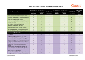
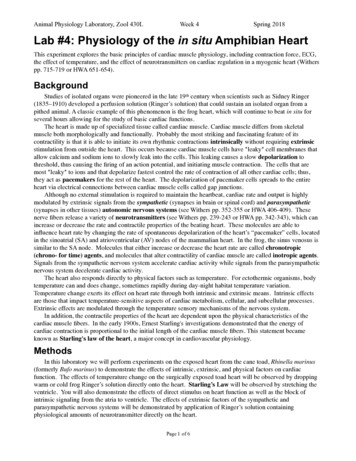
Animal Physiology Laboratory, Zool 430LWeek 4Spring 2018Required EquipmentPowerLabBridge Pod Force TransducerStimulator BarBioAmp microhook lead wiresMounting stand with micropositionerThermocouple Pod and thermocoupleSuture threadStraight pinsBarb-less hookDissection toolsEyedropperRinger’s solution, room tempCold Ringer’s (5 C)Warm Ringer’s (40 C)Ringer’s with:Acetylcholine (0.1 mg/mL)Epinephrine (1% solution)Pilocarpine (2.5% solution)Atropine sulfate (5% solution)CaffeineCadmium ChlorideKClCalcium-free Ringer’sPotassium-free Ringer’sProceduresFigure 1. Setup of dissected toad and ForceTransducer.A. Setup and calibration of equipment1. Set up your mounting stand with the Force Transducer mounted on the micropositioner (Figure 1).2. Connect:Force transducer - Bridge Pod - PowerLab (Input 1).Thermocouple Pod - Input 2.Hook electrode (micrograbber) leads into the BioAmp cable, BioAmp - Input 3.3. Software setup:“Toadheart” Settings file.Select Bridge Pod from the Force Channel Function pop-up menu.Zero the bridge pod reading by using the zeroing knob on the front of the bridge pod.B. Toad dissection procedureRefer to the toad dissection guide for diagrams of this procedure.1. Obtain a double-pithed toad from your instructor. Secure the animal ventral side up to a dissecting boardusing straight pins.2. Lift the abdominal skin with forceps, make a longitudinal incision from the thorax to the abdomen using ascalpel or scissors.3. Peel back the skin to expose the sternum and ribs.4. Using sharp scissors, cut through the sternum to expose the thoracic cavity. You should see the heart in itspericardial sac.Page 2! of 6!
Animal Physiology Laboratory, Zool 430LWeek 4Spring 20185. Grasp the pericardium with forceps and carefully cut it away, exposing the heart. Apply Ringer’s solution tothe heart every two or three minutes to prevent desiccation.6. Attach a small hook tied with thread through the toad’s heart. Push the hook through the apex of the heart.Note: Be very careful NOT to pierce the ventricular cavity! The hook is used to tie the heart to the forcetransducer to measure the force of heart contractions as they are transmitted through the string.7. Adjust the micropositioner so that the force transducer is closer to the bottom and you have room to adjustupward. Gently lift the heart away from the animal’s body cavity and tie the other end of the thread to theforce transducer using a square knot, taking up slack so that the string is straight. Trim the string.8. Raise the micropositioner to remove any additional slack and bring the heart to a vertical position. Note: Donot over-tighten the thread! Doing so can damage the heart. The thread should be vertical and the forcetransducer horizontal.9. Place the exposed tip of the thermocouple wire as close as possible to the exposed heart without touching it.10. Locate the ECG leads: Positive: right wrist, Negative: left wrist, Ground: right ankle (same as Human ECG).However, you will cut a slit in the skin overlying a large muscle and attach the micrograbber leads in themuscle belly to make good electrical contact. Keep moistened with Toad Ringer’s solution.C. Recording baseline heart rateRecord for 30 seconds. You should see a heartbeat waveform in the Force channel and an ECG in the ECGchannel. Adjust the tension on the heart with the micropositioner if you get a weak signal in the Force channel,but be careful not to over-tighten the thread. If the ECG signal is weak or noisy, try covering your toad with analuminum foil “tent” to block electrical noise from the room lights or your computer.D. Effect of temperature on cardiac function1. Record 30 seconds of baseline data at room temperature.2. Irrigate the heart in refrigerated ( 5 C) Ringer’s solution for 15 seconds (use dropper). Measure hearttemperature by placing the thermocouple in the solution as close to the heart as possible without touchingthe heart. Once the heart reaches the desired temperature, record for 30 seconds.3. Bathe the heart in room temperature Ringer’s until baseline values return.4. Record heart rate at a range of temperatures. Repeat steps 2 and 3 using Ringer’s that is 10 C warmer eachtime, up to 40 C (don’t cook the heart!). Mix the warm and cold Ringer’s together to achieve severaltemperatures spanning 5 - 40 C.5. Bathe the heart in room temperature Frog Ringer’s before continuing to Part E.E. Starling’s law of the heartStarling’s law addresses cardiac performance when cardiac muscle is stretched1. Record 10 seconds of baseline data.2. While recording, slowly increase the tension on the heart by turning the micropositioner knob. Add acomment to your data file called “stretch”. You do not need to turn the knob excessively to see a result.3. Immediately return the micropositioner to its original position to reduce the tension on the heart.4. Stop recording. Allow the heart to recover for two minutes before proceeding to Part F.F. Effects of drugs on the heartYou will be provided with Ringer’s solutions that contain drugs that affect cardiac activity, cardioactiveneurotransmitters, and vary in ion composition. Be sure to apply these drugs IN THE ORDER indicated.Page 3! of 6!
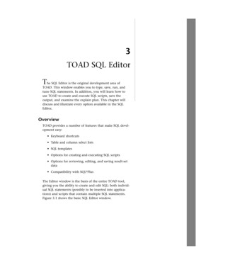
Animal Physiology Laboratory, Zool 430L1.2.3.4.5.6.7.8.9.Week 4Spring 2018Acetylcholine - released by the parasympathetic nervous system.Epinephrine - released by post-ganglionic sympathetic nerves.Pilocarpine - stimulates release of acetylcholine from parasympathetic fibers.Atropine Acetylcholine - Atropine is a plant alkaloid that blocks acetylcholine receptors in the heart.First apply atropine, and then without rinsing the heart, immediately follow the atropine application byan application of acetylcholine.Caffeine – increases potassium sensitivityPotassium-free Ringer’sCadmium Chloride – blocks calcium channelsCalcium-free Ringer’sHigh Potassium RingersFOR EACH DRUG:1. Record 30 seconds of baseline data.2. Apply two or three drops of the drug to the heart. Comment each drug application. Record for 30 seconds.3. Rinse the heart with Ringer’s and allow the heart two minutes to recover.G. Refractory period of cardiac muscle.(Understanding cardiac function through direct electrical stimulation)The refractory period is the minimum time required for the heart to regenerate the membrane potentialrequired for contraction.Setup1.2.3.4.5.Attach bipolar stimulator to the positive and negative terminals on the PowerLab analog output (2 BNCs).In Chart, select Macro: refractory period.Click Start to begin recording. Gently place the tips of the stimulating electrode on the ventricle.Click the Stimulate button in the Stimulator Panel to stimulate the heart.Stimulate the heart at each of the following times in the cardiac cycle (see diagram below):forcetimeStudy QuestionsAt what point (1–6) were you able to get a second heartbeat?What happens to the heartbeat interval when you observe a second beat? H. Heart Block1. Using a heavy thread, place a single-loop ligature around the heart at the junction of the atria and ventricle.2. Take two short pieces of thread and run each one through the ligature on opposite sides of the loop asreleasing threads. You will use these to open the ligature at the end of the experiment.3. Tighten the ligature slowly and observe the beating of the atria and ventricle for changes in rhythm. Do notPage 4! of 6!
Animal Physiology Laboratory, Zool 430LWeek 4Spring 2018tighten the ligature too much or you will cut the heart.Can you see any of the following types of heart block? First-degree block. The interval between atrial and ventricular contraction is prolonged. Second-degree block. Some impulses fail to reach the ventricle so that the ratio of atrial to ventricular beats isaltered. You may see 2:1, 3:1, 5:1, 8:1 types of heart block. Third-degree block. Impulses fail to pass through the AV node and bundle of His and the ventricle may startits own independent rhythm of beating, or you may see only the atria contracting.After producing a complete heart block (third degree), release the ligature by pulling on the release threads andsee if a normal AV beat is restored.BEFORE YOU LEAVE - CLEANUP!!!Place your toad into a ziplock bag. Rinse all of your dissection gear and set it to dry so that it will not rust.Suggestions for your Lab ReportWrite a full lab report following the lab report guidelines.IntroductionDescribe cardiac muscle and how the rhythmic activity of the heart is created (why would it be called a“myogenic heart”). Include the pacemaker. Describe some of the properties of cardiac muscle that differentiateit from skeletal muscle, in particular how signals are transmitted throughout the heart. Touch upon some of thefactors that are thought to affect cardiac activity. End with hypothesis or hypotheses constructed around theaspects of heart physiology you investigated. Keep it brief, 3 paragraphs for a 5 page report.MethodsDescribe the subject and preparation. Briefly describe the techniques and equipment used to measure heartrate and contractile force. Describe the control and experimental treatments (keeping in mind your hypothesis).Describe how you will analyze your data to address the hypothesis.ResultsRecording of the heartbeat and ECGYou may want to include a sample of your data to demonstrate data quality - a screen shot showing a cleartrace of the Force and ECG in a few cardiac cycles at rest. Indicate atrial contraction and ventricularcontraction on the Force channel.Collect data (in your notebook) such as indicated by the table below from the resting frog heart.P-wave and atrialcontractionQRS complex andventricularcontractionTime difference(sec) between:Page 5! of 6!Atrial contractions(beat to beat)Ventricularcontractions (beat tobeat)
Animal Physiology Laboratory, Zool 430LWeek 4Spring 2018Effect of temperatureInclude a graph (such as a bar plot) showing the effects of temperature on your Toad’s heart.The Q10 effectWhat is the value of Q10 for the toad heart rate?10 /( t 2 t1 )Q (HR2 / HR1 )10Where: t temperature, HR1 is heart rate @ t1; HR2 heart rate @ t2Starling’s Law of the Heart Demonstrate Starling’s Law using a scatter plot of heart rate vs. the relative stretch (% of resting length).Effect of drugs on heart rateCreate a bar graph showing the relative heart rate (% of resting heart rate -- how do you do that?) for thefollowing conditions: acetylcholine, epinephrine, pilocarpine, and atropine followed by acetlycholine.DiscussionHere are points to consider for enriching your discussion. See grading rubric for overview.1) Describe the basis for the delay between the atrial and ventricular contractions.2) How did temperature affect heart rate? What do you suppose is a consequence of being a poikilotherm?3) What is Starling’s Law of the Heart? Does your data support this law?4) Describe the mechanisms by which the following drugs affect heart rate:a) Acetylcholineb) Epinephrinec) Atropine followed by acetylcholine5) How long was the delay between the QRS complex and the observed ventricular contraction? Explain themechanism for this delay.6) Based on your results, what happens to a frog’s metabolism and heart rate in cold weather? Does ambienttemperature affect human heart in the same way? What is the significance of this?7) The effect of acetylcholine on cardiac muscle is the opposite of its effect on skeletal muscle. Can youexplain the mechanistic basis for the difference?8) Epinephrine mimics the effects of which branch of the autonomic nervous system?9) During exercise, venous return of blood to the heart increases. Based on your knowledge of Starling’s Law,what happens to stroke volume during exercise? How would this help?Page 6! of 6!
B. Toad dissection procedure Refer to the toad dissection guide for diagrams of this procedure. 1. Obtain a double-pithed toad from your instructor. Secure the animal ventral side up to a dissecting board using straight pins. 2. Lift the abdominal skin with forceps, make a longitudinal incision from the thorax to the abdomen using a scalpel or .