Transcription
Back To ChiropracticContinuing Education SeminarsLower Extremity Adjusting:Foot & Ankle Module 4 HoursWelcome:This course counts as 6 Hours of CE for Lower Extremity Adjusting for theChiropractic Board of Examiners for the state of California.There is no time element to this course, take it at your leisure. If you read slow orfast or if you read it all at once or a little at a time it does not matter.How it works:1.Helpful Hint: Print exam only and read through notes oncomputer screen and answer as you read.2.Printing notes will use a ton of printer ink, so not advised.3.Read through course materials.4.Take exam; e-mail letter answers in a NUMBERED verticalcolumn to marcusstrutzdc@gmail.com.5.If you pass exam (70%), I will email you a certificate, within 24hours, if you do not pass, you must repeat the exam. If you donot pass the second time then you must retake and pay again.6.If you are taking the course for DC license renewal you mustcomplete the course by the end of your birthday month for it tocount towards renewing your license. I strongly advise to takeit well before the end of your birthday month so you can send inyour renewal form early.7.Upon passing, your Certificate will be e-mailed to you for yourrecords.8.DO NOT send the state board this certificate.9.I will retain a record of all your CE courses. If you get auditedand lost your records, I have a copy.
The Board of Chiropractic Examiners requires that youcomplete all of your required CE hours BEFORE you submityour chiropractic license renewal form and fee.NOTE:It is solely your responsibility to complete the course by then, norefunds will be given for lack of completion.Enjoy,Marcus Strutz, DCCE ProviderBack To Chiropractic CE SeminarsCOPYRIGHT WARNINGThe copyright law of the United States (Title 17, United States Code) governs the making ofphotocopies or other reproductions of copyrighted material.Under certain conditions specified in the law, libraries and archives are authorized to furnisha photocopy or other reproduction. One of these specified conditions is that the photocopy orreproduction is not to be "used for any purpose other than private study, scholarship, orresearch." If a user makes a request for, or later uses, a photocopy or reproduction forpurposes in excess of "fair use," that user may be liable for copyright infringement.This site reserves the right to refuse to accept a copying order if, in its judgment, fulfillmentof the order would involve violation of the copyright law.
Dr. Jason W. Kelberman, D.C., BCIM, DMBBPOwner and founder of the Westside Wellness Center, a chiropracticcorporation for education, non-surgical treatment and prevention programs.PROFESSIONAL EXPERIENCE & MEMBERSHIP:Westside Wellness Center 2008-present, 2990 S. Sepulveda Blvd. Suite 203 L.A. CA 90064310-268-8008 Websites: www.losangelesneuropathycenter.com, www.drkelberman.com,email: drkelberman@gmail.comBoard Certified Integrative Medicine.Diplomate of Manipulative Therapies.Member American Association of Integrative Medicine.Masters Certification in the treatment of frozen shoulder: Neil Asher Technique.Lifetime participant Applied Kinesiology USAWestside Wellness Center was the first office in Los Angeles to offer a comprehensivetreatment program for the treatment of Peripheral Neuropathy. Treating nerve damagesuccessfully by treating neurologically and metabolically.Previous Experience:Joint Rehabilitation & Sports Medical Center 1997-2008 Partner and Director ofRehabilitation and chiropractic services, multi doctor, multi specialty clinic (MD,DC).Santa Monica Chiropractic Center from 1985 to 1997.Palisades Holistic Chiropractic Center from 1983-1985, associate Dr. Dean Raffelock, DC.The Cleveland College of Chiropractic @ Los Angeles: 1983 Graduated Cum LaudeThe City College of New York, Bachelor degree in education: 1976 Graduated Cum LaudeTeacher, Secondary School in New Haven, Connecticut 1976-1978: Woodworking ShopDr. Kelberman’s life’s work is a chiropractic specialty known as “Applied Kinesiology”.Our bodies are moved by muscles and Dr. Kelberman is a master in the diagnosis andtreatment of sport related traumas and injuries of all types. Tennis is Dr. Kelberman’sfavorite sport and Afghan Hound dogs are one of his passions. His first exposure to thewonderful world of chiropractic happened after he was struck by a car at the age oftwelve. Chiropractic care was the only treatment that enabled restoration of function. Asa result, he continues to treat motor vehicle accidents to this date.
Thank you to :Dr.’s David Leaf, Bob Blaich, Nancy McBride, Jim Blumenthal, Steven Kaufman, KirkMeier, Dean Raffelock for giving me my first associate position in 1984, Joe DiDuro forteaching me detailed neurologic examination and to my mom who was an educator,and has two sons that are chiropractors, for supporting me no matter what.
LOWER EXTREMITY: FOOT AND ANKLE MODULEINTRODUCTION:Our body is able to perform some of the most complex movements imaginable. Most ofour movements are reliant on our feet unless you are in a non weight bearing position.One of the reasons why non weight bearing rehabilitation is so effective for someinjuries. (bicycle, pool, etc.) The feet are probably one of the most overlooked areas inour body.So, today we are going to take a much closer look!Here is a mini course to help you understand, diagnose and treating foot and gaitdysfunction. I’ve been working on this information and adjusting feet in my practice for31 years.I will be showing you techniques for manual adjustments, drop adjustments andinstrument adjusting. I will discuss how to use kinesiotape and make foot orthotics tocorrect the problems that you will find.There are 24 vertebra, 206 bones in the body, 650-840 muscles, and Iwant to be the chiropractor that can get you going in the right direction.
ANATOMY: Bones firstThe human foot and ankle has 26 bones, 33 joints, 107 ligaments, 19 muscles andtendons. The 52 bones in your feet make up about 25 percent of all the bones in yourbody.
Tibia: weight bearingFibula: Non weight bearing. Attaches laterally from the tibia and makes up the anklejointTARSAL BONES:Talus, Calcaneus, Navicular, Cuneiforms (3), Cuboid These are the bonesthat you are most often adjusting.Metatarsals: (5) ForefootPhalanges: (10) ForefootHallux Sesamoid bones: 2 under great toeOthers: There may be accessory bones on the footCalcaneus: Heel bone, first point of contact in walking.Talus: Makes up the ankle joint, but also connects to the calcaneusCuboid: LateralNavicular: MedialCuneiforms: Medial, lateral and intermediate create the mid foot.
There are 3 arches in the foot.Medial and lateral Longitudinal arch and transverseWhat makes up the transverse arch (AKA metatarsal): (3) Cuneiforms,Whether you are treating sport injuries, sprains or just adjusting feet because it willmake your pelvic adjustments last longer. The better you know the anatomy the betteryou can adjust and help your patients.
FLAT FEET (pes planus)Are they fallen? Did they not develop? Are they genetically flat?Some people have flat feet because of a developmental fault during childhood, whileothers may find that the problem develops as they age, or during or after a pregnancy.When the Renalin hormone is released during the latter parts of pregnancy all ligamentshave a tendency to become lax. Foot adjustments and arch supports of all types can behelpful. There are some simple devices which may prevent the complications of flatfeet. Observing the rear foot having a significant inward distortion is a positiveHelbing’s’ Sign
Podiatrists remind me of chiropractors in that they look at x-rays of the feet anddetermine the course of action based on that data. They take measurements from thefilms and create orthotics accordingly. Doesn’t that sound familiar? How many of youstill mark x-rays?I recently had a conversation with a Doctor of Chiropractic in California who trained witha podiatrist who used to practice in the 1930’S and 40’s. Yes we are both older than wewant to admit. He related that the podiatrists used to be foot manipulators almostexclusively. They had open treatment room with patients in a circle where patients linedup to get foot adjustments!People with other foot problems may find that flat feet either contribute to them or makesymptoms worse. Examples include: Achilles tendinitisArthritis in the ankle or foot.BunionsHammertoesPlantar fasciitis (pain and inflammation in the ligaments in the soles of feet)Posterior tibialis tendinitisShin splints
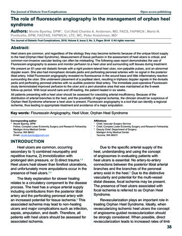
Cavus Foot (High-Arched Foot)What is Cavus Foot?Cavus foot is a condition in which the foot has a very high arch. Because of this higharch, an excessive amount of weight is placed on the ball and heel of the foot whenwalking or standing. Cavus foot can lead to a variety of signs and symptoms, such aspain and instability. It can develop at any age, and can occur in one or both feet.CausesCavus foot is can be caused by a neurologic disorder or other medical condition such ascerebral palsy, Charcot-Marie-Tooth disease, spina bifida, polio, muscular dystrophystroke or peripheral neuropathy. In other cases of Cavus foot, the high arch mayrepresent an inherited structural abnormality.If the high arch is due to a neurologic disorder or other medical condition, it is likely toprogressively worsen. On the other hand, cases of Cavus foot that do not result fromneurologic disorders usually do not change in appearance.SymptomsThe arch of a cavus foot will appear high even when standing. In addition, one or moreof the following symptoms may be present: Hammertoes (bent toes) or claw toes (toes clenched like a fist)Calluses on the ball, side, or heel of the footPain when standing or walkingAn unstable foot which can lead to ankle sprains Some people with cavus footmay also experience foot drop, a weakness of the muscles in the tibialis anteriormuscle and or extensor hallucis longus that results in dragging the foot whentaking a step. Foot drop is usually a sign of an underlying neurologic condition.HELLO! Doctors of chiropractic you are uniquely skilled to diagnose and treat thiscondition.The Computerized weight bearing pictures below will give you a better idea of thedistribution differences. The same can be done with wet feet on concrete etc
Muscles:Intrinsic Muscles of the foot: All begin and end within the footExtrinsic Muscles of the foot: Those that are on the calf that also attach to the foot.Major muscles that control our arches.Medial ArchTibialis posterior Inserts @ navicular Tibialis Anterior Inserts @ medial cuneiformLateral ArchPeroneus longus, brevis and tertius (AKA Fibularis)Transverse archThe transverse arch of the foot is maintained by the strength and integrity of the tibialisposterior and the peroneus longus. I know this is confusing but when you view theplantar surface of the foot, the tendons of these muscles cross so that contraction ofthe muscles pulls the medial and lateral aspects of the foot together supporting thetransverse arch.Extensor digitorum longusOften involved with ankle sprainsExtensor Hallucis LongusMost commonly involved muscle of the foot.Flexor Hallucis LongusFlexor Digitorum Longus Commonly with plantar fasciitis.
Types of feetYour feet can reveal many things about you. For the purpose of this lecture we will takea one minute fanciful look at what your feet reveal about your personality and then stickto the mechanical factors.RomanGermanicGreekEgyptian
Long second toe (MORTON’S FOOT)Indicates leadership qualities. Rulers from ancient Egyptian and Hawaiian royal dynasties all hadlong second toes. You need to be in charge.Last joint of third toe at an angleYou have the natural ability to deceive, as well as the propensity to be misunderstood.Frequently to be found in spies.Extra-small little toeDenotes a childlike nature, with playful sense of fun.Second toe on left foot leaning towards big toeSign of a sentimental, nostalgic nature. Shared by Hollywood actor Reece Witherspoon.Little toe pointing at an angleDenotes unconventional nature. Being able to waggle your little toe indicates restlessness and aneed for constant change.Joint MechanoreceptorsSurrounding and protecting all joints are tough, fibrous tissues which contain a variety of sensorynerve endings. The input from these specialized sensors keeps the nervous system informed as tothe location of the joint, and also the degree of stretch, compression, tension, acceleration, androtation. (6) These joint mechanoreceptors are classified by their anatomy and their neurologicalfunction. (7) Type I mechanoreceptors are found in higher densities in the proximal joints. Theysense the position of a joint by signaling the joint angle through normal ranges of motion. Thesehelp determine postural (tonic) muscle contractions. Type II nerve endings adapt to changes inposition, and are most active at onset and termination of movement. These are more denselydistributed though the distal joints, and affect phasic muscle actions. Type III mechanoreceptorsare high threshold, which means they require considerable joint stress at end ranges before firing.These receptors serve a protective function similar to the Golgi tendon organs. Type IV receptorsare free nerve endings located in the ligaments, joint capsules, and articular fat pads whichrespond to pain stimulus. They can generate intense, non-adapting motor responses in allmuscles related to a joint, resulting in the protective muscle contractions that restrict jointmovement.Foot InvolvementThese six specialized nerve sensors are found throughout the musculoskeletal system, in allskeletal muscles and in every ligament, joint capsule, and articular connective tissue. With manysmall joints, lots of connective and articular tissues, and both intrinsic and extrinsic muscles, thefeet are particularly well-supplied with proprioceptive nerve endings. Mechanoreceptors in thejoints along with the muscle spindles of the foot muscles are responsible for the positive supportreflexes and a variety of automatic reflexive reactions. (8) These include the flexor/extensorreflex, which converts the lower limb into a firm, yet compliant pillar. Weight bearing
compresses the joints and muscles, evoking reflexive activity in the extensors and inhibition ofthe flexor muscles. (9)The first research to demonstrate how altered proprioceptive input predisposes to recurringinjuries was performed on patients with chronically sprained ankles. (10) Freeman et al. calledthis phenomenon “articular de-afferentiation” to recognize the importance of inappropriateafferent signals from injured ankle and foot proprioceptors. They pointed out that, “Sincearticular nerve fibers lie in ligaments and capsules, and since these fibers have a lower tensilestrength than collagen fibers, it seems inevitable that a traction injury to a ligament or capsulewill lead to the rupture of nerve fibers as well as collagen fibers”. (11)ConclusionExcept for the spine, the foot is the anatomical region which contains the mostproprioceptive sensory receptors, and the foot has very distinctive nerve circuits whichmust be considered.Because of the magnitude of sensory input, the feet are frequently involved in clinical conditionswhich will respond to specific treatment approaches that include the proprioceptors — such ascustom orthotics. Structural support and shock absorption for the musculoskeletal system isprovided by the corrective orthotics, thereby reducing physical stressors on the muscles andjoints of the feet, legs, and pelvis.Greater understanding of the proprioceptive system of sensory receptors in the muscles andjoints has enabled us to more accurately assess and treat many complex musculoskeletalproblems. When custom-fitted orthotics are included, treatments can be more effective andresponses will be more comprehensive and longer-lasting.References1. Kuhn DR, Shibley NJ, Austin WM, Yochum TR. Radiographic evaluation of weight bearing orthotics and their effect on flexibl epes planus. J Manip Physiol Ther 1999; 22(4):221-226.2. Stude DE, Brink DK. Effects of nine holes of simulated golf and orthotic intervention on balance and proprioception in experiencedgolfers. J Manip Physiol Ther 1997; 20:590-601.3. Stude D, Gullickson J. Effects of orthotic intervention and nine holes of simulated golf on gait in experienced golfers. J ManipPhysiol Ther 2001; 24(4):279-287.4. Stude D, Gullickson J. Effects of orthotic intervention and nine holes of simulated golf on club-head velocity in experiencedgolfers. J Manip Physiol Ther 2000; 23(3):168-174.5. Gatterman MI, ed. Chiropractic Management of Spine-Related Disorders. Baltimore: Williams & Wilkins, 1990:413.6. Slosberg M. Effects of altered afferent articular input on sensation, proprioception, muscle tone and sympathetic reflex r esponses.J Manip Physiol Ther 1988; 11:400-408.7. Wyke BD. The neurology of joints. Ann R Coll Surg Engl 1967; 41:25.8. Freeman MAR, Wyke BD. Articular contributions to limb muscle reflexes. J Physiol 1964; 171:20.9. Panzer DM, Fechtel SG, Gatterman MI. Postural complex. In: Gatterman MI, ed. Chiropractic Management of Spine-RelatedDisorders. Baltimore: Williams & Wilkins, 1990:263.10. Bosien WR, Staples OS, Russell SW. Residual disability following acute ankle sprains. J Bone Joint Surg Am 1955; 37:1237.
11. Freeman MAR, Dean MRE, Hanham IWF. The etiology and prevention of functional instability of the foot. J Bone Joint Surg Br1965; 47:678-685.Examination of the feetWhen you get up from your chair, what muscle should contract first? NO it’s not yourgluts! Flexor hallucis longus You should start your evaluation of your patients whilethey are walking into your office or as they get up from the chair if you enter after theydo.I like to have the patient facing me at first. Have them march gently up and down inplace and then stop without fixing there posture. Observe everything you can at thispoint about the feet, knees, pelvis, shoulders and head posture. Walk around the patientto visualize them anteriorly, laterally and posteriorly.The most telling simple test to have the patient perform, is to ask them to go up on theirtoes and balance as long as they can. And I show them how to do it, with their feetshoulder width apart and NO toe out. After that I ask them to balance on their heels.You get to see all the weakness in their feet right away.Does the patient have a high arch, normal arch or low arch?Does he have a Morton’s foot? Of what ancestry does his feet say he belongs to?Check the ROM of the foot and determine whether the foot is hypermobile orhypomobile. Is it rigid or supple? This will help you determine what to do about theissues you find. Hypomobile require mobilization while hypermobile requirestabilization. You will usually find that as we age we become less mobile.ROM: ankle and foot (should be equal bilaterally)Ankle dorsiflexion:0-20 degrees
Ankle plantar flexion:0-50 degreesAnkle foot inversion:0-35 degreesAnkle and foot eversion 0-15 degreesWhat’s Next:In order to determine best adjustments of the feet we need to look at severalprocedures. If there is foot pain you should be able to determine how to reproduce thepain or how to reduce the pain. This should be done manually. It’s been my experiencethat if you show the patient you know how the pain was created and then show themhow to stop the pain they will follow you anywhere.If you have never tested foot muscles before, I recommend you start testing theExtensor Hallucis Longus muscle (Big toe extensor EHL for short). It’s the largest toe ofthe foot so it’s easier to test.What I initially found, was that people who had weakness of the EHL muscle also hadweakness of the arch or flat foot. And that if I strengthened the plantar flexors (restoredarch of the foot) the extensors responded in kind.What I now understand is that when you strengthen the archit increases the height of the arch. This in turn shortens thefoot. That makes it easier on the extensors to work. Shortfoot exercise is essential in balance therapy.Fascial chain connection from the extensor hallucis longuswill be medial musculature of the foot to the knee and thigh(Tibialis posterior, VMO, adductor, Sartorius, medialgastrocnemius head) as well as tibialis anterior.
If the patient had weakness of the peroneal group (lateral stabilizers) they either hadankle injuries that left them with deltoid ligament or muscular weakness or they haddisuse weakness. Very , very common as we age, since most people stop movingsideways as we age.Fascial Chain connection will be tensor fascia lata, IT band, gluteus medius andminimus.The following sequence is from Dr. David Leaf’s work. A doctor that I feel is the best Ihave ever trained with. Highly recommend you attend anytime he is on the west coast.
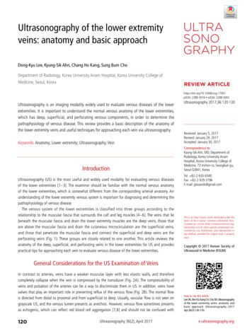
Buerger’s test.(Simple challenge for vascular problems in the lower extremity)
Patient is supine and the straight leg is raised 45 degrees and held there for threeminutes. The subject then sits with their legs over the tablePositive foot blanches and veins collapse with leg elevated OR it takes 1 - 2 minutesfor reddishcyanosis tocover thefoot and theengorge.veins to
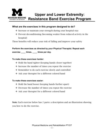
I wanted you to see the slide above because it shows you why the tibialis posteriortendon is so important to the medial arch, and where to find it. It may also mimic otherpain in the calf. You may think you have a gastroc or soleus problem when in actuallyit‘s tibialis posterior. More than 50% of the attachments for tibialis posterior in the footare in the navicular bone. When the arches are intact the body weight should besupported on a tripod.
Adjusting feet 101What is the most important area of the foot to adjust? Is it medial or lateral is your firstdecision.MAKE SURE THE PATIENT CAN TOLERATE THE ADJUSTMENT YOU WANT TOPERFORM! SELECT MANUAL VS INSTRUMENT VS DROP or SOFT TISSUE.I used to love to manipulate all metatarsals manually. Once you sprain a foot and haveto fix it for a patient that wanted to go travelling or run a race that day you willappreciate this fact. This is experience talking here! Do this by examining ROM in thearea you are going to adjust and see if the patient is already in pain or not.Calcaneal and talus adjustments are great to do as a pull move : check inversion ,eversion ROM and adjust to improve those ROM’s. As the foot elongates it tends toallow the calcaneus to drift posteriorly. Remember this later when we use tapingtechniques.A dropped navicular (most common) Does not make sense to do a pull move since itis already inferiorly displaced. Try drops or instrument adjustments since they are easyto adjust superiorly and DNFT is fine. Usually enhances Tibialis posterior but canenhance other closely related fascial chain muscles.Cuboid adjustment: subluxation either lateral or inferior, same as navicular just lateralin location. Usually enhances the peroneus longus musculature.Transverse arch: Cuneiforms: I prefer drops (use form to adjust over) or activator forthis region as well since they tend to drop inferiorly. Flexion manipulation is great also.Phalanges: Axial pull traction manipulation works fine here. Just make sure the patientcan tolerate the pressure required to hold the phalange. Older patients may not toleratethis adjustment very well.Fibula: The fibula has an arthrodial joint between the lateral condyle of the tibia andthe head of the fibula. The contiguous surfaces of the bones present flat, oval facetscovered with cartilage and connected together by an articular capsule and by anteriorand posterior ligaments. I mention this in greater detail because it is often overlooked.Usually enhances the peroneal group function.Fibula-tibia junction, unless traumatically misaligned will respond to whatever the footand associated muscle weakness pattern is occurring. So, pronators usually sublux thefibula anteriorly And Supinators usually sublux the fibula posteriorlyThe other very common issue with that joint is that it becomes hypermobile, unstableand just needs to be approximated and taped (can do with whatever adjustments youelect). As long as you apply the force in the direction all techniques are great.
Which came first? The chicken or the egg? Was the muscle weakened or tightened andthe bone became misaligned? Or was there a single or multiple traumas?Are there racial differences?You bet. Just like Asians have straighter spines meaning much less spinal curves ,African Americans have more spinal curve and lower arches. That does not mean theyall need orthotics.Compared to Caucasians, African Americans were almost 3 times more likely to havepes planus and were nearly 5 times less likely to have Tailor’s bunions or pes cavus.EXAMPLES FOR YOUR OFFICE MONDAY MORNING:1. The patient is complaining of foot pain. You test their extensor hallucis longus(EHL) and find it to be weak. Challenge the transverse arch by manuallycompressing it together with one hand while you retest the extensor hallucismuscle with the other. If this strengthens the muscle you know you have ametatarsal failure. You can adjust and tape accordingly. Think about adding ametatarsal support to facilitate the rest of the inflammation reduction. This isprobably why foot levelers became so popular.2. Raising the navicular bone strengthens the tibialis posterior muscle. Adjust thenavicular superiorly. Recommend a drop adjustment or an instrument as thetypical pull move is not optimum. Or do a prone push move.3. Lowering the navicular strengthens the EHL, means you had a trauma to thedorsal surface (patient rolled the foot underneath them) and you need to pull thenavicular inferiorly (yank and crank OK). Will usually see weakness of the toeextensors also (Extensor digiti longus). Soft tissue treatment helps a lot in thistype of trauma along with taping. Try active tissue therapy with the patientextending their foot while you rub it out.4. Pulling the calcaneus anteriorly strengthens the EHL. There is a plantar fascialissue and the calcaneus has drifted posteriorly. Adjust calcaneus anteriorly andtape accordingly. ALSO check the navicular afterward.5. The patient has pain at the lateral ankle area. You will usually find a weakperoneal (longus, brevis or tertius). Try challenging the cuboid superiorly and orthe fibula posteriorly.6. We have not yet really talked about the fibula so here goes. You should find thatwithout trauma if the foot tends toward pronation or pes planus the fibula willmove anteriorly and if the tendency is for a cavus foot or one that rolls outwardyou will find a posterior fibula. Adjustments can be done manually with drops orinstruments and if needed taped. Especially effective for knee pain.7. You perform ROM on the ankle and you find it bound completely or in one otherdirection (inversion or eversion). The Talus may be jammed and is classicallyand wonderfully released with a pull manipulation. It should create an audiblerelease. Oh this one feels so good!
8. OK, so you are new to this stuff. You know the patient has foot problems but youdon’t have time to figure it out or just need a few suggestions. Remember themillion dollar roll? Ok here’s mine.Use a rubber dowel (Theraband foot roller or equivalent shapes). Adjust thenavicular superiorly with a drop block or drop section of your table over the footieas I call them. Do the same for the cuboid and the metatarsal arch. You onlyneed to drop on them 1-2 times three is not necessary unless it’s really stubborn.Perform this gently, as if it was your mothers feet and they hurt. It should be painfree if you are gentle. They will rarely be audible but you should find somethingthat you are working on is either better, easier, or the patient feels less pain,more function, more ROM. Don’t have access to a drop block then use aninstrument while you hold the foot steady.CHECK YOUR PATIENTS SHOES!This is frequently an issue. Patients have no idea what a good shoe is! Especiallywomen! Quick Check. The two most important things to look for in a shoe are:Strong counter (rear heel structure) and Torsional stability. The shoe cannot betwisted easily especially in the forefoot. I recommend a closed rear shoe since that isthe impact point when we walk. Open toe for a woman is OK if they have a closed rear.Look at your patient from the rear while they are in their shoes. You will be surprised atwhat you see. If the shoe is broken down, you will see it with them in it. The old days ofwearing a shoe till you see your toes coming through is a very bad idea.Most common causes for weakness of the extensor hallucis longus muscle: Injury to themuscle itself, metatarsal splaying, dropped navicular, plantar fascia issues andweakness of the flexor hallucis or flexor digitorum longus.Most common causes for weakness of the peroneal group: Deltoid ligament tear,stretch, or instability, cuboid subluxation , fibula subluxation, myofascial issues, highheels, or an arch supports that is too high.Muscle Activity From heel strike to flat footTibialis anterior EccentricControls pronation of the subtalar joint and decelerates plantarflexionExtensor hallucis longus/Extensor digitorum EccentricDecelerates plantar flexion and controls motion of tibia on the talus
Foot Muscle StrengtheningWe initially utilized the typical old fashioned techniques for feet, which are really morefor keeping range of motion than strengthening (picking up marbles, towels etc.). Laterwe used the Pilates foot corrector or the Elgin ArchXerciser. These are far superior to allof the older techniques, except barefoot walking or running on the beach. Superiorbecause they are a form of resistance training. People actually get stronger in their feet.CORD THERAPYTubular cords are great for strengthening the peroneal group as well as tibialisposterior and anterior.Rocker Board Therapy(later wobble board can be used)Grea
Palisades Holistic Chiropractic Center from 1983-1985, associate Dr. Dean Raffelock, DC. The Cleveland College of Chiropractic @ Los Angeles: 1983 Graduated Cum Laude The City College of New York, Bachelor degree in education: 1976 Graduated Cum Laude Teacher, Secondary School in New Haven, Connecticut 1976-1978: Woodworking Shop