Transcription
Skenderi et al. Diagnostic Pathology 2013, /38CASE REPORTOpen AccessInfarcted fibroadenoma of the breast: report oftwo new cases with review of the literatureFaruk Skenderi1, Fikreta Krakonja2 and Semir Vranic1*AbstractIntroduction: Fibroadenomas are the most common benign breast tumors in young women. Infarction is rarelyobserved in fibroadenomas and when present, it is usually associated with pregnancy or lactation. Infarction canexceptionally occur as a complication of previous fine-needle aspiration biopsy or during lactation and pregnancy.Materials and methods: Retrospective review of 650 cases of fibroadenomas diagnosed at our institution duringthe 8-years period identified two cases of fibroadenomas with infarction (rate 0.3%).Results: Two partially infarcted fibroadenomas were diagnosed on core biopsy and frozen section in an adolescentgirl (13 years old) and in a young woman (25 years old), respectively. No preceding fine-needle aspiration biopsywas performed in these cases, nor were the patients pregnant or lactating at the time of the diagnosis.Conclusion: Spontaneous infarction within fibroadenoma is a rare phenomenon in younger patients. The presenceof necrosis on core biopsy or frozen section should be cautiously interpreted and is not a sign of malignancy.Virtual Slides: The virtual slide(s) for this article can be found here: 060549847356Keywords: Breast tumors, Benign tumors, Fibroadenoma, Infarction, NecrosisIntroductionFibroadenomas are the most common benign neoplasmsof the breast usually affecting adolescents and youngwomen [1,2]. Infarction in benign breast lesions is rareand may occur in various conditions, including fibroadenomas [1,3]. Infarction within a fibroadenoma wasfirst described by Delarue and Redon in 1949 [4] andusually affects young women during pregnancy or lactation, but may occur at any age following fine-needle aspiration biopsy (FNA) [1,5-8].Clinically, fibroadenoma typically presents as a palpable mass and may occasionally be mistaken for inflammatory lesions due to pain and tenderness or formalignancy due to hardness, fixation to the surroundingtissue or bloody nipple discharge [2,9,10].Spontaneous infarction of fibroadenomas not relatedto previously mentioned causes occurs exceptionally and* Correspondence: semir.vranic@gmail.com1Department of Pathology, Clinical Center of the University of Sarajevo,Bolnička 25, Sarajevo BA-71000, Bosnia and HerzegovinaFull list of author information is available at the end of the articleonly a few cases are described in the available literature[2,7,9,11-14].Herein, we describe a review of fibroadenomas of thebreast with two new cases of spontaneous infarction, unrelated to any known risk factor.Materials and methodsWe did a retrospective search of our database for fibroadenomas of the breast that were diagnosed at our department during the 8 years period (2005–2012). Two cases offibroadenoma with spontaneous infarction were identified.Paraffin-embedded tissue blocks and hematoxylin andeosin (H&E) slides were retrieved from the pathologyarchive and retrospectively reviewed (F.S. and S.V.).In a case #1 immunohistochemical staining against estrogen receptor (ER, clone: 1D5, Dako, Glostrup, Denmark)and progesterone receptor (PR, clone: PgR636, DAKO,Glostrup, Denmark) was performed.Clinical history was available for both cases along withradiologic images (ultrasound and magnetic resonanceimaging [MRI]). 2013 Skenderi et al.; licensee BioMed Central Ltd. This is an Open Access article distributed under the terms of the CreativeCommons Attribution License (http://creativecommons.org/licenses/by/2.0), which permits unrestricted use, distribution, andreproduction in any medium, provided the original work is properly cited.
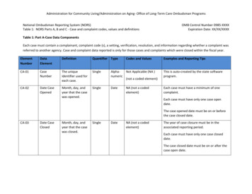
Skenderi et al. Diagnostic Pathology 2013, /38ResultsOur search revealed 650 fibroadenomas during the period2005–2012. Infarcted fibroadenomas were diagnosed intwo patients (rate 0.3%): an adolescent girl (13 years old,Case #1) and in a young woman (25 years old, Case #2).Both patients had no previous history of pregnancy, lactation or previous FNA. No hormonal disturbances werepresent in the patients. Postoperative course was uneventful in both patients.Case 1A 13-year-old pubertal girl presented with a rapidly growing, mobile and painful mass in the right breast. Radiologic findings (ultrasound and MRI) indicated thepresence of a well-circumscribed tumor of the right breastconsistent with a juvenile fibroadenoma (BI-RADS Category 2) (Figure 1A-B).Radiologist performed a core needle biopsy thatrevealed a fibroepithelial lesion exhibiting focal necrosisbut without malignant cells (classified as B3 lesion)which led to the immediate excision biopsy, performed aweek later (Figure 2A).Grossly, the tumor was an encapsulated, soft yellow,measuring 40 35 25 mm with foci of hemorrhage andnecrosis. The entire tumor was submitted for microscopic evaluation. Microscopic findings were consistentwith partially infarcted benign fibroadenoma with focalincreased stromal cellularity resembling that of benignphyllodes tumor (Figure 2B-C). Squamous metaplasia,observed in close proximity to the necrotic foci, waspresent and was reminiscent of the so-called necrotizingsyringometaplasia in the skin or necrotizing sialometaplasiain salivary glands (Figure 2D).In Case #1, both epithelial and stromal cells were focallypositive for estrogen receptor (ER) and progesterone receptor (PR) ( 20% of the cells with weak to moderate nuclear intensity).Page 2 of 5Case 2A 25-year-old gravida 0, para 0 woman presented to abreast radiologist with a short history of a rapidly growingpalpable and painful tumor in the left breast. Clinical andradiologic findings (ultrasound) were suggestive of juvenilefibroadenoma or intraductal papilloma (BI-RADS Category2) and the patient was referred to the breast surgeon. Theexcision biopsy was performed and intraoperative frozensection consultation was obtained which read as a benignfibroepithelial tumor with focal intratumoral hemorrhagewithout signs of malignancy.Gross examination revealed a nodular, circumscribedtumor, measuring 25 20 17 mm that was soft, yellowwith peripheral areas of hemorrhage. The entire tumorwas submitted for histopathologic evaluation whichrevealed juvenile (cellular) fibroadenoma with foci ofhemorrhage and necrosis (Figure 3A-B). No evidence ofmalignancy was found.DiscussionFibroadenomas are the most common benign tumors ofthe breast that usually affect premenopausal women butmay occur at any age. Diagnosis of fibroadenoma rarelyposes a diagnostic dilemma, even on core biopsy, FNAor frozen section. We presented here two rapidly growing infarcted fibroadenomas that were causing pain. Infarction within fibroadenoma is a very rare event andthe frequency of infarction in our study is in line with astudy of Haagensen [15] who found only five infarctedcases among 1,000 reviewed breast fibroadenomas (rate:0.5%). Another two studies based on the series of fibroadenomas in West African women revealed a slightlyhigher incidence of spontaneous infarction (0.9%) [16,17].The highest frequency (3.6%) was reported in a recentstudy of Al-Atrooshi [18].The presence of infarction and necrosis are usually worrisome signs in breast pathology although spontaneousFigure 1 Ultrasound examination (A) and MRI (B) of the breast (Case #1) revealed a circumscribed, hypoechoic mass, measuring40 mm.
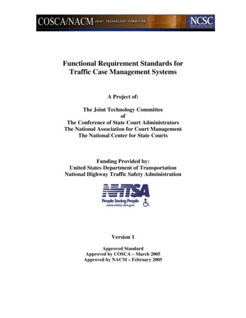
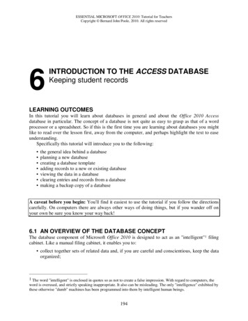
Skenderi et al. Diagnostic Pathology 2013, /38Page 3 of 5Figure 2 (A): Hematoxylin & Eosin (H&E) slide of a core biopsy showing a classical fibroadenoma with an area of necrosis (arrow) (5x);(B): An excision biopsy revealing a fibroadenoma with a focal morphologic features of benign phyllodes tumor (H&E, 10x); (C): A zoneof necrosis (H&E, 10x); (D): Squamous metaplasia, observed in close proximity of necrotic foci, reminiscent of the so-called necrotizingsyringometaplasia in the skin or necrotizing sialometaplasia in salivary glands (H&E, 10x).infarction can be seen in variety of benign breast lesionsincluding fibroadenoma, phyllodes tumor, lactating adenoma, and intraductal papilloma [3,19-23]. Spontaneous infraction in fibroadenomas is a rare phenomenon andusually associated with pregnancy, lactation or a recentFNA [5-7]. Exceptionally, spontaneous infarction mayaffect multiple fibroadenomas in the same patient [24]. Infarction may also be associated with the use of oralcontraceptives [25]. Rarely, it can be seen in youngpatients without any associated risk factors, as illustratedin our two cases. The cause and mechanism of infarctionare largely unknown. One of the possible explanations isthat infarction represents a spectrum of regressive changesthat also may include calcification and hyalinization, bothof which are much more commonly seen in fibroadenomas[26]. Newman et al. [27] also found thrombo-oclussive vascular changes as a possible cause of infarction withinfibroadenomas.In our first case, a focus of necrosis was seen on corebiopsy which prompted excision biopsy while in the second case clinical suspicion for malignancy prompted anintra-operative frozen section consultation which alsorevealed the presence of intratumoral hemorrhage andnecrosis. A meticulous histopathologic evaluation of theentire tumors, however, revealed no signs of malignancydespite the presence of necrosis. Of note, case #1 alsoFigure 3 (A): A classical cellular (juvenile) fibroadenoma (H&E, 10x); (B): An area with hemorrhage and necrosis (H&E, 5x).
Skenderi et al. Diagnostic Pathology 2013, /38showed areas of squamous metaplasia resembling socalled necrotizing syringometaplasia in the skin orsialometaplasia in salivary glands. This phenomenon hasalready been described in infarcted breast fibroadenomas[28] and can also be seen in other benign breast lesionsin a close proximity to the area of infarction (e.g. intraductalpapilloma, [20]).Fibroadenomas, particularly in older women, may beaffected by various proliferative changes including malignant epithelial lesions [29,30]. The most frequent are insitu carcinomas (both ductal and lobular), and their invasive counterparts [30-32]. Exceptionally, sarcomas mayalso develop within fibroadenomas (e.g. angiosarcoma,osteosarcoma) [33,34]. Little is known about the molecular mechanisms that drive the development and progression of malignant tumors within fibroadenomas.Comparative studies that analyzed various molecularmarkers in fibroadenomas and breast carcinomas failedhowever to identify the potential drivers [35,36]. However,a clonality study of Kuijper et al. [37] indicated that fibroadenomas possessed a potential to progress in an epithelial direction (carcinoma) or in a stromal direction(phyllodes tumor).We conclude that partial spontaneous infarction is arare event in breast fibroadenomas and may not beassociated with any known risk factor. The presence ofnecrosis on core biopsy or intra-operative frozen sectionshould be cautiously interpreted and is not itself a signof malignancy.ConsentThe case reports were shared with the local ethical committee whose policy is not to review case reports.Competing interestsThe authors declare no conflict of interest.Authors’ contributionsSV and FK have been directly involved in diagnosis and interpretation ofpatient’s diagnosis. FS and SV conceived the study design. All authors wroteand approved the final manuscript.AcknowledgmentThe authors thank Dr. Zoran Gatalica, MD, DSc, Caris Life Sciences, Phoenix,Arizona, United States of America for proof reading the manuscript andcritical comments.The preliminary results of the study were in part presented at the XXIXInternational Congress of the International Academy of Pathology, CapeTown, South Africa, 2012.Author details1Department of Pathology, Clinical Center of the University of Sarajevo,Bolnička 25, Sarajevo BA-71000, Bosnia and Herzegovina. 2Department ofRadiology, Clinical Center of the University of Sarajevo, Sarajevo, Bosnia andHerzegovina.Received: 16 November 2012 Accepted: 25 February 2013Published: 27 February 2013Page 4 of 5References1. Rosen PP: Rosen’s breast pathology. 3rd edition. Lippincott Williams &Wilkins; 2009.2. Liu H, Yeh ML, Lin KJ, Huang CK, Hung CM, Chen YS: Bloody nippledischarge in an adolescent girl: unusual presentation of juvenilefibroadenoma. Pediatr Neonatol 2010, 51:190–192.3. Fratamico FC, Eusebi V: Infarct in benign breast diseases. Description of 4new cases. Pathologica 1988, 80:433–442.4. Majmudar B, Rosales-Quintana S: Infarction of breast fibroadenomasduring pregnancy. JAMA 1975, 231:963–964.5. Vargas MP, Merino MJ: Infarcted myxoid fibroadenoma following fineneedle aspiration. Arch Pathol Lab Med 1996, 120:1069–1071.6. Pinto RG, Couto F, Mandreker S: Infarction after fine needle aspiration. Areport of four cases. Acta Cytol 1996, 40:739–741.7. Ichihara S, Matsuyama T, Kubo K, Tamura Z, Aoyama H: Infarction of breastfibroadenoma in a postmenopausal woman. Pathol Int 1994, 44:398–400.8. Lucey JJ: Spontaneous infarction of the breast. J Clin Pathol 1975,28:937–943.9. Oh YJ, Choi SH, Chung SY, Yang I, Woo JY, Lee MJ: Spontaneouslyinfarcted fibroadenoma mimicking breast cancer. J Ultrasound Med 2009,28:1421–1423.10. Deshpande KM, Deshpande AH, Raut WK, Lele VR, Bobhate SK: Diagnosticdifficulties in spontaneous infarction of a fibroadenoma in anadolescent: case report. Diagn Cytopathol 2002, 26:26–28.11. Fowler CL: Spontaneous infarction of fibroadenoma in an adolescent girl.Pediatr Radiol 2004, 34:988–990.12. Toy H, Esen HH, Sonmez FC, Kucukkartallar T: Spontaneous Infarction in aFibroadenoma of the Breast. Breast Care (Basel) 2011, 6:54–55.13. Hsu S, Hsieh H, Hsu G, Lee H, Chen K, Yu J: Spontaneous Infarction of aFibroadenoma of the Breast in a 12-Year-Old Girl. Journal of medicalsciences Taipei 2005, 25:313.14. Meerkotter D, Andronikou S: Unusual presentation and inconclusivebiopsy render fibroadenoma in two young females a diagnosticdilemma: case report. SA Journal of Radiology 2009, 13:62–65.15. Haagensen CD: Diseases of the Breast. Saunders; 1986.16. Jayasinghe Y, Simmons PS: Fibroadenomas in adolescence. Curr OpinObstet Gynecol 2009, 21:402–406.17. Onuigbo W: Breast fibroadenoma in teenage females. Turk J Pediatr 2003,45:326–328.18. Al-Atrooshi SA: Fibroepithelial tumors of female breast: a review of 250cases of fibroadenomas and phylloides tumors. The Iraqi PostgraduateMedical Journal 2012, 11:140–145.19. Verslegers I, Tjalma W, Van Goethem M, Colpaert C, Biltjes I, De SchepperAM, Parizel PM: Massive infarction of a recurrent phyllodes tumor of thebreast: MRI-findings. JBR-BTR 2004, 87:21–22.20. Flint A, Oberman HA: Infarction and squamous metaplasia of intraductalpapilloma: a benign breast lesion that may simulate carcinoma.Hum Pathol 1984, 15:764–767.21. Behrndt VS, Barbakoff D, Askin FB, Brem RF: Infarcted lactating adenomapresenting as a rapidly enlarging breast mass. AJR Am J Roentgenol 1999,173:933–935.22. Baker TP, Lenert JT, Parker J, Kemp B, Kushwaha A, Evans G, Hunt KK:Lactating adenoma: a diagnosis of exclusion. Breast J 2001, 7:354–357.23. Okada K, Suzuki Y, Saito Y, Umemura S, Tokuda Y: Two cases of ductaladenoma of the breast. Breast Cancer 2006, 13:354–359.24. Akdur NC, Gozel S, Donmez M, Ustun H: Spontaneous infarction ofmultiple fibroadenoma in a postlactational woman: case report.Turkiye Klinikleri Journal of Medical Sciences 2012, 32:1429–1432.25. Tavassoli FA: Pathology of The Breast. McGraw-Hill; 1999.26. Greenberg R, Skornick Y, Kaplan O: Management of breast fibroadenomas.J Gen Intern Med 1998, 13:640–645.27. Newman J, Kahn LB: Infarction of fibro-adenoma of the breast. Br J Surg1973, 60:738–740.28. Pandit AA, Deshpande RB: Infarction of fibroadenoma with squamousmetaplasia. Indian J Cancer 1985, 22:271–273.29. Tissier F, De Roquancourt A, Astier B, Espie M, Clot P, Marty M, Janin A:Carcinoma arising within mammary fibroadenomas. A study of sixpatients. Ann Pathol 2000, 20:110–114.30. Goldman RL, Friedman NB: Carcioma of the breast arising infibroadenomas, with emphasis on lobular carcinoma. A clinicopathologicstudy. Cancer 1969, 23:544–550.
Skenderi et al. Diagnostic Pathology 2013, /38Page 5 of 531. Diaz NM, Palmer JO, McDivitt RW: Carcinoma arising withinfibroadenomas of the breast. A clinicopathologic study of 105 patients.Am J Clin Pathol 1991, 95:614–622.32. Cole-Beuglet C, Soriano RZ, Kurtz AB, Goldberg BB: Fibroadenoma of thebreast: sonomammography correlated with pathology in 122 patients.AJR Am J Roentgenol 1983, 140:369–375.33. Babarovic E, Zamolo G, Mustac E, Strcic M: High grade angiosarcomaarising in fibroadenoma. Diagn Pathol 2011, 6:125.34. Killick SB, McCann BG: Osteosarcoma of the breast associated withfibroadenoma. Clin Oncol (R Coll Radiol) 1995, 7:132–133.35. Bal A, Joshi K, Logasundaram R, Radotra BD, Singh R: Expression of nm23in the spectrum of pre-invasive, invasive and metastatic breast lesions.Diagn Pathol 2008, 3:23.36. Xu X, Jin H, Liu Y, Liu L, Wu Q, Guo Y, Yu L, Liu Z, Zhang T, Zhang X, DongX, Quan C: The expression patterns and correlations of claudin-6, methyCpG binding protein 2, DNA methyltransferase 1, histone deacetylase 1,acetyl-histone H3 and acetyl-histone H4 and their clinicopathologicalsignificance in breast invasive ductal carcinomas. Diagn Pathol 2012, 7:33.37. Kuijper A, Buerger H, Simon R, Schaefer KL, Croonen A, Boecker W, van derWall E, van Diest PJ: Analysis of the progression of fibroepithelial tumoursof the breast by PCR-based clonality assay. J Pathol 2002, 197:575–581.doi:10.1186/1746-1596-8-38Cite this article as: Skenderi et al.: Infarcted fibroadenoma of the breast:report of two new cases with review of the literature. DiagnosticPathology 2013 8:38.Submit your next manuscript to BioMed Centraland take full advantage of: Convenient online submission Thorough peer review No space constraints or color figure charges Immediate publication on acceptance Inclusion in PubMed, CAS, Scopus and Google Scholar Research which is freely available for redistributionSubmit your manuscript atwww.biomedcentral.com/submit
Arizona, United States of America for proof reading the manuscript and critical comments. The preliminary results of the study were in part presented at the XXIX International Congress of the International Academy of Pathology, Cape Town, South Africa, 2012. Author details 1Department of Pathology, Clinical Center of the University of Sarajevo,