Transcription
This is a text version of a podcast from Pedscases.com on “Pediatric Eye Exam in Primary Care.” These podcasts aredesigned to give medical students an overview of key topics in pediatrics. The audio versions are accessible on iTunes orat www.pedcases.com/podcasts.The Pediatric Eye Exam in Primary CareDeveloped by Jennifer Ling, Harry (Chaocheng) Liu, and Dr. Ian McDonald for PedsCases.comOctober 10, 2018IntroductionWelcome to “The Pediatric Eye Exam in Primary Care”, a podcast made for PedsCases.com atthe University of Alberta. I am Jennifer Ling, a medical student at the University of BritishColumbia, and I am Harry Liu, a medical student at the University of Alberta. This podcast willprovide an organized approach on performing the pediatric eye exam highlighting somecommon clinical findings. We will also provide indications for a referral to a pediatricophthalmologist. We’d like to thank Dr. Ian MacDonald, an accomplished ophthalmologist andprofessor at the University of Alberta for developing this podcast with us.Accurately performing a pediatric eye exam is vital to catching eye problems and preventingtheir progression during a child’s development. The Centre for Disease Control has found thatover a given lifetime, visual disorders are the most disabling pediatric illnesses. For example,approximately 15-20% of children are at risk of developing amblyopia, also known as lazy eye,where one eye does not have normal visual acuity. Visual assessment by a healthcare provideris most vital at birth and during the first 6 months of life when the visual system is highlysusceptible to interference. The main goal of vision screening is to identify children who have orare at risk for developing amblyopia or strabismus, which can lead to permanent visualimpairment unless treated in early childhood. Other conditions that can be detected by visionscreening include: cataracts, glaucoma, ptosis, refractive errors (e.g., myopia, hyperopia, andastigmatism), and other serious conditions, such as tumors and neurological diseases. If any ofthe conditions don’t sound familiar, don’t worry! We will cover them in a little bit.Learning ObjectivesAfter listening to this podcast, the learner should be able to:1) Discuss the importance of pediatric eye examination in primary care2) Develop an understanding of vision developmental milestones and age-appropriate eyeexaminations3) Demonstrate a focused eye history and accurately perform various eye examinations4) List common signs and symptoms in pediatric eye conditions5) Discuss the indications for referral to a pediatric ophthalmologist and what to include inreferral lettersDeveloped by Jennifer Ling, Harry (Chaocheng) Liu, and Dr. Ian McDonald for PedsCases.comOctober 10, 2018
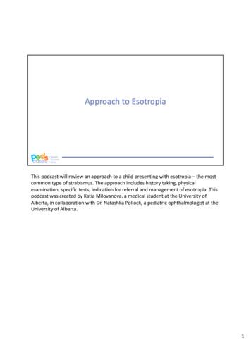
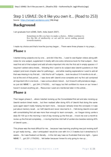
CaseFirst, we’d like to present a case. It is your first day in a family clinic in Red Deer as a fourthyear medical student and 9-month old Karl, has come in for his well-child visit. Karl appears welltoday, and his parents have no complaints on history. You are trying to recall which tests mustbe performed at this visit to ensure his eyes are developing normally.Vision Development MilestonesFirst of all, let’s quickly review some key vision developmental milestones. Here is a chart thatsummarizes the important milestones. Let’s go over them. At birth, newborns have a visualacuity of 20/400. They can focus on objects that are 10 inches in front of their face, and blink inresponse to a bright light. They also respond to movement. From birth to 2 months, infantsshould be able to maintain stable eye contact when initiated by the caregiver. It is typical fortheir eyes to appear to cross or wander at times. However, it’s concerning if their eyesconsistently turn in or out. At 3 months, infants can see as far as 8-15 inches. The infantbecomes aware of facial expression. You will find they are able to fixate on and follow objects,as well as look for objects. One clue is they will start watching their hands and bring their handsto midline and to their mouths. By 5 months, they will watch and copy the hand movements ofother children and adults. Their eyes should be straight and do not appear to cross or drift bythis point. When infants are 9 months old, they should be able to recognize family and caregiverfaces, and throw things with better accuracy as now they have better depth perception. By 2years, they should have decent hand-eye coordination. After that, they will continue improvingtheir visual acuity. It is expected to be at least 20/40 at 3 years old, 20/30 by 4 years old, and20/20 by 5 years old.Age-Appropriate Eye ExaminationsYou probably have heard of Rourke Baby Record, which is an evidence-based healthsupervision guide for primary healthcare practitioners of children in the first five years of life. Itcontains guidelines and information for comprehensive well baby and child visits.Here is a chart with relevant physical examination parameters for the eyes. For the newborn,you need to check for glaucoma, infections, and structural abnormalities including cataracts,corneal opacity, and ptosis. You need to pay special attention to preterm neonates who weregiven oxygen for an extended period of time and infants with multiple medical issues. For thewell-baby check up at 1 month, check for the red reflex. For visits from 2 to 18 months of age, inaddition to the red reflex, you need to check for the corneal light reflexes and ocular mobility, aswell as to perform cover-uncover tests. This will be the case for our baby Karl. The tests for thevisits from 2 to 5 years old are red reflex, corneal light reflex, cover-uncover test, and visualacuity. Great, now you know what eye tests to perform for different visits. Let’s now talk abouthow to perform those physical examinations as well as taking a concise eye history whencaregivers have eye concerns.Developed by Jennifer Ling, Harry (Chaocheng) Liu, and Dr. Ian McDonald for PedsCases.comOctober 10, 2018
Eye History and the Physical ExamMany eye and vision problems are difficult to detect on a short single visit so it is important toelicit a detailed history from the child’s caregivers and carefully examine the child’s eyes for anyabnormalities, especially for preterm neonates or infants with other medical comorbidities.First, you will begin with taking a history from the child’s caregivers. If it’s the first time you seethe child, ask them about the child’s overall health and maternal illnesses during the pregnancy,the route of delivery, and the babies gestational age at delivery. Early gestational age before 28weeks, low birth weight and oxygen supplementation after birth is associated with retinopathy ofprematurity. These cases should be checked at birth and at 4-6 weeks of age, and referred toan ophthalmologist to be followed; many cases resolve spontaneously, but further treatmentmay be necessary. Also note any neonatal complications and/or interventions during historytaking. Note any medication or substance use, especially smoking, by the parents. Multiplemeta analyses have identified maternal smoking as a significant risk factor for childhoodhyperopia, amblyopia, and strabismus. Make sure to ask about child’s eye contact with theircaregivers. If a child has poor eye contact after 2 months of age, this requires furtherassessment and referral to a pediatric ophthalmologist.Second, you need to ask about the recent history of the child’s eyes. Has there been chronictearing or discharge? In the morning, is there crusting in and around the eyes? Any persistentredness or irritation should be noted. These suggest the possibility of blocked tear ducts, eyeinfections, allergies, or allergic conjunctivitis. Also inquire whether there is a past history of eyeinfections and allergies, in the child and in the family. This is important at every visit. In thenewborn, blocked nasolacrimal ducts are common and may present as persistent tearing ordischarge, as well as debris on the eyelashes. The vast majority of these cases are resolved inthe first 6 months of development, but to promote the unblocking of the tear ducts, you shouldbe able to teach parents how to use a clean finger and massage downward from the innercorner of the eye to the nose. Parents should do this 2-3 times per day until the symptomsresolve.Next, inquire about eye and vision problems in the family. Was anyone born with eyemalformations such as strabismus or aniridia? Did anyone develop glaucoma or amblyopia? Arethere any eye diseases that run in the family? Is anyone currently or previously blind? A familyhistory of eye disorders will inform your physical exam to ensure you rule out similarabnormalities in your patient. Lastly, in conjunction with inspecting the infant, ask if the infanthas demonstrated an eye turning inwards or outwards frequently or if one eye appears lazy.Also ask the caregiver if they have ever noted one eyelid more drooped compared to the other.Correlate those concerns to the child’s vision developmental milestones because themanagement for the same concern will be very different depending on the child’s age. Manyfactors besides a positive family history increase the risk for an eye or visual problem. Theseinclude: Premature babies that have been given oxygen for significant periods of time Congenital infections (such as Toxoplasmosis and CMV) History of congenital cataracts Down syndrome Cerebral palsyMoving on to the physical exam, you need a child that is cooperative, alert and engaged.Sometimes, poor interest is indicative of poor vision by the child. It’s important to watch theDeveloped by Jennifer Ling, Harry (Chaocheng) Liu, and Dr. Ian McDonald for PedsCases.comOctober 10, 2018
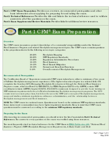
infant’s visual interaction during the visit to see if there are any concerns or not. Usually, a childis most comfortable and agreeable to examination in the parent’s lap, though children over theage of 4 are more cooperative. Talk to the child at an appropriate level and include play so thatthe child is more engaged and cooperative during the exam.First, you will inspect the eyes of the child. Notice any structural abnormalities, such as ptosis,corneal opacity or cataract, that may affect vision later. The eyelids should move together whenthe child blinks or shifts their focus. Check the periorbital skin for redness, inflammation,streaking or excess warmth that is indicative of infections such as periorbital cellulitis. Whenbabies are first born, it’s important to look for signs of congenital infections.Then, you will observe the irises and pupils. There may be defects in the shape of the eyes,pupil irregularities, and differences in size between the two pupils. Normal pupils should beround, equal and reactive to light. Small variations 0.5mm in pupil size difference are tolerated,but if it’s observed, it should be documented and checked at every visit. Symmetry should beobserved in both light and dim light.You need to inspect the sclera next. Normally, the sclera is clear, white, with a tinge of blue orgray. Some light-brown pigment spots can be seen near the iris at birth, especially in Asians,African-Americans, and dark-skinned races. An abnormal sclera may have lesions,discolorations, or one or more dark freckles; these findings require a referral to a specialist. Ayellow-tinged sclera may indicate elevated bilirubin in the circulation or liver abnormalities; thisfinding requires blood work and further follow up until it is resolved. Also check if there is scleralinjection, or, excess prominence of the scleral blood vessels. No lacrimal duct discharge shouldbe present and the eyes should move together during the inspection.Now you will ask the child to help you perform the next parts of the exam. First, let’s examinewhether the child can fix and follow objects. Newborns cannot fix and follow, but at 6 weeks,some babies will respond to the examiner’s face. Most infants are able to fix and follow by 3months old, and during this time, intermittent strabismus may be present but should disappearby 2-3 months postpartum. Each eye should be tested separately for sustained fixation andfollow. Failure to fix and follow strongly indicates visual impairment, and if it is observedbinocularly after 3 months, infants can have significant eye-brain abnormalities. This requires areferral to a pediatric ophthalmologist.To perform the red reflex exam, focus the ophthalmoscope light on each pupil from about 12 to18 inches away. The light transmitted should reflect off the fundus and transmit back throughthe ophthalmoscope, giving the pupil a red appearance. This is what is often seen in picturestaken with flash photography. A normal finding would be equal, bright and symmetric. Abnormalfindings include dark spots in the red reflex, reduced red reflex, white reflex, asymmetry ofreflexes and an absent reflex. A white reflex is called leukocoria, which is discussed in one ofour other PedsCases podcasts. Congenital cataracts result in increased corneal opacity so thered reflex cannot be observed; cataracts must be removed in the first 2-3 months of life aspermanent loss of sight may occur. Any abnormal findings should be referred urgently. Movingfurther away, the Bruckner test can be performed at 1 meter away from the child. Both eyesshould now show a red reflex. If you see inferior crescents as shown in the diagram, it meansthe child is myopic. Superior crescents indicate hyperopia. Be aware that most children havesome degree of hyperopia as their eyes are still immature. The Bruckner test assess binocularvision and provides an indirect evaluation of refractive error. An abnormal finding would be ifthere is white in the pupil of one eye or whitening in both eyes; this is indicative of strabismus(misalignment of the eyes) or anisometropic amblyopia. In an abnormal finding, the opticalDeveloped by Jennifer Ling, Harry (Chaocheng) Liu, and Dr. Ian McDonald for PedsCases.comOctober 10, 2018
power in each eye should be evaluated separately. Strabismus can be assessed by either thecover test or the Hirschberg test.In the Hirschberg test, the child stares at a penlight held 1 arm length away. The corneal lightreflex should be centered on each pupil and be sharp. If there is strabismus, the light reflex willbe off-center. If the child refuses to look at the light, the cover/uncover test may be performed.The child should focus on a target object, and the examiner will then cover each eye to look fora shift in alignment as the child looks at the object with each eye. Shifts in alignment suggeststrabismus, but sometimes this is pseudostrabismus, which is common if the child hasepicanthal folds or a wide nasal bridge. In pseudostrabismus, the corneal light reflex during theHirschberg test should be symmetrical.Last, let’s briefly touch on visual acuity testing. This test indicates how well the child sees. Inthis test, the child must be cooperative and be able to read the vision chart. If the child cannotread, picture charts can be used. Optotype tests are the gold standard when testing visualacuity. Each eye needs to be tested separately.Findings in Common Pediatric Eye ConditionsNo matter if it is taking a history or performing the physical examination, it is always beneficial tokeep the common causes or differentials in mind so you won’t miss any important findings.Next, we will go over some important eye conditions in primary care settings, including myopia,hyperopia, strabismus, amblyopia, and leukocoria.First are myopia and hyperopia. Myopia is nearsightedness, which means objects appear clearwhen close but blurry when far away. Light is focused in front of the retina. Hyeropia isfarsightedness, which is completely the opposite. Distant objects are much more clear than nearones and light focuses behind retina. There is 1.5 increase in risk of developing hyperopia whenwomen smoke during pregnancy. In terms of presentation, children may complain ofheadaches, eye strain, and blurry vision. The caregiver may notice squinting. The fast and easyway is to measure the visual acuity with the Snellen chart. When a child cannot read the eyechart, one quick method to determine if they are myopic or hyperopic is to perform the Bruckner(Brookerner) test that was mentioned earlier.Next, strabismus means misalignment of the eyes and if untreated, may result in permanentvision loss from impaired depth perception or amblyopia. The risk factors for strabismus includerefractive errors, Down syndrome, retinopathy of prematurity, head injury, and maternal smokingduring pregnancy. When strabismus is severe, it is easily noticeable as the children’s eyes donot look in the same direction at the same time or move together. Alternatively, the child couldbe squinting or closing one eye at times, tilting or turning their head to look at an object, andcomplaining of double vision. They could even bump into things.Amblyopia is the inability to focus one eye. To simplify the pathophysiology, one eye has poorvision compared to the other, so the brain tries to use the good eye for adaption and ignoresimages from the eye with worse vision. The visual development of the bad eye will be affectedand permanent vision loss may occur. Minor refractive error and cataracts are risk factors foramblyopia. One eye or both may wander inwards or outward with poor depth perception.Developed by Jennifer Ling, Harry (Chaocheng) Liu, and Dr. Ian McDonald for PedsCases.comOctober 10, 2018
Last but not least is leukocoria. Possible causes include congenital cataract, retinoblastoma,retinopathy of prematurity, and so on. Again, there is a podcast called “Approach to Leukocoria”by Dr. Novak for Pedscases. You can get more information from there.When and How to Refer to a Pediatric OphthalmologistThe following issues noted on history warrant ophthalmologic referral: retinopathy ofprematurity, premature birth congenital infections such as CMV and Toxoplasmosis, a diagnosisof Down syndrome and a positive family history for childhood cataracts, retinoblastoma orglaucoma. Infants who do not track by 3 months of age should also be referred to a pediatricophthalmologist. If the parents suspect the child has eye problems they should be referred toophthalmology or optometry. Substance use or infections during pregnancy may result incongenital developmental defects that need to be investigated by pediatricians, neurologists andophthalmologists.Referral is warranted if inspection reveals: corneal asymmetry, unilateral ptosis, and pupillaryasymmetry greater or equal to 1 mm. Corneal asymmetry is suggestive of glaucoma, andpupillary asymmetry is suggestive of neurological issues. During your physical exam, anabnormal red reflex, strabismus, nystagmus or amblyopia also needs to be referred. Anabnormal red reflex should be referred as it may indicate cataract, glaucoma, retinoblastoma,strabismus or a significant refractive error difference between the two eyes. Finally, eyepreference or a visual acuity difference of two lines or more between eyes should be referred toa pediatric ophthalmologist. In children 3-5 years old, visual acuity shouldn’t be worse than20/40, and in children over 6 years old, vision shouldn’t be worse than 20/30. If visual acuityfalls below that, the child should be examined by a pediatric ophthalmologist.Take Home PointsWe hope you find our podcast helpful and you learn something new. There are some quick takehome messages for you:1) For well-baby/child check-up, refer to Rourke Baby Record for the indicated eyeexamination maneuvers.2) The eye and surrounding area should be carefully inspected for any signs of infection,trauma or abnormal structure.3) The red reflex should be normal in both eyes of the child, and any abnormalities shouldbe referred.4) Infants should be able to fix and follow with their gaze, and failure to do so after a fewmonths of age requires an urgent referral to a pediatric ophthalmologist.5) The alignment of the eyes should be checked with Bruckner (Brook-ner) test or thecover-uncover test, and strabismus should be referred to prevent long termconsequences such as amblyopia.6) Visual acuity testing is important to identify myopia or hyperopia early. It is important toinvolve either ophthalmologist or optometrist when the visual acuity is poor or whenthere is difference of 2 lines or more between eyes.Quiz QuestionsWe are not done yet. We have some quiz questions for you to show us how smart you are.Developed by Jennifer Ling, Harry (Chaocheng) Liu, and Dr. Ian McDonald for PedsCases.comOctober 10, 2018
1) A mother brought in her 18-month-old son because her neighbor thinks her baby’s eyeslook “crooked”. Which physical maneuver you can perform to measure ocular alignmentin this child? The answer is corneal light reflex.2) What are some risk factors for developing retinopathy of prematurity? The answer isgestational age less than 32 weeks, birth weight under 1.5 kg, and systemic hypoxemia.3) What should you check for during an eye exam in a newborn? The answer is glaucoma,infections, and structural abnormalities including cataracts, corneal opacity, and ptosis.This concludes our discussion. Thank you for listening to PedsCases Podcasts!Developed by Jennifer Ling, Harry (Chaocheng) Liu, and Dr. Ian McDonald for PedsCases.comOctober 10, 2018
References1. Martin EF. Performing pediatric eye exams in primary care. The Nurse Practitioner. 2017Aug; 42(8):41-47.2. Rourke L, Leduc D, Rourke J. Rourke baby record: evidence-based infant/child healthmaintenance. 2018 Apr 09. Available %20National%20English%20-%20Black%20170926.pdf3. David DK. Visual development and vision assessment in infants and children. UptoDate.2018 Apr 09. Available from: rch pediatric%20ophthalmology&source search result&selectedTitle 4 101&usage type default&display rank 44. Coats DK, Paysse EA. Amblyopia in children: management and outcome. UpToDate.2018 Apr 08. Available from: ldrenmanagement-andoutcome?search child%20eye&source search result&selectedTitle 2 150&usage type default&display rank 2#references5. Coats DK, Paysse EA. Evaluation and management of strabismus in children.UpToDate. 2018 Apr 08. Available from: nagement-of-strabismus-inchildren?search child%20eye%20history&source search result&selectedTitle 7 150&usage type default&display rank 76. Amit M. Vision Screening in Infants, Children and Youth. Canadian Paediatric Society.2016 Feb 01. Available from: visionscreeningDeveloped by Jennifer Ling, Harry (Chaocheng) Liu, and Dr. Ian McDonald for PedsCases.comOctober 10, 2018
The Pediatric Eye Exam in Primary Care Developed by Jennifer Ling, Harry (Chaocheng) Liu, and Dr. Ian McDonald for PedsCases.com October 10, 2018 Introduction Welcome to "The Pediatric Eye Exam in Primary Care", a podcast made for PedsCases.com at the University of Alberta. I am Jennifer Ling, a medical student at the University of British