Transcription
NeuroscienceApplications Booklet
Harvard Apparatus and affiliatesare cited over 232,037 times in‘Google Scholar’, and over10,000 times in 2012 alone.*The following booklet containsexamples of Harvard Apparatusproducts used in NeuroscienceResearch, along withsummarizations of theexperiments and relatedpublished articles.Thank you for consideringHarvard Apparatus productsfor your NeuroscienceResearch needs.*Data compiled November 2014.
Dear Scientist,The importance of your research workcannot be overstated. Everyone’s life isimpacted by a family member or friend thathas a neurodegenerative disorder, a learningdisability, a traumatic injury or a psychologicaldisorder. Your work is our only hope thatfuture generations will be relieved of suchconditions. We at Harvard Apparatuscongratulate and thank you for devotingyour career to neuroscience research.Application Briefs2 - 25Bibliography26 - 27Harvard Apparatus and affiliated entities,have been supplying tools for neuroscienceresearch for over 100 years. We provideresearchers with an extensive productoffering for animal, organ, tissue and cellularresearch. Whether you need to executeprecise infusion into cells or animals, sampleextraction, behavior analysis, or control andmeasure physiological parameters – HarvardApparatus is ready to meet your needs. Ourproduct quality, dedicated support andneuroscience expertise have made HarvardApparatus the leader in this field. Ourproducts have been extensively used forneuroscience experiments for decades andhave been cited in thousands of publications.The enclosed is a sampling of publishedarticles that cited the use of products fromone or more of our affiliated companies. Wehope that these abstracts and thebibliography at the end will assist you in yourimportant work and that you considerHarvard Apparatus as your primary sourcefor neuroscience research apparatus.November 2014
Ventral Tegmental AreaNeuronal ActivityCorrelates to Animals’Behavioral Response toChronic MethylphenidateRecorded from AdolescentSD Male RatsZ. Jones et al (2014)Journal of Behavioral and Brain ScienceVol 4(4); ID 45258, 22IntroductionMethylphenidate (MPD) is considered as the firstline pharmacotherapy to treat ADHD. Morerecently, MPD has also been used as a cognitiveenhancement recreationally. Its therapeuticeffects are not fully understood, nor are the longterm effects of the drug on brain development. Theventral tegmental area (VTA) neuronal activity wasrecorded from freely behaving adolescent ratsusing a wireless recording system.2EXPERIMENT SUMMARIZATIONMaterials & MethodsMale Sprague-Dawley rats (N 142 at post natal day ofabout 32 days were purchased (Harlan, Indianapolis, IN,USA) and were allowed 3-4 days of acclimation in thevivarium on a 12 hour light/dark schedule prior to theelectrode implantation. Food and water were given atlibitum. The animals were housed individually in clearacrylic standard cages that served as both home cageand test cage for this study.Electrodes were implanted during stereotaxic surgery onanesthetized subjects that had their heads shaved priorto the procedure. Once electrodes were implanted,subjects were allowed 3-4 days recovery in their homecages and were connected to the wireless head stagetransmitter (Triangle BioSystems (TBSI); Durham, NC,USA) for acclimation for at least 2 hours/day to thebehavioral and electrophysiological recording systems.At first experimental day, the animal’s age was aboutpost natal 39-41 days.Five groups were used: saline, 0.6, 2.5, 5.0 and 10.0mg/kg MPD. Behavioral and neuronal activityrecordings were resumed for an additional hour postdrug injection. The wireless TBSI head stage sentneuronal activity signals from the 4 recording electrodesto a receiver that was connected to an analog to digitalconverter where data was collected and stored foranalysis. The experiment lasted for 10 days. On days 2through 6, subjects received either saline or a daily MPDinjection, but without neuronal recording depending onthe group. On days 7 through 9, the subjects underwenta washout period in which no injections were given, andthen on day 10, a saline injection was delivered.Following the saline injection on day 10, the neuronaland behavioral baseline activity was recorded for anhour followed by a rechallenge administration of eithersaline or MPD dose similar to day 1, and recordings wereresumed for an additional hour.phone 508.893.8999 toll free 800.272.2775 fax 508.429.5732
Harvard Apparatus Neuroscience Application GuideLocomotor activity was recorded concomitantly withneuronal activity, using an open field activity monitoringsystem. The system evaluated data to horizontal activitycounts, total distance travelled and the number ofstereotypic movements over 10 minute bins. These bincounts were used to produce temporal graphs andhistograms for total activity/hour for both the salebaseline activity and the activity after MPDadministration for the experimental days 1 and 10.TBSIProducts fromused in this study:Results & ConclusionsThe study examined the effects of MPD on adolescentVTA neuronal firing rates and behavioral activity offreely behaving non-anesthetized SD rats. The majorityof VTA neurons did not respond to acute low andmedium MPD dosing (i.e 0.6 and 2.5 mg/kg MPD), whilehigher MPD doses of 5.0 and 10.0 mg/kg elicited mainlyan increase in neuronal firing rates in response to MPD.Additional studies will need to be done in order tofurther understand the effects of MPD on the VTA andother brain areas of adolescent rats and how they aresimilar and different from adult rats. Knowledge of thiswill help those affected with ADHD to be better treatedand will help to further comprehend the role thatpsychostimulants play in the brain.S2W WirelessStimulatorPart #DescriptionN50-4113-G3 S2W Wireless Stimulation HeadsetKS-020S2W Wireless Stimulation Accessory Kitwww.trianglebiosystems.comSimilar locomotor activitymonitoring systems usedin this study:TRU SCANPart #DescriptionTRU SCANControl and Data AcquisitionL18-16XHS-10A TRU SCAN Expander Box (for 2-10 stations)E63-10HSTRU SCAN LincU90-11USB InterfaceE63-10E63-12E63-91TRU SCAN Arena for MouseSensor Ring for MouseElevation Rod Kitwww.coulbourn.comweb www.harvardapparatus.com3
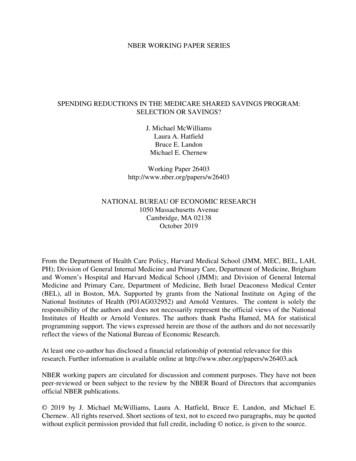
Step-By-StepInstructions for RetinaRecordings withPerforated MultiElectrode ArraysKatja Reinhard, Alexandra Tikidji-Hamburyan,Hartwig Seitter, Saad Idrees, Marion Mutter, BorisBenkner, Thomas A. MünchWerner Reichardt Centre for Integrative Neuroscienceand Bernstein Center for ComputationalNeuroscience, University of Tübingen,Tübingen, Germany(2014) PLoS ONE 9:e106148Introduction4Multi-electrode arrays are a state-of-the-arttool in retina research. The structure of theretina has allowed the use of multi-electrodearrays for high-throughput, parallel recordingsof retinal responses to presented visual stimuli,and has led to significant new insights intoretinal organization and function. However,using conventional arrays can be associatedwith three main problems: (1) low signal-tonoise ratio due to poor contact betweenelectrodes and tissue, especially in the case ofstrongly curved retinas from small animals, e.g.rodents; (2) insufficient oxygen and nutrientsupply to cells located on the bottom of therecording chamber; and (3) displacement of thetissue during recordings. Perforated multielectrode arrays (pMEAs) have been found toalleviate all three issues in brain slicerecordings. Over the last years, we have beenusing such perforated arrays to study lightevoked activity in the retinas of various speciesincluding mouse, pig, and human. In this article,we provide detailed step-by-step instructionsfor the use of perforated MEAs to record visualresponses from the retina, including spikerecordings from retinal ganglion cells and invitro electroretinograms (ERG).EXPERIMENT SUMMARIZATIONMaterials & MethodsThe setup for pMEA recordings consists of two perfusionloops: An upper loop to supply the tissue with freshsolution and a lower loop to adjust the proper negativepressure. We provide an overview of this dual perfusionsystem and a detailed list of the components we used tobuild our setup.The procedure on how to use the system is described indetail. Except for the constant vacuum pump theamplifier baseplate that allows vacuum application, andsome small components, no additional material isneeded compared to conventional MEA recordings. Theupper perfusion system supplies the retina with freshsolution during the recordings. The lower perfusionsystem is only used before the experiment to fill the MEAchamber with solution without introducing air bubblesinto the vacuum system. The vacuum system providesnegative pressure to pull the retina towards theelectrodes. This negative pressure needs to be constantto avoid fluctuations, and high enough to ensure goodtissue-electrode contact without tearing the tissue.Constant negative pressure is provided by a ConstantVacuum Pump (CVP) and is further reduced by anadditional fine flow control between the CVP and theMEA baseplate. The most important step for ensuringreliable negative pressure is the removal of air bubbles:any air bubble in the vacuum system will degrade thestable negative pressure. Setting up for pMEA recordingstakes approximately 40–60 minutes. Except for the stepsinvolving the vacuum system and preparation of thefilter paper, all steps are very similar to conventionalMEA recordings. Further, no coating of the MEA isnecessary for pMEA recordings. Overall, pMEArecordings require about 10 minutes more preparationtime than conventional MEAs.Results & ConclusionspMEAs provide good signal-to-noise ratiosThe vacuum applied through the perforation of pMEAsgreatly enhances the contact between the tissue and theelectrodes. On good recording electrodes, we can detectphone 508.893.8999 toll free 800.272.2775 fax 508.429.5732
Harvard Apparatus Neuroscience Application Guideand properly spike-sort one to three cells per electrode.To get an estimate of the number of recorded cells thatone might expect in such experiments, we counted thenumber of extractable cells in 153 recordings frommouse retina and found on average 38 18 cells. Pig andhuman retina recordings often had even better signal-tonoise ratios and therefore lead to more sortable cells. Inpig retina, we found on average 48 31 cells and inhuman retina 51 32 cells.Products fromused in this study:pMEAs allow stable long-term recordingsInsufficient oxygen and nutrient supply to the ganglioncell might limit the recording time. We showed variouslight stimuli to a mouse retina on a pMEA during 6 hoursand recorded ganglion cell responses. A very simplestimulus was presented over 120 times during these 6hours. The cell responded to every repetition of thestimulus, also after 6 hours of continuous recording.MEA2100-60-SystempMEAs prevent movement of retinaWe recorded ganglion cell responses to binarycheckerboard stimuli to calculate receptive fields and tovisualize tissue movement. Location and shape of thecalculated receptive fields were very stable during an 8hrecording period.Although the preparation and adjustment of theadditionally required perfusion and vacuum systemmight seem complicated at a first glance, the additionaltime required for perforated compared to conventionalMEA recordings amounts to only around 10 minutes.Further, little additional material is needed whenswitching from standard to perforated MEA recordings.Especially when isolating single cell responses fromMEA recordings, the user will appreciate the resultinghigh signal-to-noise ratio in pMEA art #DescriptionMEA2100-60-System-EMEA Recording System forone 60-electrode MEA withperfusion cannulaMEA2100-PE60/120Perfusion Element forMEA2100-headstage60pMEA200/30iR-Ti-grPerforated MEA with interelectrode distance of 200µm,electrode diameter of 30µm,internal reference electrodeand glass ringCVPConstant Vacuum PumpMEA setup consists of two perfusion loops. Solution is supplied tothe MEA chamber from the top through the upper perfusion (A)and excessive solution is removed by the suction (B). Thenecessary negative pressure is supplied by the additionalperfusion, consisting of the lower perfusion (C) and a vacuum (D).web www.harvardapparatus.com5
An immunoblot fordetection of Taeniasaginata cysticercosisEXPERIMENT SUMMARIZATIONMaterials & MethodsSameh Abuseir,Uschi Nagel-Kohl,Sonja Wolken& Christina StrubeIntroductionFood-borne zoontic diseases are significant andwidespread throughout the globe. It is contractedby food consumption contaminated by pathogenicorganisms between species. One particularorganism, Taenia Saginata, more commonlyknown as beef tapeworm is common in Africa,some parts of Eastern Europe, the Philippines, andLatin America. However, this organism is foundanywhere beef is consumed. Humans are the finaland bovines are the intermediate hosts to thisinfection. Infection with the adult T.saginata, orhuman taeniosis, is characterized by the presenceof a tapeworm that is 3.5 – 20 meters long in thesmall intestine. Bovine cysticercosis is theinfestation of the intermediate host and may sufferfrom cysts of T.saginata in their muscles andtissues. Light infestations are unlikely to bedetected during meat inspections, overall thereneeds to be more sensitive methods for detection.Developing an immunoblot for detection ofT.saginata infected cattle may lead to a potentialvaccine against cysticercosis and cysticechinococcosis in cattle.6SDS-PAGE was conducted using Hoefer Mighty Small IISE260 Mini Vertical Electrophoresis Unit in 6, 8, 10, 12,and 15% gels. 30-40µg of protein of the cyst sampleswere loaded per lane. Duplicate gels were done, one setwas for transfer and the other set was stained withCoommassie Blue dye to visualize proteins. Afterelectrophoresis, crude antigens from the one set of gelswere transferred using the Hoefer TE22 Mighty SmallTransfer Tank onto nitrocellulose membrane.Results & ConclusionsT. saginata cyst antigen SDS-PAGE and ImmunblotT. saginata cyst antigen showed ten visible bandsranging from 260 to 14kDa at approximately 260, 150,130, 67 (major band), 60, 55, 50, 23, 18, and 14 kDa. Allprotein bands reacted with T. saginata-positive sera(n 10) from experimentally infected cattle. Proteinbands of T. saginata was tested against positive E.granulosus sera (n 10) and showed only 18-14kDaproteins of T. saginata were immunoreactive. Noantigenic reactions were seen with any of the extractedproteins of the tested cestodes from serum samples(n 10) of infected animals with F. hepatica. All negativeserum samples (n 100) showed no reaction with anyextracted proteins.E. granulosus cyst antigen SDS-PAGE and ImmunblotE. granulosus cyst antigen showed visible bands rangingfrom 260 to 23 kDa at approximately 260, 250, 150, 130,120, 80, 67, 60, 55, 35, and 23kDa. All proteins bandsreacted with positive E. granulosus sera (n 10) except for260 and 35 kDa bands. All proteins bands reacted withpositive T. saginata sera (n 10) except for 260, 30, and 35kDa bands.phone 508.893.8999 toll free 800.272.2775 fax 508.429.5732
T. hydatigena cyst antigen SDS-PAGE and ImmunblotConclusion:Harvard Apparatus Neuroscience Application GuideT. hydatigena cyst antigen showed visible bands rangingfrom 290 to 14 kDa at approximately 290, 270, 260, 150,130, 80, 67, 55, 35, 23, and 14kDa. All proteins bandsreacted with positive T. saginata sera (n 10) except for290 and 14 kDa bands. All proteins bands reacted withpositive E. granulosus sera (n 10) except for 290, 270,260, and 14 kDa bands.Products fromHoeferused in this study:There is a wide range of antigenic similarity between thedifferent helminthes as seen in the results. This mayhelp develop a possible vaccine against the family ofTaeniidae that are affecting cattle. Two T. saginataprotein bands were identified as potential antigens.However, substantial research on additional Taeniaspecies still needs to be done to confirm results andantigen-antibody interactions.Mighty Small II MiniVertical ElectrophoresisMighty Small Transfer TankPart #DescriptionSE260-10A-.75Mighty Small II Mini VerticalElectrophoresis, CompleteSE260-10A-1.0SE260-10A-1.5TE22Mighty Small Transfer Tankwww.hoeferinc.comweb www.harvardapparatus.com7
Implementation of aTwo-dimensional BehaviorMatrix to DistinguishIndividuals with DifferentialDepression States in aRodent Model of DepressionJin-Young Park et al. (2014)Exp Neurobiol. Sep; 23(3); 215-223IntroductionAnimal models of depression are used to studypathophysiology of depression and to advancetherapeutic strategies. Stress-induced depressionmodels in rodents are widely used. However,amenable behavior criteria and experimentalprocedures that are suitable for animal modelshave not been established. Given that depression isclinically diagnosed by multiple symptomaticcriteria and stress effects are imposed to the brainnon-specifically in stress-induced depressionmodels, analyses of depression states in rodentsusing multiple symptomatic criteria may providemore power than any methods relying on a singlesymptomatic criterion.EXPERIMENT SUMMARIZATIONMaterials & MethodsMale C57BL/6 inbred mice at seven weeks of age (Eumsung,Chungbuk, Korea) were used in this investigation. Subjectswere restrained for 2h daily for 14 days and depressionstates of individual mice were assessed using the U-fieldtest, behavioral assessment developed to measure animal’ssociability and the tail suspension test and/or forced swimtest, which are the typical methods that measurepsychomotor withdrawal states.The U-field test was monitored using a computerizedvideo tracking system (SMART, Panlab, Spain).A webcam was used to record behavioral performancesin the TST and FST. Behavioral tests were performedduring the light cycle (9AM-3PM). The behavior testingroom was lit with indirect illumination by 20 lux for theU-field test and 250 lux for TST and FST, and sound wasmasked with 65 dB of white noise. Subjects were given20 minutes to adapt to the behavior testing room priorto the start of each behavioral test.Results & ConclusionsThe majority of these mice showed severe depressivebehaviors in both tests, a significant proportion of them,which were all inbred mice and received the sameamount of restraints, expressed differential depressionstates in the sociability test and psychomotorwithdrawal tests. To easily readout differentialdepression states of individuals in two different tests, astandard method and basic parameters required toconstruct two-way behavior matrix were introduced.The two-way behavioral matrix can be used not only tomeasure behavioral performance of a group of animals,but also to differentiate different depression states ofindividuals. For example, the two-way matrixdifferentiates behavioral states with normal sociabilityand psychomotor withdrawal, which was referred to as(i) RST-RSL individuals, (ii)behavioral states with normalsociability and high psychomotor withdrawal,(iii)behavioral states with low sociability and normalpsychomotor withdrawal and (iv)behavioral states withlow sociability and high psychomotor withdrawal,referred to “RST-DD” (Fig 3A and B)The RST-DD and88phone 508.893.8999 toll free 800.272.2775 fax 508.429.5732
Harvard Apparatus Neuroscience Application GuideProducts fromPanlabused in this study:SMART Video Tracking SoftwareRST-RSL individuals may have some similarity tosusceptible and resilient mice, respectively, which wereisolated on the basis of sociability alone [15].Cylinder for ForcedSwim Test ExperimentsPart #Description76-0696SMART Video Tracking Software76-0471Cylinder for Forced Swim Testswww.panlab.comIndividual animals determined to be depressive by theTST and FST are not necessarily defective inpsychomotor activity alone, but presumably in othersymptoms as well. Similarly, individuals determined tobe depressive by the U-field test are not necessarilydefective in sociability alone, but presumably in othersymptoms as well. Based on the same rationale, the twoway behavioral matrix may be expanded to the analysesof any two different behaviors, and should haveadvanced resolving power to distinguish individualswith different depression states compared with thosemeasured by a single behavioral symptomatic criterion.web www.harvardapparatus.com9
Protective effects ofremote ischemicconditioning againstischemia/reperfusion-induced retinal injury in ratsXuxiang Zhang et al. (2014)Visual Neurocience 31, 03, 246-262IntroductionLimb remote ischemic conditioning (LRIC) providesa physiologic strategy for harnessing the body’sendogenous protective capabilities against injuryinduced by ischemia-preperfusion in the centralnervous system. This study seeks to determine ifLRIC played a role in protecting the retina fromischemia-reperfusion injury. The role of astrocytesand cellular pathways was analyzed, which influencethe severity of ischemia/reperfusion injury (Eng etal., 2000). Retinal ischemia promotes increasedproliferation and differentiation of astrocytes.The increase in astrocyte activity following ischemiacauses an increase in glial fibrillary acidic protein(GFAP) transcription and translation.10EXPERIMENT SUMMARIZATIONMaterials & MethodsA total of 81 adult male Sprague-Dawley rats wererandomly assigned to ischemia/reperfusion with orwithout remote LRIC arms. Subjects were anesthetized,orally intubated and mechanically ventilated (RodentVentilator Model 683, Harvard Apparatus). The retinalischemic model was generated through right middlecerebral artery occlusion (MCAO) and pterygopalatineartery occlusion for 60 minutes followed by 1, 3, and 7days of subsequent reperfusion.Paraffin sections were stained to quantify the numberof cells in the retinal ganglion cells (RGCs) layerthroughout the duration of the study. Cellularexpression of glial fibrillary acidic protein (GFAP) wasdetected and examined through immunohistochemistry.Protein expression was analyzed via Western blot.phone 508.893.8999 toll free 800.272.2775 fax 508.429.5732
Results & ConclusionsThe study concluded that limb remote ischemicconditions (LRIC) protect the rat retinal fromischemia/reperfusion injury. Although themechanism(s) involved in retinal ischemic conditioningneed to be further examined, the result of this study andcurrent literature support the idea that the Nrf2/HO-1pathway is directly involved in retinal protectioninduced by LRIC. The results exhibited may provideclues to help elucidate the protective capabilities ofLRIC. The study suggests that LRIC could serve as avaluable tool for seeking therapeutics in enhancingneurological outcomes following ischemia/reperfusioninjury, and suggest three direction s in whichtherapeutics could be designed.Harvard Apparatus Neuroscience Application GuideAnalysis demonstrated that the loss of cells in the RGClayer were attenuated by LRIC treatment at 3 and 7 daysfollowing reperfusion (P 0.05). Immunohistochemistrystudies depicted a gradual increase (P 0.05 in GFAPlevels from day 1 through day 7 following ischemia andsubsequent reperfusion, whereas LRIC reduced GFAPlevels at 1, 3 and 7 days postreperfusion. The study alsoshowed that LRIC increased the expression of Nrf2 andHO-1 at day 1 and 3 following ischemia/reperfusion.Products fromHarvard Apparatusused in this study:Rodent Ventilator, Model 683Part #Description55-0000Rodent Ventilator, Model 683www.harvardapparatus.comweb www.harvardapparatus.com11
Intrahippocampalglucocorticoids generatedby 11β-HSD1 affect memoryin aged miceJ. Yau et al (2014)Neurobiology of Agingavailable online July 15, 2014IntroductionBoth aging and stress induce memory impairmentsnotably in hippocampus-associated cognitivefunctions. Stress activates the hypothalamicpituitary-adrenal (HPA) axis causing acute elevationof blood glucocorticoid levels, whereas in a subsetof aged individuals, plasma glucocorticoid levelsare elevated, and this correlates with hippocampalassociated memory deficits (Lupien et al., 1998)In the present study, in vivo microdialysis was usedin freely behaving mice to examine (1) the effects ofaging and/or stress on intro-hippocampal CORTlevels and memory during learning and retrieval;and (2) the role of 11β-HSD1 in these processes.EXPERIMENT SUMMARIZATIONMaterials & MethodsMale mice homozygous for a targeted disruption ofhsd11b1, congenic on the C57BL/6J genetic backgroundand wild-type (WT) C57BL/6J mice were bred in-houseunder standard conditions (12 hours light-dark cycle)with food and water ad libitum until experimentation ateither 6-8 months old (young) or 24-26 months old(aged) (Carter et al., 2009 and Kotelevtsev et al., 1997).A 2-trial Y-maze task was used to assess hippocampaldependent spatial recognition memory (Conrad et al.,1996 and Yau et al., 2011). The size of the maze allowedfor in vivo microdialysis in freely behaving mice.Mice were anesthetized and fixed within a stereotaxicframe for microdialysis guide implantation at a positionjust entering the dorsal hippocampus. Mice wereallowed to recover for 4 days before microdialysis began.A CMA 7 microdialysis probe was introduced via thepre-implanted guide cannula. Subjects wereindividually housed in the CMA 120 system for freelymoving animals with free access to food and water. Theprobe was perfused continuously with a sterile artificialcerebrospinal fluid at 0.3 µl per minute using aninfusion pump and samples were collected via theCMA470 Refrigerated Fraction Collector.Histology was performed and samples werehomogenized for CORT and 11β-HSD1 activity assays.12phone 508.893.8999 toll free 800.272.2775 fax 508.429.5732
Results & ConclusionsThis study highlighted the direct role of 11β-HSD1 inage-and stress-associated impairment of spatialmemory. Evidence is presented that in aged mice as wellas stressed young mice, a component part of theelevations in intra-hippocampal CORT levels during Ymaze testing is because of local 11β-HSD1 activity andthat this 11β-HSD1 component can cause impairedspatial memory. Importantly, 11β-HSD1 mice, thatcannot reactivate hippocampal CORT and, henceamplify levels to those found in aged WT controls,resisted both the adverse effects of age and acute stresson spatial memory. Such memory effects are not aconsequence of altered hippocampal MR and GR levelsthat do not differ between WT and 11β-HSD1 mice (Yauet al. 2011).Local intracellular regeneration of active glucocorticoidsby 11β-HSD1in the hippocampus is dynamicallyregulated during learning. This regenerativecomponent is even greater with aging reflecting asassociated increase in 11β-HSD1 expression in thehippocampus. The data presented that it is this rise inhippocampal CORT levels amplified by 11β-HSD1activity during the retrieval phase of spatial learning thatappears to impair memory in aged WT controls orstressed young WT mice. Also, data suggests that whenselective 11β-HSD1 inhibitors are used to treat chronicage-related memory deficiencies, there may be theadded advantage of protection from acute stressinduced memory impairments.Harvard Apparatus Neuroscience Application GuideThe study found that higher intrahippocampal CORTlevels associate with impaired spatial memory retentionduring the behavioral tasks for the aged, WT group, butnot the 11β-HSD1 mice. Acute stress was found toelevated plasma CORT levels and impair spatialmemory, and also its retrieval, in the WT, but not the11β-HSD1 mice. Pharmacologic inhibition of 11β-HSD1reduced intrahippocampal CORT levels in the agedsubjects and improves spatial memory.Products fromCMA Microdialysisused in this study:CMA 120 SystemPart #CMA 470 RefrigeratedFraction CollectorDescriptionCMAP000137 CMA 7 Guide CannulaCMAP000082 CMA 7 Microdialysis Probe, 1mm length (6kDa)CMA8309049 CMA120 System for Freely Moving AnimalsCMA400400 CMA4004 Syringe PumpCMA8002770 CMA470 Refrigerated Fraction Collector59-7316Artificial Cerebral Spinal Fluidwww.microdialysis.comSimilar products used in thisstudy can be found from ourother brands:Part #Description76-0681SMART Video Tracking76-0691SMART T/Y Maze Preconfigured Software Module76-0079 Y-Maze72-6335web www.harvardapparatus.comStereotaxic Frame, Mice and Rats13
Back to basics:Comprehensive DNAquantitation using BioDrop inthe UV spectrophotometryrange, why is it worth it?Boesenberg-Smith, K. A., Pessarakli, M. M. & Wolk, D. M.,2012. Assessment of DNA yield and purity: an overlookeddetail of PCR troubleshooting.Clinical Microbiology Newsletter, 34(1), pp. 2-5.Haque, K. A. et al., 2003. Performance of high-throughputDNA quantification methods. BMC Biotechnology, pp. 1-10.Wilson, I. G., 1997. Inhibition and Facilitation of Nucleic AcidAmplification. Applied and environmental microbiology,October, 63(10), pp. 3741-3751.EXPERIMENT SUMMARIZATIONMaterials & MethodsSample preparationLyophilized pure genomic DNA sample (dsDNA) fromcalf thymus from Sigma (PN# D4764) was weighed witha precision scale (d 0.1mg) and diluted to 1000mg/ml inTris-EDTA buffer solution pH7.4.The master sample was subsequently diluted forconstituting new samples. Each sample was vortexedand centrifuged.The samples were measured using 1.5μl of solution eachtime. The micro-volume sample port was wiped usinglint free tissue after each sample measurementResults & ConclusionsIntroductionWith the advent of new technologies aiming at moreclearly discerning subtle and complex informationfrom little amount of samples, the requirement forsetting quality control processes within a particularworkflow has never been greater.Examples of areas where sample concentrationdetermination has seemed to expand the most areproteomics and genomics which require lengthysample preparation and data analysis steps. Set asan intermediary step, protein or nucleic acidquantitation plays a very determinant role in theoutcome of many downstream experiments.The way the quantitation is performed is equallyvery important: the quality control process that canpote
Harvard Apparatus and affiliates are cited over 232,037 times in 'Google Scholar', and over 10,000 times in 2012 alone.* The following booklet contains examples of Harvard Apparatus products used in Neuroscience Research, along with summarizations of the experiments and related published articles. Thank you for considering