Transcription
8The Role of Molecular Imaging Technologies inBreast Cancer Diagnosis and ManagementAnne Rosenberg, Douglas Arthur Kieper, Mark B. Williams,Nathalie Johnson and Leora LanzkowskyJefferson University, Hampton University, University of Virginia, Legacy GoodSamaritan, Nevada Imaging CenterUSA1. IntroductionAnatomic breast imaging techniques such as mammography and ultrasound are very usefulin the detection of breast cancer, but can have limited sensitivity and positive predictivevalue, particularly in patients with dense breasts (Kolb et al., 2002). These limitations haveprovided the impetus for adjunctive technologies such as nuclear medicine and PET baseddiagnostic imaging procedures. The nuclear medicine based technique is referred to asBreast-Specific Gamma Imaging (BSGI) or molecular breast imaging (MBI) while thepositron-emission tomography (PET) based technique is referred to as Positron EmissionMammography (PEM). Both have demonstrated good results in clinical studies and areincreasingly being adopted into clinical practice. Although these imaging techniques havesimilarities, they are different in several aspects. This chapter is designed to provide anoverview of these imaging technologies and their potential roles in patient management.2. RadiotracersBoth BSGI/MBI and PEM are physiologic imaging modalities conducted through theinjection of a pharmaceutical, called a tracer, which is tagged with a radioactive isotope andthe resulting molecule is called a radiotracer. Each radiotracer is designed to bind to aspecific target (organ, tissue, physiologic process, cell receptor or protein) while the isotopetag emits radiation that is detected by cameras placed near the patient. The cameras providean image of the distribution of the radiotracer tracer and thus measure a specific physiologicprocess in the area being imaged.2.1 IsotopesThere are two types of radioactive isotope tags used in medical imaging: single gammaemission isotopes and positron emission isotopes. Single gamma emission isotopes release agamma ray from the nucleus. There are a variety of single gamma isotopes used in nuclearmedicine. The most common isotopes used in diagnostic imaging are referred to as lowenergy isotopes with gamma-ray energies ranging from 80 – 200 kiloelectron volts (keV).The gamma ray is a photon with sufficient energy to exit the body and be captured byspecially designed detectors called gamma cameras. Positron emission isotopes emit awww.intechopen.com
180Imaging of the Breast – Technical Aspects and Clinical Implicationpositron, an antimatter particle with the same mass as an electron, but with a positivecharge. This positron travels a short distance from the nucleus prior to colliding with anelectron. Since the positron is a particle traveling through the tissue until this collision, thepatient radiation dose associated with positron emission isotope studies is generally higherthan that from single gamma emission isotope examinations. This collision results inannihilation of both particles converting the mass of the two particles into energy andproducing a pair of 511 kiloelectron volt (keV) gamma rays traveling approximately 180 from each other. In positron emission tomography imaging (PET) these gamma rays exit thebody and are captured by a pair of opposed gamma cameras.The units for measuring the activity of radiotracer delivered to the patient are the Becquereland the Curie. The Becquerel (symbol Bq) is the SI-derived unit of radioactivity. One Bq isdefined as: one decay (emission) per second. The curie (symbol Ci) is a unit of radioactivity,defined as 3.7 1010 decays per second. In breast imaging, the megabecquerel (MBq) andmillicurie (mCi) are the most common units used; one millicurie equals 37 Megabecquerel.These units only describe the number of decays per second for a given sample and are not tobe confused with the units used to describe the radiation dose they deliver to a patient. Amore detailed discussion of radiation dose is provided in Section 4.2.1.1 Pharmaceuticals for BSGI/MBIBSGI/MBI is a single photon imaging technique that has been conducted using a variety ofimaging agents, but the most common is 99mTc-hexakis-2-methoxyisobutylisonitrile alsoreferred to as 99mTc-Sestamibi or MIBI. MIBI is a 140 KeV gamma ray emitting isotope in alipophilic cation molecule. It was originally cleared by the US FDA for use as a cardiacperfusion agent; breast imaging was subsequently added subsequently breast imaging wasadded as an indication following additional clinical studies to determine its efficacy in thisapplication. It is injected intravenously and is retained in cells likely by electronegativecellular and mitochondrial membrane potentials (Piwnica-Worms et al., 1990). Studies showthat its accumulation is roughly proportional to blood flow, desmoplastic activity andcellular proliferation and therefore it accumulates preferentially in breast cancers comparedwith surrounding tissues (Cutrone et al., 1998). It is a lipophilic substrate for the Pglycoprotein (Pgp), a cellular efflux pump for various compounds (Ballinger et al., 1995).Therefore, Sestamibi exhibits rapid tumor wash-in (within about 2 minutes) followed by aslow tumor washout (over the course of several hours) (Sciuto et al., 2002). Based on thesefactors, imaging can begin within minutes after injection and can continue for up to about 90minutes post injection, providing ample time for all required views to be conducted beforethe washout cycle negatively impacts lesion-to-background tracer concentration ratio. Inaddition, since the level of Pgp expression correlates with tumor response to cytotoxicchemotherapy, a comparison of immediate and delayed images (4 hours post injection) maybe used to quantify the radiotracer washout as a measure of Pgp expression and theprobability of multi-drug resistance.There are no known contraindications for use. Reactions to Sestamibi are generally minoraccording to the Cardiolite drug data sheet. In the analysis of potential reactions, 3068patients (77% men, 22% women, and 0.7% not recorded) were documented from the cardiacclinical trials and 673 were recorded from the breast imaging trials. Of the 673 breastimaging patients, all of whom were women, the most common reported reaction was tastewww.intechopen.com
The Role of Molecular Imaging Technologies in Breast Cancer Diagnosis and Management181perversion with most of those patients reporting a metallic taste at the time of injection. Theother minor reactions are listed in table 1. More serious reactions were reported in less than0.5% of patients and included: signs and symptoms consistent with seizure occurringshortly after administration of the agent; transient arthritis, angioedema, arrythmia,dizziness, syncope, abdominal pain, vomiting, and severe hypersensitivity characterized bydyspnea, hypotension, bradycardia, asthenia, and vomiting within two hours after a secondinjection of Technetium Tc99m Sestamibi. However the list of serious reactions is from thetotal population of patients including men and women undergoing a cardiac stress test.Body SystemBody as a wholeHeadacheCardiovascularAnginaST segment changesDigestive SystemNauseaSpecial SensesTaste PerversionParosmiaN 67321 (3.1%)11 (1.6%)9 (1.3%)0 (0%)0 (0%)8 (1.2)4 (0.6%)132 (19.6%)129 (19.2%)8 (1.2%)Table 1. Reactions to Sestamibi from 673 breast imaging patients.2.1.2 Pharmaceuticals for PEMPEM is a positron emission imaging technique conducted with 2-[fluorine-18] fluoro-2deoxy-D-glucose (FDG), a modified glucose molecule with a positron-emitting isotope.Breast cancers exhibit a greater uptake of FDG than the surrounding breast tissue due totheir hyperglycolytic rate. For some malignant lesions, although they possess an elevatedGLUT-1 transmembrane transport function, however this does not necessarily result inincreased tracer uptake (Smith, 1999). Studies have established that the uptake of FDG isprimarily dependent on blood flow, the type of breast malignancy and the microstructure ofthe lesion (nodular vs. diffuse) (Avril et al., 2001). For example, lobular carcinoma exhibits aroughly 30% lower uptake than ductal carcinoma (Avril et al., 2001). For the PEMprocedure, patients must fast 4 – 6 hours prior to the injection of FDG. It is important to notethat patients with compromised glucose metabolism should have their glucose levelchecked prior to the administration of FDG and at least one study reports that alteredglucose metabolism can affect the sensitivity of this procedure (Berg et al., 2006). FDG isadministered intravenously and imaging is conducted approximately 60-90 minutes postinjection in order to allow sufficient time for glucose uptake into the tissue. Patients shouldbe requested to sit quietly in a dark, calm room to avoid manipulating the distribution ofFDG. A dual-phase imaging technique may be used to improve specificity of the study(imaging at both 60 and 90 minutes post injection).The emitted positron has a mean free path of approximately 1 mm in the breast tissue beforeannihilation with an electron resulting in the emission of two 511 KeV gamma rays. Therandom nature of the displacement between the positron and gamma ray points of originwww.intechopen.com
182Imaging of the Breast – Technical Aspects and Clinical Implicationhas some impact on the lower limit of spatial resolution in studies using positron-emittingisotopes (Turkington, 2001).2.2 Comparison of BSGI/MBI and PEM RadiotracersBoth Sestamibi and FDG demonstrate increased accumulation in breast cancers, althoughthe mechanism for this accumulation is better understood for FDG. In addition, although thebreast tissue typically has a homogeneous uptake of both tracers, they can accumulate innormal glandular tissue resulting in a diffuse heterogeneous uptake pattern, especially inpre-menopausal women who are in the luteal phase of their menstrual cycle (Lin et al.,2007). This is not surprising as it reflects the heterogeneous nature of the breast tissue andthe impact of blood hormone levels on the breast parenchyma. This heterogeneity does notgenerally impact cancer detection, but may complicate interpretation. The intensity of thispattern can be reduced for both tracers by imaging outside of the luteal phase and severalstudies report that day 2 – 14 of the menstrual cycle is optimal. Neither tracer is linked tonephrogenic systemic fibrosis, a sometimes fatal condition that is associated withgadolinium contrast agents used in breast MRI. MIBI has some minor pharmacologicconsiderations and rare reactions occurring in less than 0.5%of patients. FDG imagingrequires fasting for a minimum of 4 hours prior to injection and as mentioned in theprevious section, it is beneficial to check the blood glucose level prior to FDG administrationas the tumor uptake of FDG is reduced in hyperglycemic states (Schelbert et al., 1999) andthis results in some potential for complications and reduced sensitivity for the procedure indiabetic patients. In comparison, MIBI does not require fasting and imaging can beconducted within minutes of the injection.Some of the physical and clinical differences between FDG and MIBI imaging aresummarized in Tables 2 and 3 respectively.FDGMIBIEnergy ofEmission photon511 KeV140 KeVHalf Life(minutes)110360Table 2. gamma-ray emission information for radiotracers.Pre-study FastingFDG4 - 6 hoursMIBINoneInjection toImaging time60 – 90minutes5 minutesRecommendedPre-procedural testingBlood glucoseNoneTable 3. Radiotracer administration and imaging considerations.3. Imaging systemsBoth the single gamma and positron isotopes described in section 2.1 ultimately emitgamma rays (the positron isotopes through the annihilation and the conversion of thepositron) that exit the patient’s body and can be detected with external detectors. The goal ofwww.intechopen.com
The Role of Molecular Imaging Technologies in Breast Cancer Diagnosis and Management183these detector systems is to reconstruct the gamma ray emissions into an image that allowsthe physician to visualize the distribution of the radiotracer in the body. There are twomodes of image reconstruction, plannar and tomographic. The planar method results in asingle 2-dimensional image per acquisition, similar to the mammogram. Tomographicreconstruction provides a 3-dimensional reconstruction of the breast similar to MRI. Thesingle gamma detector systems used for BSGI/MBI can be planar or tomographic and canbe constructed of a single or multiple detectors. The positron imaging systems used for PEMby their design provide tomographic imaging only and since the detection of the pair ofgamma rays is required for image reconstruction, positron systems must consist of either apair of opposed detectors or a ring detector design.3.1 Gamma-ray imaging basicsThe detection of an abnormality in BSGI and PEM imaging is based on the ability of theimaging system to depict the variations of uptake in the tissue. Unlike anatomical imagingwhere high spatial resolution is needed to visualize the detailed morphology used toprovide differential diagnosis, the molecular imaging system must provide sufficient imagecontrast in order to visualize the variations in radiotracer uptake; the contrast betweenradiotracer concentration in the tumor and the uptake of the surrounding breast tissue.While this is partially a function of resolution, there are several other factors impactingimaging. This contrast based imaging requires a careful balance between spatial resolution,image noise and photon sensitivity. Generally, as spatial resolution increases, image noiseincreases and photon sensitivity decreases proportionally to some degree thus if spatialresolution is increased to a level where the detector has poor photon sensitivity and theresulting image noise is too high, the ability of the system to visualize the contrasting tissueuptake is diminished. A detailed discussion of the balance between these factors is beyondthe scope of this text, but it is important to realize that in molecular imaging is a contrastbased imaging and spatial resolution is not the only parameter affecting the visualization oflesions. For example, it is possible to detect a 1 mm cancer using a system with a 4 mmspatial resolution if the uptake of that lesion is sufficiently enough higher than thebackground to overcome the partial volume effect. Conversely, a 40 mm cancer could bemissed by the same imaging system if the lesion uptake is not sufficiently higher than thesurrounding tissue.As an illustration, nearly all commercially available large field-of-view gamma cameras,typical to the nuclear medicine department, have a variable matrix setting, including 512 x512, 256 x 256 and 128 x 128. Although the 512 x 512 setting produces the highest spatialresolution, nearly all nuclear medicine studies are conducted on the 256 x 256 or 128 x 128settings because the resulting image noise at the 512 x 512 setting diminishes image qualityfor the majority of studies.System photon sensitivity is another important parameter in BSGI/MBI and PEM imaging.As photon sensitivity increases, the amount of radiotracer, the length of time the image isacquired, or some combination of both parameters can be decreased. For Example: a givendetector system is providing good clinical images using a dose of 300 MBq and anacquisition time of 10 minutes. If the photon sensitivity of this system can be increased 50%the clinician would have three possible options. First, they could reduce the patient’sradiation exposure by reducing the amount of radiotracer delivered to the patient by 50%, towww.intechopen.com
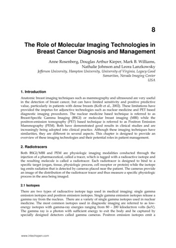
184Imaging of the Breast – Technical Aspects and Clinical Implication150 MBq and maintain the same imaging time, 10 minutes. Second, they could reduce theimaging time by 50% to 5 minutes using a 300 MBq dose. Or third, they could reduce boththe time and the dose by roughly 25% resulting in a dose of 225 MBq and an imaging time of7.5 minutes. It is important to remember that in molecular imaging techniques such asBSGI/MBI and PEM, the imaging time and the dose delivered can be manipulated, butreducing both to any large degree is not possible unless significant improvements in photonsensitivity are obtained.3.1.1 BSGI/MBI ImagingBSGI/MBI imaging is conducted with a single-head or dual-head detector system (seeFigure 1). Only one detector equipped with a collimator is required for imagereconstruction. Generally, gentle breast compression (normally less than 12 lbs or 53newtons) is used to provide breast immobilization. This compression is noticeably lowerthan that used in mammography for two reasons. First, the typical imaging time for a singleBSGI/MBI image is significantly longer than that needed for a mammographic projection, 5– 10 minutes, thus lower pressures are better tolerated by patients and second, the 140keVgamma ray emitted in BSGI/MBI has sufficiently higher tissue penetration than the 8 – 35keV x-ray used in mammography therefore these images benefit less from highercompression. As shown in figure 1, in the single-head design a paddle is used to providecompression and in the dual-head system, the breast is compressed between the detectors.The compression paddle used in the single head system can be exchanged for a fenestratedpaddle to allow biopsy. Biopsy is currently not available on the dual-head design.Fig. 1. A single and dual head imaging system for BSGI/MBI.3.1.2 BSGI/MBI detectorsAs mentioned in the previous section, there is generally an inverse relationship betweenphoton sensitivity and spatial resolution however both are important to imaging. Theseparameters are determined by several aspects of the detector design, especially that of thecollimator. The most commercially available systems have an extrinsic spatial resolution ofbetween 1.9 and 3.3 mm at the surface of the detector however it is important to note thatthe spatial resolution of planar, single gamma imaging systems decreases with increasingsource-to-collimator distance, thus the spatial resolution of a lesion near the detector isbetter than that of one deep in the breast tissue, relative to the detector face. For example, ifthe breast is being imaged in the cranio-caudal position (detector inferior to the breasttissue), lesions in the inferior portion of the breast tissue will be somewhat more visible thanwww.intechopen.com
The Role of Molecular Imaging Technologies in Breast Cancer Diagnosis and Management185those in the superior portion of the breast. This resolution loss is, at least in part, the drivinglogic behind the dual-head opposed detector design. In theory, if the breast is imaged usinga dual-head system in the cranio-caudal position, the upper detector would maximizevisualization of the superior portion of the breast while the lower detector optimizesvisualization of inferior lesions.Although there is a theoretical benefit to the dual-head design, it is interesting to note thatclinical data from the dual-head and single-head systems shows similar performance interms of lesion sensitivity (see section 5 below). This is likely due to the two-view imagingprotocol adopted from mammography that is standard in BSGI/MBI protocols. Just as inmammography, the optimal coverage of breast tissue is obtained by acquiring an MLO andCC image of each breast and additional images are obtained as needed. Since all patientshave a minimum of two views obtained, the likelihood of a lesion being deep to bothprojections is quite small. In addition, similar to mammography, when a lesion is seen inonly one image, additional images are obtained in order to determine the location of thelesion in the breast tissue. As long as this two-view protocol remains the standard, it isunlikely that the dual-head system will result in significant improvements in the sensitivityof the detection of breast malignancies. However, provided the two detector images arefused properly, it may be possible to reduce either the injected dose or the acquisition timeto facilitate low-dose imaging or higher throughput on the imaging system.Single photon emission computed tomography (SPECT) is a recent development inBSGI/MBI. Currently these devices are only available in the research setting (Williams et al.,2010). Additional research is needed to determine if dual-head image combinationtechniques or the implementation of SPECT imaging will provide a clear benefit toBSGI/MBI in terms of breast cancer detection. Such studies are underway, but the data isnot yet available for analysis.Fig. 2. Left and Center - a single-head BSGI/MBI system with a compression paddle usedfor positioning. The left image illustrates the cranio-caudal (CC) position and the centerimage illustrates the medial lateral oblique position (MLO). The right image illustrates theMLO position with the dual-head system where the compression paddle is replaced by asecond detector.www.intechopen.com
186Imaging of the Breast – Technical Aspects and Clinical ImplicationFig. 3. A typical 4-view BSGI study.3.1.3 Detectors for PEMPEM imaging is conducted with either a dual head or ring style gamma-ray detector. Bothsystems are designed to detect the coincident gamma rays which, are travelingapproximately 180 from each other after the annihilation reaction (see figure 4). UnlikeBSGI imaging devices, PEM devices do not use a collimator to help determine the location ofeach event. In PEM imaging, since there are two gamma-rays traveling 180 apart, the eventlocation is calculated as a line of response between the location that each gamma-ray strikesthe pair of opposed detectors. One advantage to PEM is that it does not have the same lossof resolution with distance that BSGI/MBI systems experience. As mentioned in theprevious section, the mean free path of the 18F positron within the breast tissue isapproximately 1 mm and commercially available PEM systems report an in-plane spatialresolution of about 2 mm. One limitation of the dual-head PEM detector design is that, dueto the limited angle of acquisition, it has limited resolution in the Z-axis (depth). Ringdetectors do not suffer from this limitation as they provide a 360 acquisition forreconstruction however there is currently no biopsy capability on the ring detector systems.A needle biopsy localization device was recently introduced for the opposed dual-headdetector system.Fig. 4. The left image provides an example of an opposed dual-head imaging system whilethe system on the right is an example of a ring detector system.www.intechopen.com
The Role of Molecular Imaging Technologies in Breast Cancer Diagnosis and Management187One limitation to PEM image reconstruction is that the detector photon sensitivity is notlinear across the field-of-view with lower sensitivity along the detector edges. This causes ahigher level of noise to be present in the breast images, along the chest wall. Figure 5provides a schematic representation of factors affecting the photon sensitivity in a PEMdetector. The maximum angle of reconstruction (MAR) is a setting used in PEM softwareand it is defined as the maximum angle away from the detector normals (90 from thedetector face) for which coincident gamma ray detections are included in tomographicimage reconstruction. Larger MAR values yield greater overall photon sensitivity but withpotential loss in spatial resolution due to the depth-of-interaction (DOI) blur. The blue linesin Figure 1 show the angular range over which gamma rays emitted from two points in thebreast are accepted. Figure 1A shows a point near the nipple, and Figure 1B shows one nearthe chest. For events that occur in the center of the field-of-view (FOV), all of the eventswithin the MAR are captured by the detector system. However a significant fraction ofevents occurring near the FOV edges go uncounted for because one of the paired gammarays traveling outside the edges of the detector is not detected. As a recent study found, thisloss of photon sensitivity along the edges limits the ability of the PEM system to detectlesions located near the chest wall (Rosen et al, 2005).Detector 1Detector 1Detector 2Detector 2Fig. 5. A and B: a schematic example of the maximum angle of reconstruction near the centerof the detector field-of-view and then near the chest wall respectively.Fig. 6. a typical PEM study with multiple slice reconstruction for each projection.www.intechopen.com
188Imaging of the Breast – Technical Aspects and Clinical ImplicationPEM detectors are tomographic imaging devices, an example image from the opposeddetector system is provided in Figure 6. Note the noise level along the chest wall and theZ-resolution affect is expressed as a blurry, low intensity focus in the reconstruction planesoutside of the plane the lesion is located in. In this particular case, it is most noticeable in theMLO projection images. There is noticeable residual blur in the area of the largest lesion inall of the projections, including those outside of the lesion.4. Radiation doseAs with all nuclear medicine procedures, the detector systems do not emit radiation. Theradiation dose delivered to the patient in these procedures comes from the radiotracer and isdependent on both the activity of the radiotracer injected and the biologic distribution of thetracer in the organs.4.1 BSGI/MBI radiation doseSestamibi (MIBI) was cleared by the US FDA in 1991 for cardiac perfusion studies. In 1997,breast imaging was added as an indication to the drug package insert following a clinicaltrial conducted with standard gamma cameras equipped with high-resolution collimators.According to the drug package insert, the patient whole-body radiation dose is 4.8 milligrayat 1110 megabecquerels (0.5 rads at 30 millicuries), see Table 4. According to the Dosageand Administration section of the drug package insert, breast imaging is to be conductedusing a dose of 740 – 1110 MBq (20 – 30 mCi).At the time of the US FDA approval, the breast imaging studies were being acquired withstandard, large field-of-view gamma cameras, typical to a nuclear medicine department andthe dose required for imaging was determined largely by the low photon sensitivity of theseimaging systems when equipped with high-resolution collimators (Khalkhali et al., 2004)Since that time, several breast optimized gamma camera systems have been developed withsignificantly higher photon sensitivity and several studies indicate that it is possible to lowerthe injected dose of MIBI required for breast imaging with these systems.A recent clinical trial was conducted to examine breast tissue uptake as a function ofinjected dose. The results of this analysis indicate that breast tissue uptake of MIBIappears to be linear relative to the injected dose thus implying there is no physiologiclimitation to using lower doses (Böhm-Vélez et al., 2011). According to additional studies,conducted by the Mayo Clinic, the new, breast optimized detector systems provide aphoton sensitivity roughly 3 times higher than that of the older imaging systems (Hruskaet al., 2008).From the available data, it is evident that these new detector technologies can reduce thedose required to conduct breast imaging with MIBI. Reducing the dose MIBI from 740 –1110 MBq (20 – 30 mCI) to 259 – 370 MBq (7 – 10 mCi) reduces patient radiation exposure bynearly a factor of 3. The radiation exposure from a 259 MBq injection of MIBI isapproximately 2 millisieverts (mSv) and is approximately equivalent to the radiation dosediagnostic breast patients receive from the combination of screening and diagnosticmammograms (Hendrick, 2010; Valinten, 2007).www.intechopen.com
The Role of Molecular Imaging Technologies in Breast Cancer Diagnosis and Management189Table 4. Radiation dosimetry of Sestamibi.Graph 1. The relative photon sensitivity of commercially available, breast-optimizedimaging systems compared to that of the standard gamma camera.4.2 PEM radiation doseF-18 fluorodeoxy-D-glucose (FDG) was cleared by the US FDA in 2000 for a variety of usesincluding tumor localization. The total body radiation dose in FDG PET is 39 mrads per mCiinjected activity (Table 5). According to the clinical literature, the typical FDG dose used forimaging with the standard whole body PET detectors ranges between is approximately 370 740 MBq (10 - 20 mCi).www.intechopen.com
190Imaging of the Breast – Technical Aspects and Clinical ImplicationTable 5. Radiation dose for FDG based on 1 mCi injection.The dose of FDG used for PEM studies has generally followed the guidelines establishedwith the lager systems, typically using approximately 444 MBq (12 mCi) (Berg et al, 2006).However, more recent studies have demonstrated that doses of 111 – 185 MBq (3 – 5 mCi)are possible with the breast-optimized imaging systems (MacDonald et al, 2010). Theresulting radiation dose to the patient is 1.9 – 3.1 mSv using a low dose protocol, nearlyidentical to that of low dose BSGI/MBI (O’Connor et al., 2010).5. Clinical evidenceThere is a substantial history of clinical literature on imaging breast cancers with nuclearmedicine techniques. One of the first reports of breast imaging using MIBI was provided byCampeau and his colleagues in 1992 while the first report of breast cancer imaging usingFDG was reported by Wahl the previous year (Campeau et al., 1992; Wahl et al,. 1991) Sincethat time, hundreds of articles have been published on breast imaging using theseradiotracers. However until recently, these imaging studies were conducted with largegamma cameras typical in the nuclear medicine department. The development of breastoptimized detector systems used in BSGI/MBI and PEM is more recent and the primaryadvantage of these systems is that they provide higher sensitivity for the detection of breastlesions than their predecessors.5.1 Clinical evidence for BSGI/MBIThere have been several clinical studies evaluating BSGI/MBI in breast cancer det
Samaritan, Nevada Imaging Center USA 1. Introduction Anatomic breast imaging techniques such as mammography and ultrasound are very useful in the detection of breast cancer, but can have limited sensitivity and positive predictive value, particularly in patients with dense br easts (Kolb et al., 2002). These limitations have