Transcription
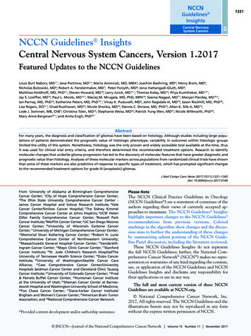
NCCN1331Guidelines InsightsCECentral NervousSystem CancersNCCN Guidelines InsightsCentral Nervous System Cancers, Version 1.2017Featured Updates to the NCCN GuidelinesLouis Burt Nabors, MD1,*; Jana Portnow, MD2,*; Mario Ammirati, MD, MBA3; Joachim Baehring, MD4; Henry Brem, MD5;Nicholas Butowski, MD6; Robert A. Fenstermaker, MD7; Peter Forsyth, MD8; Jona Hattangadi-Gluth, MD9;Matthias Holdhoff, MD, PhD5,*; Steven Howard, MD10; Larry Junck, MD11,*; Thomas Kaley, MD12; Priya Kumthekar, MD13,*;Jay S. Loeffler, MD14; Paul L. Moots, MD15,*; Maciej M. Mrugala, MD, PhD, MPH16; Seema Nagpal, MD17; Manjari Pandey, MD18,*;Ian Parney, MD, PhD16; Katherine Peters, MD, PhD19,*; Vinay K. Puduvalli, MD3; John Ragsdale III, MD19; Jason Rockhill, MD, PhD20;Lisa Rogers, DO21,*; Chad Rusthoven, MD22; Nicole Shonka, MD23; Dennis C. Shrieve, MD, PhD24; Allen K. Sills Jr, MD15;Lode J. Swinnen, MB, ChB5; Christina Tsien, MD25; Stephanie Weiss, MD26; Patrick Yung Wen, MD27; Nicole Willmarth, PhD28;Mary Anne Bergman29,*; and Anita Engh, PhD29,*AbstractFor many years, the diagnosis and classification of gliomas have been based on histology. Although studies including large populations of patients demonstrated the prognostic value of histologic phenotype, variability in outcomes within histologic groupslimited the utility of this system. Nonetheless, histology was the only proven and widely accessible tool available at the time, thusit was used for clinical trial entry criteria, and therefore determined the recommended treatment options. Research to identifymolecular changes that underlie glioma progression has led to the discovery of molecular features that have greater diagnostic andprognostic value than histology. Analyses of these molecular markers across populations from randomized clinical trials have shownthat some of these markers are also predictive of response to specific types of treatment, which has prompted significant changesto the recommended treatment options for grade III (anaplastic) gliomas.J Natl Compr Canc Netw 2017;15(11):1331–1345doi: 10.6004/jnccn.2017.0166From 1University of Alabama at Birmingham ComprehensiveCancer Center; 2City of Hope Comprehensive Cancer Center;3The Ohio State University Comprehensive Cancer Center James Cancer Hospital and Solove Research Institute; 4YaleCancer Center/Smilow Cancer Hospital; 5The Sidney KimmelComprehensive Cancer Center at Johns Hopkins; 6UCSF HelenDiller Family Comprehensive Cancer Center; 7Roswell ParkCancer Institute; 8Moffitt Cancer Center; 9UC San Diego MooresCancer Center; 10University of Wisconsin Carbone CancerCenter; 11University of Michigan Comprehensive Cancer Center;12Memorial Sloan Kettering Cancer Center; 13Robert H. LurieComprehensive Cancer Center of Northwestern University;14Massachusetts General Hospital Cancer Center; 15VanderbiltIngram Cancer Center; 16Mayo Clinic Cancer Center; 17StanfordCancer Institute; 18St. Jude Children’s Research Hospital/TheUniversity of Tennessee Health Science Center; 19Duke CancerInstitute; 20University of Washington/Seattle Cancer CareAlliance; 21Case Comprehensive Cancer Center/UniversityHospitals Seidman Cancer Center and Cleveland Clinic TaussigCancer Institute; 22University of Colorado Cancer Center; 23Fred& Pamela Buffet Cancer Center; 24Huntsman Cancer Instituteat the University of Utah; 25Siteman Cancer Center at BarnesJewish Hospital and Washington University School of Medicine;26Fox Chase Cancer Center; 27Dana-Farber Cancer Institute/Brigham and Women’s Cancer Center; 28American Brain TumorAssociation; and 29National Comprehensive Cancer Network.*Provided content development and/or authorship assistance.Please NoteThe NCCN Clinical Practice Guidelines in Oncology(NCCN Guidelines ) are a statement of consensus of theauthors regarding their views of currently accepted approaches to treatment. The NCCN Guidelines Insightshighlight important changes to the NCCN Guidelines recommendations from previous versions. Coloredmarkings in the algorithm show changes and the discussion aims to further the understanding of these changesby summarizing salient portions of the NCCN Guideline Panel discussion, including the literature reviewed.These NCCN Guidelines Insights do not representthe full NCCN Guidelines; further, the National Comprehensive Cancer Network (NCCN ) makes no representation or warranties of any kind regarding the content,use, or application of the NCCN Guidelines and NCCNGuidelines Insights and disclaims any responsibility fortheir applications or use in any way.The full and most current version of these NCCNGuidelines are available at NCCN.org. National Comprehensive Cancer Network, Inc.2017, All rights reserved. The NCCN Guidelines and theillustrations herein may not be reproduced in any formwithout the express written permission of NCCN. JNCCN—Journal of the National Comprehensive Cancer Network Volume 15 Number 11 November 2017
CE1332NCCN Guidelines InsightsCentral Nervous System Cancers, Version 1.2017NCCN: Continuing EducationTarget Audience: This activity is designed to meet the educational needs of physicians, nurses, and pharmacists involved inthe management of patients with cancer.Accreditation StatementPhysicians: National Comprehensive Cancer Network is accreditedby the Accreditation Council for Continuing Medical Education(ACCME) to provide continuing medical education for physicians.NCCN designates this journal-based CE activity for a maximumof 1.0 AMA PRA Category 1 Credit . Physicians should claimonly the credit commensurate with the extent of their participation in the activity.Nurses: National Comprehensive Cancer Network is accreditedas a provider of continuing nursing education by the AmericanNurses Credentialing Center s Commission on Accreditation.NCCN designates this knowledge-based continuing educationactivity for 1.0 contact hour (0.1 CEUs) of continuing educationcredit. UAN: 0836-0000-17-011-H01-PAll clinicians completing this activity will be issued a certificateof participation. To participate in this journal CE activity: 1) review the educational content; 2) take the posttest with a 66%minimum passing score and complete the evaluation at http://education.nccn.org/node/81831; and 3) view/print certificate.Release date: November 10, 2017; Expiration date: November10, 2018Learning Objectives:Upon completion of this activity, participants will be able to:NCCN designates this educational activity for a maximum of1.0 contact hour.Pharmacists: National Comprehensive Cancer Network isaccredited by the Accreditation Council for Pharmacy Education as a provider of continuing pharmacy education. Integrate into professional practice the updates to theNCCN Guidelines for Central Nervous System Cancers Describe the rationale behind the decision-makingprocess for developing the NCCN Guidelines for CentralNervous System CancersDisclosure of Relevant Financial RelationshipsEditor:Kerrin M. Green, MA, Assistant Managing Editor, JNCCN—Journal of the National Comprehensive Cancer Network, has disclosed that she has no relevant financial relationships.JNCCN:Kimberly Callan, MS, Senior Director, Professional and Patient Publications, NCCN, has disclosed that she has no relevant financial relationships.Genevieve Emberger Hartzman, MA, Journal Production Specialist, NCCN, has disclosed that she has no relevant financial relationships.CE Authors:Deborah J. Moonan, RN, BSN, Director, Continuing Education, NCCN, has disclosed that she has no relevant financial relationships. (Employed by NCCNuntil 2/17/17.)Karen Kanefield, Manager, Continuing Education Accreditation and Program Operations, NCCN, has disclosed that she has no relevant financial relationships.Kathy Smith, Manager, CE Grant Writing & Project Management, NCCN, has disclosed that she has no relevant financial relationships.Kristina M. Gregory, RN, MSN, OCN, Vice President, Clinical Information Operations, NCCN, has disclosed that she has no relevant financial relationships.Rashmi Kumar, PhD, Director, Clinical Information Operations, NCCN, has disclosed that she has no relevant financial relationships.Individuals Who Provided Content Development and/or Authorship Assistance:Louis Burt Nabors, MD, Panel Chair, has disclosed that he serves as a scientific advisor for Bristol-Myers Squibb Company and ZioPharm. He also receivesconsulting fees/honoraria from and is on the product/speakers bureau for Merck & Co., Inc.Jana Portnow, MD, Vice Chair, has disclosed that she has no relevant relationships.Matthias Holdhoff, MD, PhD, Panel Member, has disclosed that he serves as a scientific advisor for Celgene and has received honoraria from Arbor.Larry Junck, MD, Panel Member, has disclosed that he serves as a scientific advisor for Ignyta, Inc., PRA Health Services, and Tocagen.Priya Kumthekar, MD, Panel Member, has disclosed that she serves as a scientific advisor for AbbVie Inc. and Vivacitas Oncology, Inc.Paul L. Moots, MD, Panel Member, has disclosed that he has no relevant financial relationships.Manjari Pandey, MD, Panel Member, has disclosed that she has no relevant financial relationships.Katherine Peters, MD, PhD, Panel Member, has disclosed the she serves as a scientific advisor for AbbVie Inc. and Novocure. She also received grant/research support from Argos Therapeutics, Inc.; BioMimetix Pharamaceutical, Inc.; Eisai Inc.; Genentech, Inc.; Merck & Co., Inc.; and VBL Therapeutics.Lisa Rogers, DO, Panel Member, has disclosed that she has served as a scientific advisor for Novocure.Mary Anne Bergman, Guidelines Coordinator, NCCN, has disclosed that she has no relevant financial relationships.Anita Engh, PhD, Oncology Scientist/Medical Writer, NCCN, has disclosed that she has no relevant financial relationships.This activity is supported by educational grants from Astellas, AstraZeneca, Celldex Therapeutics, Clovis Oncology, Genomic Health, Inc., Kyowa HakkoKirin, Jazz Pharmaceuticals, Novartis Pharmaceuticals Corporation, and NOVOCURE. This activity is supported by an independent educational grantfrom Merck Co., ehensiveComprehensiveCancerCancerNetworkNetwork VolumeVolume1515 NumberNumber1111 NovemberNovember20172017 JNCCN—Journal
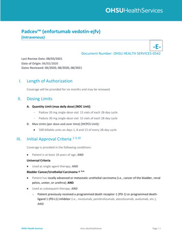
CENCCN Guidelines Insights1333Central Nervous System Cancers, Version 1.2017ANAPLASTIC GLIOMAS(See GLIO-3/GLIO-4 for GBM)PATHOLOGYdADJUVANT TREATMENTAnaplastic oligodendroglioma(1p19q codeleted)1p19q codeleted:Anaplastic oligoastrocytomaFractionated external beam RT l and neoadjuvant or adjuvantmPCV chemotherapy (category 1) norFractionated external beam RT l with concurrent and adjuvanttemozolomide chemotherapyn1p19q uni-or non-deleted:Anaplastic oligodendrogliomaAnaplastic astrocytomaAnaplastic oligoastrocytomakAnaplastic gliomasaPoor performancestatus (KPS 60)FOLLOW-UPbFractionated external beam RT l with concurrent andadjuvant temozolomide chemotherapynorFractionated external beam RT l neoadjuvant oradjuvantm PCVorPCV or temozolomide chemotherapyorFractionated external beam RT l (category 1)Brain MRI 2–6 wks afterRT, then every 2–4 mofor 3 y, then every 6months indefinitelyoSeeRecurrence(GLIO-5)Fractionated external beam RT l(hypofractionated [preferred] or standard)orPCV or temozolomide chemotherapy (category 2B)norPalliative/Best supportive careaThispathway includes the classification of mixed anaplastic oligoastrocytoma (AOA),anaplastic astrocytoma (AA), anaplastic oligodendroglioma (AO), and other rareanaplastic gliomas.bSee Principles of Brain and Spine Tumor Imaging (BRAIN-A).dSee Principles of Brain Tumor Pathology (BRAIN-F).kNOS WHO 2016 has deleted this category, although it may continue to be used for somepatients.lSeePrinciples of Brain and Spinal Cord Tumor Radiation Therapy (BRAIN-C).panel recommends that PCV be administered after RT (as per EORTC 26951) sincethe intensive PCV regimen given prior to RT (RTOG 9402) was not tolerated as well.nSee Principles of Brain and Spinal Cord Tumor Systemic Therapy (BRAIN-D).oWithin the first 3 months after completion of RT and concomitant temozolomide, diagnosisof recurrence can be indistinguishable from pseudoprogression on neuroimaging.mTheVersion 1.2017 National Comprehensive Cancer Network, Inc. 2017, All rights reserved. The NCCN Guidelines and this illustrationmay not be reproduced in any form without the express written permission of NCCN .GLIO-2OverviewNCCN Categories of Evidence and ConsensusCategory 1: Based upon high-level evidence, thereis uniform NCCN consensus that the interventionis appropriate.Category 2A: Based upon lower-level evidence, thereis uniform NCCN consensus that the intervention isappropriate.Category 2B: Based upon lower-level evidence, thereis NCCN consensus that the intervention is appropriate.Category 3: Based upon any level of evidence, thereis major NCCN disagreement that the interventionis appropriate.All recommendations are category 2A unless otherwisenoted.Clinical trials: NCCN believes that the best managementfor any patient with cancer is in a clinical trial. Participation in clinical trials is especially encouraged.Estimates based on recent population analyses indicate that nearly 24,000 people in the United Statesare diagnosed with primary malignant brain or othercentral nervous system (CNS) neoplasms each year.1,2In adults, the annual incidence of malignant primary brain and other CNS tumors is 8.7 per 100,000.1These cancers are a leading cause of death in adults,especially for those 40 years old, and are estimated tobe responsible for 16,700 deaths in the United Statesin 2017.1,2 High-grade gliomas are the most commontype of brain cancer, accounting for more than half ofall malignant primary tumors of the brain and CNS.1Although the prognosis for glioblastoma (grade IVglioma) is grim (5-year survival rates between 1% and19%, depending on age), outcomes for anaplastic gliomas (grade III gliomas) are typically better, dependingon the molecular features of the grade III glioma.1 Clinicians are learning how to better predict survival andselect treatments for patients with high-grade gliomas JNCCN—Journal of the National Comprehensive Cancer Network Volume 15 Number 11 November 2017
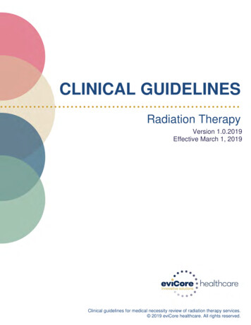
CE1334NCCN Guidelines InsightsCentral Nervous System Cancers, Version 1.2017PRINCIPLES OF BRAIN TUMOR PATHOLOGY (1 OF 3)Standard Histology Histologic subgrouping of CNS neoplasms provides valuable prognostic information, as isencompassed in the WHO classification of gliomas.1 Inter-observer differences in histologic diagnosis and grading are a recognized issue. Even so, the traditional histologic distinction of CNS neoplasms into primary neuroectodermalneoplasms (eg, glial, neuronal, embryonal) from other primary CNS neoplasms (eg, lymphoma, germcell, meningeal), metastatic neoplasms, and non-neoplastic conditions mimicking tumors, remainsfundamental to any pathologic assessment.Molecular/Genetic Characterization The development of sophisticated genetic and molecular characterization of CNS neoplasms hasshown that histologically similar neoplasms can be characterized more accurately for prognosisand in some instances for response to different therapies.2-6 Molecular characterization of primary brain tumors/gliomas has had a substantial impact onstratification and eligibility in clinical trials for CNS neoplasms over the last 10 years, and isincreasingly becoming a common part of standard neuro-oncology management. Molecular/genetic characterization should not be used in lieu of standard histologic assessment, butserves as a complementary approach to provide additional diagnostic and prognostic informationthat may aid in treatment selection. There are no identified targeted agents with demonstrated efficacy in glioblastoma. Assessment ofEGFR may lead practitioner to consider EGRF-targeted therapies in some patients.ContinuedVersion 1.2017 National Comprehensive Cancer Network, Inc. 2017, All rights reserved. The NCCN Guidelines and this illustrationmay not be reproduced in any form without the express written permission of NCCN .based on the increasing amount of information obtained from molecular profiling of these tumors.These NCCN Guidelines Insights focus on the molecular analyses of gliomas that prompted the additionof a section titled “Principles of Brain Tumor Pathology” (see BRAIN-F, pages 1334 and 1335) to providebackground and recommendations for histologic characterization and molecular testing for gliomas. This article also describes data from clinical trials with available molecular information that have led to revisions orrefinements in NCCN Clinical Practice Guidelines inOncology (NCCN Guidelines) recommendations, particularly for the treatment of newly diagnosed anaplasticgliomas (see GLIO-2, page 1333).Molecular Profiling forGlioma ClassificationClassification of Gliomas Based on HistologyAlthough molecular tests for subtyping gliomas havebeen in use at some institutions since the late 1980s,BRAIN-F1 OF 3histologic features, as observed by pathologists’ review, have for many years been the primary basis forglioma grading and subtyping, and were the basis forthe 2007 WHO classification system for gliomas.3Supplemental eTable 1 (available online with thisarticle at JNCCN.org) shows the 2007 WHO categories of gliomas covered in the first 2 sections of theNCCN Guidelines for CNS cancers (see ASTR andGLIO pages in the full version of these guidelinesat NCCN.org). In the 2007 WHO system, grade IIand III gliomas were further categorized based oncell types; the most commonly observed histologicsubtypes are astrocytoma, oligodendroglioma, andoligoastrocytoma.3 Although the 2007 WHO systemof grading and subtyping based on histology has beenshown to provide some prognostic separation (foroverall survival [OS] and progression-free survival[PFS]) in a few studies of larger populations ( 300 patients) with brain tumors,4,5 results from other studiesare less convincing and more variable.6–13 Categorizing gliomas based solely on histology has also been JNCCN—Journal of the National Comprehensive Cancer Network Volume 15 Number 11 November 2017
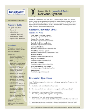
CENCCN Guidelines Insights1335Central Nervous System Cancers, Version 1.2017PRINCIPLES OF BRAIN TUMOR PATHOLOGY (2 OF 3)MOLECULAR MARKERSThe following molecular markers are often used by neuropathologists to facilitate characterization of gliomas and/or by neuro-oncologists toguide treatment decisions:Codeletion of 1p and 19q Description: This codeletion represents an unbalanced translocation(1;19)(q10;p10). Detection: The codeletion of 1p and 19q is detectable by FISH or PCR. Diagnostic value: It is strongly associated with oligodendroglialhistology and helps confirm the oligodendroglial character of tumorswith equivocal or mixed histologic features.7 Prognostic value: The codeletion confers a favorable prognosis andis predictive of response to alkylating chemotherapy and combinationtherapy with radiation and alkylating chemotherapy.8,9Isocitrate Dehydrogenase 1 and 2 (IDH1 and IDH2) Mutation Description: IDH1 and IDH2 are metabolic enzymes. Specific mutationsof these enzymes are linked to the formation of D-2-hydroxyglutarate,an oncometabolite that causes epigenetic modifications. Detection: The most common IDH1 mutation (R132) is detectable byimmunohistochemistry. Additional IDH1 as well as IDH2 mutations aredetectable by PCR or pyrosequencing. Diagnostic value: Very common in grade II and III gliomas. Much lesscommon in glioblastoma, but can help identify a glioblastoma as beinga secondary glioblastoma (one that transformed from a lower gradeglioma and generally does not behave as aggressively as a primary [denovo] glioblastoma).10,11 Prognostic value:IDH mutations are commonly associated with codeletion of 1p and19q, and with MGMT promoter methylation.4IDH1 or 2 mutations are associated with a favorable prognosis and areimportant in stratification for clinical trials.12In grade II or III gliomas, wild-type IDH1 or 2 is associated withincreased risk of aggressive disease.4IDH1 or 2 mutations are associated with a survival benefit for patientstreated with radiation or alkylator chemotherapy, but not for untreatedpatients.13,14MGMT Promoter Methylation Description: MGMT (O6-methylguanine-DNA methyltransferase)is a DNA repair enzyme that reverses the DNA damagecaused by alkylating agents, resulting in tumor resistanceto temozolomide and nitrosourea-based chemotherapy.Methylation of the MGMT promoter silences MGMT, makingthe tumor more sensitive to treatment with alkylatingagents.15 Detection: Methylation of the MGMT promoter is detectableby methylation-specific PCR16 or pyrosequencing.17 Prognostic value:MGMT promoter methylation is strongly associated withIDH status and genome-wide epigenetic changes (G-CIMPphenotype).4MGMT promoter methylation confers a survival advantagein glioblastoma and is used for risk stratification in clinicaltrials.18MGMT promoter methylation is particularly useful intreatment decisions for elderly patients with high-gradegliomas (grades III-IV).19,20Patients with glioblastoma that are not MGMT promotermethylated derive less benefit from treatment withtemozolomide compared to those whose tumors aremethylated.18References available online, inthe full version of this guideline,at NCCN.org [BRAIN-F 3 of 3]Version 1.2017 National Comprehensive Cancer Network, Inc. 2017, All rights reserved. The NCCN Guidelines and this illustrationmay not be reproduced in any form without the express written permission of NCCN .shown to be limited by interobserver variability, especially for certain categories.14–21 The variability inoutcomes for oligoastrocytoma is likely related to thehigh interobserver variability (between pathologists)in assigning this particular category.22–25 These challenges prompted searches for molecular characteristics to help better distinguish glioma subtypes andimprove estimation of prognosis for patients withbrain tumors.Revised WHO Classification System for GliomasFindings from molecular analyses (described in moredetail later) have prompted revision of the WHOclassification system to incorporate molecular features that have been shown to have better prognostic value than standard pathology.26 SupplementaleTable 1 shows the categorization of patients withgrade II/III gliomas according to the WHO classification system versions 2007 and 2016.3,26 Key changesfor grade II/III gliomas are as follows: (1) oligodendrogliomas include only tumors with 1p19q codele-BRAIN-F2 OF 3tion (1p19q-codel) and IDH mutation (IDH-mut),unless molecular data are not available and cannotbe obtained, in which case designation can be basedon histology; (2) anaplastic gliomas are further subdivided according to IDH mutation status; and (3) oligoastrocytoma is no longer a valid designation unlessmolecular data (1p19q deletion and IDH mutationstatus) are not available and cannot be obtained, orthere is phenotypic and genotypic evidence of spatially distinct oligodendroglioma (1p19q-codel) andastrocytoma (1p19q intact or deletion of only 1p or19q) components in the same tumor.Grade II/III Glioma: Search for MolecularSubgroupsMultiple independent studies on glioma tissue removed from the brain have conducted genome-wideanalyses evaluating an array of molecular features(eg, mutations, DNA copy number, DNA methylation, mRNA, microRNA, protein expression)in large populations of patients with grade II–IV JNCCN—Journal of the National Comprehensive Cancer Network Volume 15 Number 11 November 2017
CE1336NCCN Guidelines InsightsCentral Nervous System Cancers, Version 1.2017disease.5,9,27–29 Unsupervised clustering analyses, anunbiased method for identifying molecularly similartumors, have been used to identify subgroups of gliomas with distinct molecular profiles.5,9,27,29 Remarkably, further analysis showed that these molecularsubgroups could be distinguished based on only ahandful of molecular features, including mutationof IDH1 or IDH2 (IDH-mut) and 1p19q-codel, biomarkers independently verified by many studies ashallmarks for distinguishing molecular subgroups ingrade II/III glioma.4–7,9,13,28,30–39 Using these markersalone, most grade II/III tumors can be divided into3 molecular subtypes: (1) mutation of either IDH1or IDH2 with 1p19q codeleted (IDH-mut 1p19qcodel), (2) IDH-mut without deletion of 1p or 19q(1p19q intact) or with isolated deletion of 1p or 19q,and (3) no mutation of IDH1 or IDH2 (IDH-wt).5Multiple studies have shown that codeletion of1p and 19q is strongly associated with IDH-mut,such that 1p19q codeletion in IDH-wt tumors israre.7,11,13,34,35Analyses of large molecular databases have alsosuggested a number of other molecular markers asbeing potential characteristic/prognostic featuresof specific molecular subgroups.7,9,11,28,35,37,40 Molecular features suggested by more than one study asmarkers for subtyping grade II/III gliomas include(1) mutations in the TERT promoter, NOTCH1,CIC, FUBP1, PIK3CA; (2) mutation in or overexpression of TP53; (3) PTEN loss or promoter methylation; (4) loss/deletion of ATRX and CDKN2A/B;(5) amplification of EGFR; and (6) chromosome 7gain, chromosome 10 loss.5,9,27,28,34–37,40 Due to variability in results across studies, these molecularmarkers are not currently widely accepted as usefulfor classifying gliomas. Supplemental eTable 2 showsthe molecular, phenotypic, and demographic characteristics associated with the 3 molecular subgroups ofgrade II/III gliomas defined by IDH and 1p19q status.It is important to note that the “associated molecular markers” listed in supplemental eTable 2 are notnecessarily present in all tumors of a particular molecular subgroup and have not been fully validatedas markers for assigning molecular subtype. Someof these markers can be found (not infrequently) inmore than one molecular subgroup, but are merelymore prevalent in one particular subgroup; othermarkers are found almost exclusively in one particular molecular subgroup.5,11,28,35,37 It is important tonote that correlations between the molecularly defined 2016 WHO categories and the histology-based2007 WHO categories are limited and vary acrossstudies.5,7,34,37 Thus the change from 2007 WHO to2016 WHO reclassifies a significant proportion ofgrade II–IV gliomas.Grade II/III Glioma: Prognostic Relevance ofMolecular SubgroupsMost importantly, the specific markers used todefine molecular subgroups among grade II/IIIgliomas have been shown to have prognosticvalue. Numerous large studies of patients withbrain tumors have shown that among grade II/IIIgliomas, 1p19q-codel is significantly correlatedwith improved PFS and OS.4,6,7,9,10,13,30,32,41,42 Manyof these studies used heterogenous populationstreated with a variety of approaches, but a fewshowed statistically significant correlation between1p19q-codel and outcome within a population ofpatients who received the same treatment. Theindependent prognostic value of 1p19q status wasalso confirmed through multivariate analyses frommultiple studies in patients with grade II/III glioma.4,7,13,41,42 For IDH mutation status, although a fewanalyses did not find a significant correlation withPFS,7,32 many more studies found that IDH-mutwas associated with improved PFS, including several multivariate analyses.4,9,10,42,43 Numerous largestudies, including many multivariate analyses, allfound that IDH-mut is significantly associatedwith improved OS in patients with grade 44 Analyses withinsingle-treatment arms showed that the IDH status is prognostic for outcome across a variety ofpostoperative adjuvant options. For example, inthe NOA-04 phase III randomized trial in newlydiagnosed anaplastic gliomas, IDH-mut was associated with improved PFS, time to treatment failure,and OS in each of the 3 treatment arms: standardradiation therapy (RT; n 160); combination therapy with procarbazine, lomustine, and vincristine(PCV RT upon progression; n 78); and temozolomide (TMZ; RT upon progression; n 80).10Multiple independent studies have shownthat subdividing grade II/III gliomas by IDH- and1p19q-based molecular subtype yields greater prognostic separation (for PFS and OS) than subdivision based on histology (as defined by WHO 2007). JNCCN—Journal of the National Comprehensive Cancer Network Volume 15 Number 11 November 2017
CENCCN Guidelines Insights1337Central Nervous System Cancers, Version 1.2017These include very large studies covering multiplegrades and histology-based subtypes of gliomas,4,5,9,27as well as smaller studies limited to 1 to 2 gradesor histologic subtypes.6–8,12,28 Multiple studies havealso shown that among patients with grade II/IIIgliomas, the IDH-mut 1p19q-codel group has thebest prognosis, with significantly better PFS andOS than the IDH-wt group, which has the worstprognosis of the 3; and outcomes for the groupwith IDH-mut and 1p19q intact or deletion ofonly 1p or 19q usually lie somewhere in betweenthat of the IDH-mut 1p19q-codel and IDH-wtgroups.4,5,7,10,13,27,37,41 Analyses within single treatment arms have confirmed this trend in prognosisacross a variety of postoperative adjuvant treatment options.7,8,10,41 Table 1 shows a few examplesof prospective phase II/III trials reporting outcomesfor the 3 molecular subtypes within the following(postoperative) treatment arms: RT, TMZ, PCV orTMZ, and PCV then RT.Molecular Features Relevantto Treatment Selection forAnaplastic GliomaSupplemental eTable 1 shows the histologic andmolecular features used to determine the recommended treatment pathway for grade III gliomasin the NCCN Guidelines (version 1.2017). Eachpathway for anaplastic gliomas (grade III) has several recommended adjuvant treatment options (seeGLIO-2, page 1333, and GLIO-5, available in thecomplete version of these guidelines at NCCN.org),and the recommended treatment pathway is also determined by performance status.45 In addition, the“Principles of Brain Tumor
Central Nervous System Cancers, Version 1.2017 Featured Updates to the NCCN Guidelines . CE Grant Writing & Project Management, NCCN, has disclosed that she has no relevant financial relationships . Priya Kumthekar, MD, Panel Member, has disclosed that she serves as a scientific advisor for AbbVie Inc. and Vivacitas Oncology, Inc .