Transcription
Molecular Characterization of msp2/p44 of Anaplasmaphagocytophilum Isolated from Infected Patients andHaemaphysalis longicornis in Laizhou Bay, ShandongProvince, ChinaYong Wang1,2, Chuangfu Chen1*, Lijuan Zhang2*1 College of Animal Science & Technology, Shihezi University, Shihezi, Xinjiang Province, People’s Republic of China, 2 Department of Rickettsiology, NationalInstitute for Communicable Disease Control and Prevention, China CDC, Beijing, People’s Republic of ChinaAbstractMolecular characterization of the MSP2/P44 protein of Anaplasma phagocytophilum may determine not only if thebacterium is capable of invading hosts but also whether it generates antigenic variation for the purpose of escapingthe host immune response, resulting in various pathologic injuries and serious clinical outcomes. Chineseanaplasmosis patients usually present with serious manifestations, and the fatality rate is as high as 26.5%. In thisstudy, we amplified, cloned and sequenced the msp2/p44 genes of three Chinese A. phagocytophilum isolates fromLaizhou Bay, Shandong Province, where human granulocytic anaplasmosis (HGA) patients present severe clinicalmanifestations, and analyzed their genetic characterization and structural features. We also compared them with theHZ and Webster A. phagocytophilum strains. The sequences for both strains are available in GenBank. Analysesindicated that Chinese A. phagocytophilum isolates were significantly different from the HZ and Webster strains interms of nucleotide sequences, amino acid sequences and protein secondary and tertiary structures. Moreover, thenumber of immunologic B-cell epitopes (19) of the MSP2 protein of the Chinese isolates was higher than that of theA. phagocytophilum strains HZ (16) and Webster (9). This genetic diversity of the MSP2/P44 protein of Chinese A.phagocytophilum isolates might be relevant and might have serious clinical outcomes. This observation could providea clue to further understand the pathogenesis of Chinese A. phagocytophilum.Citation: Wang Y, Chen C, Zhang L (2013) Molecular Characterization of msp2/p44 of Anaplasma phagocytophilum Isolated from Infected Patients andHaemaphysalis longicornis in Laizhou Bay, Shandong Province, China. PLoS ONE 8(10): e78189. doi:10.1371/journal.pone.0078189Editor: Wenjun Li, National Center for Biotechnology Information (NCBI), United States of AmericaReceived May 20, 2013; Accepted September 9, 2013; Published October 22, 2013Copyright: 2013 Wang, Chen. This is an open-access article distributed under the terms of the Creative Commons Attribution License, which permitsunrestricted use, distribution, and reproduction in any medium, provided the original author and source are credited.Funding: This study was supported by the National Basic Research Program of China ( 973 Program, 2010CB530206) and China Mega-Project forInfectious Disease (2011ZX10004-001) and National Key Science and Technology Projects of China (Project no.2012ZX10004215). The funders had norole in study design, data collection and analysis, decision to publish, or preparation of the manuscript.Competing interests: The authors have declared that no competing interests exist.* E-mail: zhanglijuan@icdc.cn (LJZ); ccf-xb@163.com (CFC)Introductionsuggesting that the pathogen was transmitted through contactwith blood or respiratory aerosols from infected patients [14].The manifestations of human granulocytic anaplasmosis (HGA)infection include fever, chills, headache, myalgia, leukopeniaand thrombocytopenia, as well as elevated levels of liveraminotransferase [15]. The number of HGA cases hasincreased annually because of certain natural and socialfactors, such as global warming, increases in outdoor activities,globalization of the economy and worldwide trade. The numberof A. phagocytophilum infection cases reached 1,161 in theUnited States in 2009, and the HGA case-fatality rate in theMidwestern United States is 0.6%- 0.7%. This rate may be onthe rise, however, due to misdiagnosis [13]. Additionally, tickborne ruminant fever (for example, cattle and sheep) causedby A. phagocytophilum infection is common in Europe [16]. InAsian countries, including China, Japan and South Korea, HGAAnaplasma (A) phagocytophilum (APH) is a Gram-negativeand obligate intracellular pathogen that infects mammal hostsworldwide and is transmitted by ticks [1–3]. A. phagocytophilumis an important zoonotic pathogen in that it infects not onlyhumans but also some domestic animals, including horses,dogs, cattle and sheep [4,5]. The life cycle of APH is related toits natural hosts, such as rodents and ruminants, as well as itstransmission vectors, which include some members of thegenera Ixodes and Haemaphysalis [6-12]. Humans are a deadend host for A. phagocytophilum and are thus not part of thelife cycle of the bacteria [13]. The transmission of A.phagocytophilum into mammals mainly relies on infected tickvectors. However, in 2006, the nosocomial transmission of A.phagocytophilum was proven in the Anhui Province in China,PLOS ONE www.plosone.org1October 2013 Volume 8 Issue 10 e78189
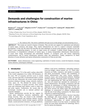
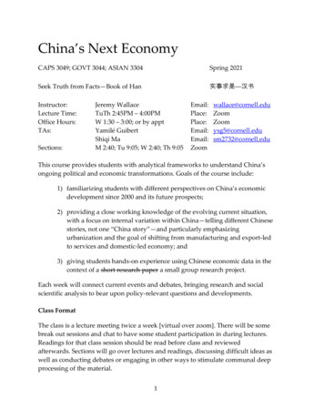
msp2/p44 of A. phagocytophilum Isolates in Chinacases and HGA agents have been continuously discoveredand detected in the last few years [7-12,17-20].A growing number of medical reports indicate that the clinicalmanifestations of Chinese HGA patients are significantlydifferent than those of patients from Western countries. TheHGA occurring in China is usually accompanied by several lifethreatening complications, including systemic inflammationresponse syndrome (SIRS) and multiple organ dysfunctionsyndrome (MODS). Moreover, the fatality rate of Chinese HGApatients has been reported to be as high as 26.5% [21].Therefore, studies that genetically characterize virulencefactors and examine the pathogenesis of native Chinese A.phagocytophilum isolates have major clinical and public healthsignificance in China. The members of the outer membraneprotein OMP1/MSP2/P44 superfamily are regarded asimportant virulence factors of A. phagocytophilum pathogens.Genetic variation of the MSP2/P44 protein may not onlydetermine if the bacterium is capable of invading the host butalso whether it can generate antigenic variation to allow forescape from the host immune response, resulting in variouspathologic injuries and serious clinical outcomes [21-24].Therefore, A. phagocytophilum pathogenesis, which is relatedto the genetic characteristics of MSP2/P44, has recentlybecome a topic of growing interest. Given the severe clinicalmanifestations of HGA in China, we focused on the analysis ofthe genetic variation of the msp2/p44 genes of three nativeChinese A. phagocytophilum isolates from Laizhou Bay,Shandong Province, where 100% of patients had severeclinical manifestations.Figure 1. Sequences and positions of the msp2/p44 PCRprimers and the predicted size of the PCR product. ThePCR product and its size are shown in the larger box. The twosmaller boxes inside the larger box indicate the p44 ORF andmsp2 ORF, respectively. Inter-genic sequences (ITS) areshown using dotted lines under letters. Ellipses inside the PCRproduct indicate the omission of some letters. The PCRproducts of the primers msp2-F and msp2-R are indicatedusing green letters under the green arrowhead. SC: startcodon; TC: terminal codon.doi: 10.1371/journal.pone.0078189.g001specificity of the PCR primers was also assessed using primerblast/). The names and relative sites of the PCR primers andthe predicted size of the PCR products are shown in Figure 1.PCR amplification analyses and sequencingGenomic DNA (gDNA) was separately prepared from threeChinese native A. phagocytophilum isolates (LZ-HGA-agent-3,LZ-HGA-agent-4 and LZ-HGA-agent-T1) using a DNeasy Blood & Tissue Kit (QIAGEN, Cat No. 69506) and was thenused as a PCR template. The PCR primers were as follows:msp2-F 5’-ACTTATGGTGTTCGGGAGTCTTC-3’ and msp2-R5’-AATAATAGGAACGGTCACGGAG-3’, and the predicted sizeof the PCR product was 2,486 bp. Briefly, 3.0 μL of gDNA wasused as a template in a 25-μL reaction mixture systemcontaining 2.5 μL 10 Taq Buffer (SDS Genetech Co., Ltd,China, Cat# ET-500), 1.0 μL of each primer: msp2-F andmsp2-R (0.4 μM final concentrations of each primer), 0.5 μL ofdeoxynucleoside triphosphates (dNTPs, 10 mM), 2.5 μL of dye,0.5 μL of Taq DNA polymerase (5 U/μL, SBS Genetech Co.,Ltd, China, Lot#042512) and 14 μL of ddH2O. PCR wasperformed using a SensoQuest LabCycler standard plus(SensoQuest GmbH, Goettingen, Germany) with a predenaturation at 94 C for 5 min, followed by 35 cycles of adenaturation step at 94 C for 40 seconds, an annealing step at57 C for 40 seconds and an extension step at 72 C for 3minutes. There was a final extension at 72 C for 10 minutes.The PCR amplification products were analyzed using 1.0%agarose gel electrophoresis. To obtain the entire sequences ofthe msp2/p44 genes and to avoid the loss of some sequenceinformation at the ends of both primers, we cloned the PCRproducts as follows: the PCR product was purified using amulti-function DNA purification kit (BioTeke Corporation,Cat#DP1502). Purified msp2/p44 was cloned into a pEASY-T1Cloning vector (Beijing TransGen Biotech Co., Ltd.,Lot#G30716), and the recombinant plasmid was designatedMaterials and MethodsEthics statementThe use of pathogenic DNA isolated from patients wasapproved by the ethics committee of the Chinese CDC (No.201103), and all samples were anonymized.Bacteria strainsThree native Chinese A. phagocytophilum isolates, includingtwo human isolates (LZ-HGA-agent-3 and LZ-HGA-agent-4)from HGA patients and one tick isolate (named LZ-HGA-agentT1) from infected Haemaphysalis (H) longicornis, were isolatedat Laizhou Bay in Shandong Province in 2009 - 2010. All threepathogenic isolates were cultured and conserved in HL-60 celllines in our laboratory. The two human pathogens wereisolated from patients with severe clinical manifestations; theank A genes from the samples had 100% identity with eachother and were 100% homologous to the tick isolate (LZ-HGAagent-T1) [25].PCR primer designThe msp2/p44 genes of A. phagocytophilum usually containtwo open reading frames (ORFs) with msp2 and p44 [26,27].To obtain msp2/p44, PCR primers were initially designed withthe bio-software Primer Premier 5.0, according to the Websterstrain sequence (accession number AY164491) of A.phagocytophilum, published in the GenBank database. ThePLOS ONE www.plosone.org2October 2013 Volume 8 Issue 10 e78189
msp2/p44 of A. phagocytophilum Isolates in ChinapEASY-msp2/p44. The recombinant plasmid pEASY-msp2/p44was transformed into E. coli DH5a competent cells. Positiveclones were screened by PCR using the primers msp2-F andmsp2-R. The recombinant plasmid was extracted from positiveclones using a high-purification plasmid mini-preparation kit(BioTeke Corporation,Cat#DP1002) and then directlysequenced by two separate commercial sequencingcompanies in China: Beijing Tsingke BioTech Co., Ltd. andSangon BioTech (Shanghai) Co., Ltd.Table 1. Selected msp2/p44(p44ESup1/omp-1)sequences for the phylogenetic analysis.AccessionNo.GeographicBacteria strainsHostsoriginRemarkLZ-HGA-AgenthumanChinap44 msp2APH-HZhumanUSAOmp-1N msp2humanUSAp44ESup1 msp2humanUSAp44ESup1 msp2KC128828/KC430333/KC430334CP000235Data analysisAY164491The sequencing was performed with universal primers fromthe pEASY-T1 Cloning Kit (Beijing TransGen Biotech Co., Ltd.,Lot#G30716) and using the Sanger sequencing method. Thesequences of msp2/p44 were processed through manualsplicing and proofreading and were also analyzed using thenucleotide blast program (http://blast.ncbi.nlm.nih.gov/). For theanalysis of the msp2/p44 sequences, the DNASTAR package(Lasergene, Madison, WI) was used. The msp2/p44 nucleotidesequences and their coded amino acid sequences were editedwith the EditSeq program of the package. The msp2/p44nucleotide sequences and their coded amino acid sequenceswere then aligned with the MegAlign program of the packageby comparison with the corresponding sequences from the A.phagocytophilum HZ and Webster strains. For the purpose ofdelineating genetic evolution information of the native ChineseA. phagocytophilum isolates, a phylogenetic tree wasconstructed with 10 sequences, including Chinese A.phagocytophilum isolates and another 9 varying Anaplasmastrains (Table 1), which were identified in different hosts fromdifferent geographic regions. The msp2 sequences for A.phagocytophilum HZ (CP000235) and Webster (AY164491)were used for outgroup comparisons. The phylogeneticanalysis of the msp2/p44 gene sequences was conductedusing the program MEGA 5.05 (Arizona State University), aspreviously described [6]. In general, the sequences werealigned using CLUSTAL W of MEGA 5.05, with the applicationof the IUB matrix for nucleotide sequences and the Gonnetmatrix for protein sequences. Tree construction was achievedusing the neighbor-joining method with the complete deletionoption, using the Jukes-Cantor matrix for nucleotide sequencesand the Dayhoff matrix for protein sequences. Bootstrapanalysis was conducted with 1,000 replicates.AY164492APH-Webster-varAAPH- HGE2-varII1AY164493APHhumanUSAp44ESup1 msp2AY137510APH-NY-37humanUSAp44 omp-1Japanp44 omp-1Japanp44 usDQ519565APH-NORSHESsheepNorwayp44ESup1 msp2DQ519566APH-SWDOGESdogSwedenp44ESup1 msp2Note: omp-1 may stand for omp-1N, omp-1X or both; APH: A. phagocytophilumdoi: 10.1371/journal.pone.0078189.t001Accession numbers of the full-length msp2/p44 genesequences of LZ-HGA-Agent-3, LZ-HGA-Agent-4 and LZ-HGAAgent-T1 have been deposited in the GenBank database underthe accession numbers KC128828, KC430333 and KC430334,respectively.ResultsPCR amplification and msp2/p44 gene sequencingPCR amplification revealed that the predicted 2.5-kbfragments of the msp2/p44 genes were successfully amplified,using the msp2-F/msp2-R primer pair and the genomic DNA ofthree native Chinese A. phagocytophilum isolates, namely LZHGA-Agent-3 (KC128828), LZ-HGA-Agent-4 (KC430333) andLZ-HGA-Agent-T1 (KC430334), as templates. The sequencinganalysis indicated that the sequences of msp2/p44 in all of theisolates were 100% identical to each other at the nucleotidelevel and were mainly composed of one p44 (825 bp) openreading frame (ORF), one msp2 (1323 bp) ORF and a fewintergenic sequences (ITS) (Figure 1). Therefore, the threenative Chinese A. phagocytophilum isolates mentioned abovewere designated LZ-HGA-Agent (KC128828/KC430333/KC430334) for simplifying the description in the study, wherethe name collectively stands for LZ-HGA-Agent-3, LZ-HGAAgent-4 and LZ-HGA-Agent-T1.The p44 ORF sequence of LZ-HGA-Agent is 100% and99.6% identical to the p44ESup1 sequence of the Websterstrain of A. phagocytophilum and the omp-1N sequence of theHZ strain, respectively. There is a 3-bp difference in the p44ORF sequence of LZ-HGA-Agent compared with the omp-1NBioinformatics analysis of the MSP2/P44 proteinThe structural information for the MSP2/P44 protein waspredicted and delineated using online software and/orprograms. In particular the ProtParam tool (http://web.expasy.org/protparam/) was used for the primary structureof the protein and the Predict Secondary Structure (PSIPREDv3.0) (http://bioinf.cs.ucl.ac.uk/psipred/) for the secondarystructures. TMHMM Server v.2.0 (http://www.cbs.dtu.dk/services/TMHMM/) was used for the transmembrane domains,BepiPred 1.0 Server (http://www.cbs.dtu.dk/services/BepiPred/)was used for B-cell epitope-bearing regions, and Galaxy TBM(http://galaxy.seoklab.org/) was used for the protein tertiarystructure prediction from sequences obtained with templatebased modeling.PLOS ONE www.plosone.orggene3October 2013 Volume 8 Issue 10 e78189
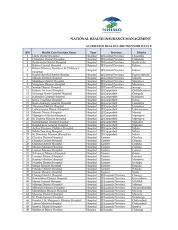
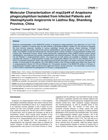
msp2/p44 of A. phagocytophilum Isolates in Chinasequence of the HZ strain at the nucleotide level. The detailsof the differences are as follows: A to G nucleotide sequencechange at positions 270 and 720 and G to A at position 484 inthe HZ strain sequence (data not shown). In contrast, the msp2ORF sequence of LZ-HGA-Agent displays 87.7% and 45.1%identity to that of the msp2 sequences of both the A.phagocytophilum Webster and HZ strains, respectively. TheLZ-HGA-Agent msp2 ORF sequence displays relatively littlehomology to the msp2 sequences of the A. phagocytophilumHZ strain (45.1%) when contrasted with the A.phagocytophilum Webster strain (87.7%) because of theoccurrence of large-scale diversity at the nucleotide level (seeFigure S1).The P44 amino acid sequence, which is encoded by the p44ORF in the LZ-HGA-Agent, is 100% and 99.6% similar to thep44ESup1-coded product sequence of the Webster strain andthe omp-1N-coded product sequence of the HZ strain at theamino acid level, respectively. The LZ-HGA-Agent and the HZstrains have identical P44 sequences, except for a difference inthe amino acid at position 162, namely V to M in the HZ strainsequence (data not shown). The MSP2 amino acid sequence,which is encoded by msp2 ORF in LZ-HGA-Agent, displays84.6% and 27.9% homology to the MSP2 amino acidsequences of the A. phagocytophilum Webster and HZ strain,respectively. It is of note that, for the msp2 ORFs and thecoding amino acid sequences that were analyzed in this work,the identities and similarities between the different msp2sequences in various strains demonstrate that coding aminoacid similarities are lower than nucleotide identities, suggestingthat the msp2 nucleotide exchanges of LZ-HGA-Agent wereextremely nonsynonymous substitutions (see Figure S1 andFigure S2). Thus, we conclude that extreme differences in thegenetic variation of the msp2 ORF sequence and its aminoacid sequence in LZ-HGA-Agent exist, but such extremedifferences do not exist with the p44 ORF sequences.In this study, we also compared our results with anotherChinese sequence of msp2/p44 (EU 008082) identified inrodents in the southeast of China [28], and the results indicatedthat the identity of the msp2 nucleotide sequence (from nt 1475to 2352) and the amino acid sequences of both the A.phagocytophilum LZ-HGA-Agent strain and the rodent(EU008082) strain were 48.4% and 27.7%, respectively.Figure 2. Phylogenetic tree based on the msp2/p44nucleotide sequences (a) and MSP2/P44 amino acidsequences (b) generated using the neighbor-joiningmethod. a. Bootstrap values ( 60%) are shown next to thenodes of the tree, and the scale bar indicates the number ofnucleotide substitutions per site; b. The MSP2/P44 amino acidsequences were obtained by msp2/p44 gene sequencetranslation, and the bootstrap values ( 50%) are shown next tothe nodes of the tree. The scale bar indicates the number ofamino acid substitutions per site. The Chinese isolate LZ-HGAAgent (red) and the international reference strains for APHWebster and APH-HZ (blue) are highlighted. APH: Anaplasmaphagocytophilum.doi: 10.1371/journal.pone.0078189.g002United States, sheep NORSHES strain (DQ519565) fromNorway, canine NORSHES strain (DQ519566) from Swedenand tick strain Tick-176-5ES-Iwate-Ip (FJ600595 andFJ600601) from Japan, but was less related to the human HZstrain (CP000235) from the United States (Figure 2a and 2b).Bioinformatics analyses of the MSP2 proteinBecause the p44 ORF sequence of LZ-HGA-Agent displayeda higher homology to the p44ESup1 sequence (100%) of A.phagocytophilum Webster (APH-Webster) and the omp-1Nsequence (99.6%) of A. phagocytophilum HZ (APH-HZ), theirbioinformatics analyses were not conducted in detail in thisstudy. In contrast, we focused on the analyses of the MSP2proteins of the different strains.Phylogenetic analysisTo assess the relationship between LZ-HGA-Agent and otherstrains of A. phagocytophilum investigated in this study,another 9 sequences identified in different host species fromdifferent geographic regions were used to construct aphylogenetic tree (Table 1). Specifically, we constructed aneighbor-joining (NJ) tree. As shown in Figure 2a, all majorbranches referring to the gene sequences used in the workwere supported by bootstrap values 60%. Using amino acidsequences to construct the tree, similar results were obtained(Figure 2b). From Figure 2a and b, we noticed that the A.phagocytophilum Chinese isolate LZ-HGA-Agent (KC128828/KC430333/KC430334) was very closely related to the humanA. phagocytophilum Webster strain (AY164491) from theUnited States, human strain NY-37 (AY137510) from thePLOS ONE www.plosone.orgMSP2 amino acid compositionThe amino acid composition of the MSP2 proteins belongingto LZ-HGA-Agent, APH-HZ and APH-Webster were analyzedusing the ProtParam tool (http://web.expasy.org/protparam/),and the results are shown in Table 2. The more abundantamino acids in MSP2 from LZ-HGA-Agent include Gly (12.5%),Ala (9.5%) and Val (9.5%), and the percent of the Gly content(12.5%) of LZ-HGA-Agent-MSP2 was higher than that of APHHZ-MSP2 (10.1%) but lower than that of APH-Webster-MSP24October 2013 Volume 8 Issue 10 e78189
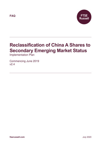
msp2/p44 of A. phagocytophilum Isolates in ChinaTable 2. Amino acid composition of MSP2 proteins fromLZ-HGA-Agent, APH-HZ and APH-Webster.LZ-HGA-AgentAPH-WebsterAPH-HZAmino acid Number Percentage Number Percentage Number PercentageAla (A)429.50%215.80%409.20%Arg (R)163.60%226.00%163.70%Asn (N)184.10%174.70%173.90%Asp (D)317.00%133.60%327.40%Cys (C)40.90%20.50%40.90%Gln (Q)61.40%92.50%71.60%Glu (E)194.30%226.00%235.30%Gly (G)5512.50%3710.10%6013.80%His (H)71.60%92.50%51.10%Ile (I)173.90%215.80%173.90%Leu (L)317.00%4211.50%306.90%Lys (K)296.60%205.50%337.60%Met (M)102.30%92.50%102.30%Phe (F)163.60%195.20%153.40%Pro (P)163.60%92.50%122.80%Ser (S)327.30%277.40%276.20%Thr (T)296.60%174.70%255.70%Trp (W)20.50%20.50%20.50%Tyr (Y)184.10%154.10%184.10%Val (V)429.50%328.80%429.70%Figure 3. Predicted transmembrane domain for LZ-HGAAgent-MSP2 (A), APH-HZ-MSP2 (B) and APH-WebsterMSP2 (C). The red legend (transmembrane —), blue legend(inside —) and pink legend (outside —) in panels A, B and Cindicate the transmembrane domain, interior domain andexterior domain, respectively, for the MSP2 protein aspredicted by the TMHMM program. The number on thehorizontal abscissa in panels A, B and C indicates the aminoacid (AA) residual site and size.doi: 10.1371/journal.pone.0078189.t002(13.8%). The Val content (9.5%) of LZ-HGA-Agent-MSP2 wasnearly equal to that of APH-HZ-MSP2 (8.8%) and APHWebster-MSP2 (9.7%). The percentage of the Ala content(9.5%) of LZ-HGA-Agent-MSP2 was roughly equal to that ofAPH-Webster-MSP2 (9.2%) but was obviously higher than thatof APH-HZ-MSP2 (5.8%). The MSP2 isoelectric point analysis(pI) indicated that the pI (5.87) of LZ-HGA-Agent-MSP2 fromChina was approximately equal to the pI (5.59) of human APHWebster-MSP2 from the United States but was different thanthe pI (9.20) of human APH-HZ-MSP2 from the United States.MSP2 secondary structure. The secondary structures ofeach MSP2 protein were predicted using the program PredictSecondary Structure (PSIPRED v3.0) (http://bioinf.cs.ucl.ac.uk/psipred/), and the results are shown in Figure S3, Figure S4and Figure S5. As shown in these figures, all MSP2 proteinstructures from LZ-HGA-Agent, A. phagocytophilum Websterand A. phagocytophilum HZ included a main random coilstructure, a β-strand structure dispersed to the two ends of theprotein and a few α-helices. However, there was a greaternumber of α-helices in the LZ-HGA-Agent-MSP2 protein than inAPH-HZ-MSP2 and APH-Webster-MSP2. In particular, therewere six α-helices in the LZ-HGA-Agent-MSP2 from China butonly three α-helices in the APH-HZ-MSP2 and five α-helices inAPH-Webster-MSP2 from the United States. In contrast, therewere 17 β-strands in the LZ-HGA-Agent-MSP2 protein but 20in APH-HZ-MSP2 and 17 in APH-Webster-MSP2, suggestingthat the secondary structure of the LZ-HGA-Agent-MSP2protein from the Chinese isolate was obviously distinct from thestructures of APH-HZ-MSP2 from the United States but notfrom APH-Webster-MSP2 from the Unites States.PLOS ONE www.plosone.orgdoi: 10.1371/journal.pone.0078189.g003MSP2 transmembrane domainsThe transmembrane domains of each MSP2 protein werepredicted using TMHMM Server v.2.0 (http://www.cbs.dtu.dk/services/TMHMM/) (Figure 3). The predicted results indicatedthat the proteins were hardly different in terms of the location ofthe transmembrane domains of MSP2, when comparing theLZ-HGA-Agent isolate, APH-HZ and APH-Webster strains. Thetransmembrane domains of MSP2 were basically located in the5th to 24th amino acid sequence region.B-cell epitope analysis of MSP2B-cell epitope-bearing regions of each protein were predictedusing BepiPred 1.0 Server (http://www.cbs.dtu.dk/services/BepiPred/), and the results are shown in Table 3. Nineteen Bcell epitopes were predicted (the number of amino acids 3 atan epitope) in the MSP2 protein of the LZ-HGA-Agent isolate,which was higher than that of the MSP2 proteins from both theAPH-HZ and APH-Webster strains, which only had 16 and 9predicted B-cell epitopes (the number of amino acids 3),respectively.MSP2 tertiary structures.Protein tertiary structureprediction was performed using the program GalaxyTBM(http://galaxy.seoklab.org/), and the predicted results are5October 2013 Volume 8 Issue 10 e78189
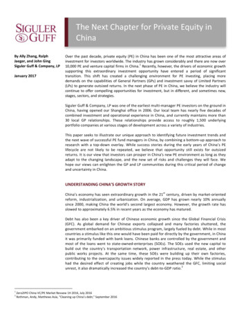
msp2/p44 of A. phagocytophilum Isolates in ChinaTable 3. Predicted epitopes for LZ-HGA-Agent-MSP2, APH-Webster-MSP2, and APH-HZ-MSP2.No. Start/End positionOligo peptide for epitopePeptide length123/38( DVSALETGGA1621/30( APH-Webster)TSAHADNDKS1048/50( LZ-HGA-Agent)SPA348/50(APH-HZ)SPA350/56( APH-Webster)IDDGGET760/68( 3( APH-Webster)YWGPEVA773/76( LZ-HGA-Agent)LKDG473/76(APH-HZ)LKDG492/98( APH-Webster)NTTFGGS778/80( LZ-HGA-Agent)SVK378/80(APH-HZ)SVK3125/132( APH-Webster)HKGRKGGG886/96( G12191/193 ( APH-Webster)LKR3109/115( 9( APH-Webster)PRNRS5132/144( KEDEAD13273/276( APH-Webster)CAGI4158/164( 5( APH-Webster)DISPTNSVREK11170/175( LZ-HGA-Agent)KTSGKD6169/174(APH-HZ)AKTSGK6185/193( 210( GKNWPTGYVNDGDNVNVLGDTNGNAEAV33214/226( T8233/246( 57/287( 398(APH-HZ)GVYDDLP7295/302( T1417312/315( LZ-HGA-Agent)EGGE418398/404( LZ-HGA-Agent)VYDDLPA719408/421( 6APH-HZ: A. phagocytophilum HZ strain, APH-Webster: A. phagocytophilum Webster strain, LZ-HGA-Agent: Chinese A. phagocytophilum isolate.doi: 10.1371/journal.pone.0078189.t003Discussionshown in Figure 4. The predicted results indicated that theMSP2 protein tertiary structures of the LZ-HGA-Agent isolate,the APH-HZ and the APH-Webster strains are very different(Figure 4).PLOS ONE www.plosone.orgThe A. phagocytophilum HZ strain was first isolated from apatient with human immunodeficiency disease in New York,USA, in 1995 [29]. This strain caused typical clinicalmanifestations of HGA and could be cultured by HL60 cells andformed morulas in the cytoplasm of the culture cells. Moreover,6October 2013 Volume 8 Issue 10 e78189
msp2/p44 of A. phagocytophilum Isolates in ChinaFigure 4. Predicted tertiary structures of MSP2 proteins based on the Nspa (PDB: 1P4T-A) template. The proteinssequences were copied into GalaxyTBM (http://galaxy.seoklab.org/) to compute their tertiary structures. The β-sheet (green), β-turn(blue) and random coil (gray) are highlighted.doi: 10.1371/journal.pone.0078189.g004this strain is highly cross-reactive with other HGA agents andE. chaffeensis. The genomic size of the A. phagocytophilumHZ strain is approximately 1.47 Mbps, consisting of 1,369ORFs, over 100 p44 (msp2) genes, type IV secretion (T4S)and numerous repeats [13,30]. The human HZ strain infectsgranulocytes by subverting its powerful innate antimicrobialdefenses, which also makes infected humans and animalsmore susceptible to opportunistic infection and causes theresulting endothelial cell adhesion, transmigration, motility,degranulation, respiratory burst and phagocytosis [13]. Thechanges in these functions are to increase bacterialdissemination into the neutrophil.The A. phagocytophilum Webster (Wisconsin) strain wasisolated from a patient in northwestern Wisconsin in 1996 [31],where the seroprevalence of HGA among permanent residentsis as high as 14.9% [32], the prevalence of A. phagocytophilumin deer was 8.9%-11.5%, and the infection rate in ticks was5.6%-26% [33]. It is noteworthy that serological cross-reactionassays indicated that there was a striking antigen differencebetween the A. phagocytophilum strains Webster and HZ [34].The LZ-HGA-Agent isolates were isolated from patients andtick-vectors from Laizhou Bay, Shandong Province, during2009-2010 [25], which is the largest wetland in northern Chinaand a famous migratory bird post across Asia and the WestPacific. As seen in a recent clinical report [21], these two caseswere characterized by severe clinical manifestations, includingsystemic inflammatory response syndrome (SIRS) and multipleorgan dysfunction syndrome (MODS). In addition, nearly 100%of Chinese HGA patients in these areas had severe clinicalfeatures including SIRS and MODS, significantly lower WBCcounts and PLT counts, as well as significantly elevated levelsof LDH, CK, BUN, ALT and AST.The pathogenesis of A. phagocytophilum is an issue ofworldwide significance because of the effects of the bacteria onhuman public health. Currently, the members of the major outermembrane protein superfam
* E-mail: zhanglijuan@icdc.cn (LJZ); ccf-xb@163.com (CFC) Introduction Anaplasma (A) phagocytophilum (APH) is a Gram-negative and obligate intracellular pathogen that infects mammal hosts worldwide and is transmitted by ticks [1-3]. A. phagocytophilum is an important zoonotic pathogen in that it infects not only