Transcription
(2021) 21:408Wang et al. BMC 986-0Open AccessRESEARCHBoth the serum AFP test and AFP/GPC3/SALL4 immunohistochemistry are beneficialfor predicting the prognosis of gastricadenocarcinomaBingzhi Wang1†, Yibin Xie2†, Li Zheng3, Xiaohao Zheng2, Jia Gao4, Xiuyun Liu1, Yanling Yuan1, Zhuo Li1,Ning Lu1 and Liyan Xue1*AbstractBackground: Both gastric adenocarcinoma with primitive enterocyte phenotype (GAPEP) (including hepatoidadenocarcinoma) and alpha-fetoprotein (AFP)-producing gastric adenocarcinoma have poor prognoses. However, thevalue of the serum AFP test and AFP/glypican-3 (GPC3)/spalt-like transcription factor 4 (SALL4) immunohistochemistry is still not clear, and these two methods have not yet been thoroughly compared.Methods: We collected 421 consecutive non-neoadjuvant surgically or endoscopically resected gastric adenocarcinoma patients with serum AFP results before surgery (group A). We divided these cases into serum AFP-high(sAFP-H) and serum AFP-normal (sAFP-N) by serum AFP levels, and into GAPEP (expressing AFP, GPC3, or SALL4) andnon-GAPEP (nGAPEP) by AFP/GPC3/SALL4 immunohistochemistry results. We also collected 12 non-resected gastricadenocarcinoma patients with serum AFP 7 ng/mL before treatment (group B). We analyzed these patients’ clinicopathological characteristics and prognoses.Results: Seventeen (4.04%) patients in group A were sAFP-H. These patients were younger and mainly had tubularadenocarcinoma with later pT (P 0.014) and pN (P 0.047) categories and more lymphovascular invasion (P 0.001),perineural spread (P 0.008), and metastases or recurrence (P 0.001). For immunohistochemistry, 34 (8.08%) caseswere GAPEP, and GAPEP cases also had later pT categories than nGAPEP cases (P 0.001). Most group B patientswith elevated serum AFP (especially 1000 ng/mL) had simultaneous metastases, mainly liver metastases. Both theserological method and immunohistochemical method were useful for predicting prognosis (AUC sAFP 0.625, AUCA/G/S-IHC 0.723, z statistic 1.726, P 0.084). The serum AFP level (especially 1000 ng/mL) is more specific (100%),and immunohistochemistry is more sensitive (50%).Conclusion: Both the serum AFP level and immunohistochemical expression of AFP/GPC3/SALL4 can be used toindicate a poor prognosis for gastric adenocarcinoma.Keywords: Gastric cancer, Pathology, Biomarker, Metastases*Correspondence: xuely@cicams.ac.cn†Bingzhi Wang and Yibin Xie have contributed equally1Department of Pathology, National Cancer Center/National ClinicalResearch Center for Cancer/Cancer Hospital, Chinese Academy of MedicalSciences and Peking Union Medical College, Beijing 100021, ChinaFull list of author information is available at the end of the articleBackgroundGastric cancer is one of the most common malignanttumors and a heterogeneous malignant tumor [1].Although most gastric cancers are adenocarcinomas,their biological behavior and prognosis are significantly The Author(s) 2021. Open Access This article is licensed under a Creative Commons Attribution 4.0 International License, whichpermits use, sharing, adaptation, distribution and reproduction in any medium or format, as long as you give appropriate credit to theoriginal author(s) and the source, provide a link to the Creative Commons licence, and indicate if changes were made. The images orother third party material in this article are included in the article’s Creative Commons licence, unless indicated otherwise in a credit lineto the material. If material is not included in the article’s Creative Commons licence and your intended use is not permitted by statutoryregulation or exceeds the permitted use, you will need to obtain permission directly from the copyright holder. To view a copy of thislicence, visit http:// creat iveco mmons. org/ licen ses/ by/4. 0/. The Creative Commons Public Domain Dedication waiver (http:// creat iveco mmons. org/ publi cdoma in/ zero/1. 0/) applies to the data made available in this article, unless otherwise stated in a credit line to the data.
Wang et al. BMC Gastroenterol(2021) 21:408different. The detection of serum biomarkers is helpfulfor predicting the prognosis of gastric adenocarcinomas[2, 3]. One of the most widely studied markers is serumalpha-fetoprotein (sAFP). In 1970, Bourreille et al. proposed the concept of hepatoid adenocarcinoma (HAC)based on morphology and found that this subtype wasoften accompanied by elevated sAFP and more likelyto have liver metastases [4, 5]. Subsequent studies alsofound that gastric adenocarcinoma with enteroblasticdifferentiation (GAED) and yolk-sac tumor-like carcinoma had similar characteristics [6, 7]. Successive studiesconcluded that regardless of whether there was a specialpathological morphology, cases of positive AFP immunohistochemistry or elevated sAFP had a suggestive risk ofprogression, collectively referred to as "AFP-producinggastric adenocarcinoma" [8, 9]. This concept suggeststransformation from morphology to molecular biology. With the application of more immunohistochemical markers, Yamazawa et al. found that AFP, glypican-3(GPC3) and spalt-like transcription factor 4 (SALL4)immunohistochemistry (A/G/S-IHC) outcomes hadsimilar effects. Patients expressing at least one of theseproteins (AFP, GPC3 or SALL4) had a poor prognosisand frequently exhibited liver metastases regardless ofmorphology. They called this subtype “gastric adenocarcinoma with primitive enterocyte phenotype (GAPEP)”[10].However, several unresolved problems remain in previous studies. The relationship between GAPEP and sAFPlevel is not clear, and no studies have compared the sAFPlevel and A/G/S-IHC results in gastric adenocarcinoma.The cutoff value for sAFP elevation is not uniform, andthe specific significance of the degree of sAFP elevationis not clear [11]. In most hospitals, the sAFP test andA/G/S-IHC test are not routine tests for gastric carcinoma patients, though both methods are easy to performwidely.In this study, we collected gastric adenocarcinomacases with definite sAFP levels and then analyzed theirclinicopathological characteristics and immunohistochemical results of AFP, GPC3, and SALL4. We tried todetermine the value of sAFP and A/G/S-IHC in the diagnosis and treatment of gastric adenocarcinoma.Materials and methodsEnrollment criteriaFor enrollment, patients must have met the followingconditions: admission to the National Cancer Center/National Clinical Research Center for Cancer/Cancer Hospital, Chinese Academy of Medical Sciencesand Peking Union Medical College between December2016 and December 2018 and without history of germcell tumors, primary hepatocellular carcinoma or activePage 2 of 10hepatitis. We also excluded the cases with probable livercirrhosis according to imaging and blood biochemistryreports. Two groups were established. Group A included421 cases consecutive non-neoadjuvant surgically (392cases) or endoscopically (29 cases) resected gastric adenocarcinoma patients with sAFP results before resection.Group B included 12 non-resected gastric adenocarcinoma patients with serum AFP 7 ng/mL before treatment (group B).Clinicopathological characteristics and follow‑upThe clinicopathological information collected from allpatients included sex, age, concurrent metastases, primary sAFP levels, Lauren classification and detailed histological classification (based on the Japanese GastricCancer Association classification [12]: tub1 well differentiated tubular type, tub2 moderately differentiatedtubular type, por1 solid type, por2 poorly cohesivetype, sig signet-ring cell type, muc mucin type, andother types). For group A, we also collected macroscopicclassification, tumor location, tumor and node categories(American Joint Committee on Cancer (AJCC) 8th edition [13]), lymphovascular invasion (LVI), and perineuralspread (PNS). Follow-up information included the location of postoperative recurrence in group A cases and thelocation of tumor progression in group B cases. The dayof last follow-up was October 1, 2020.Serum AFP testSerum AFP was measured by an electrochemiluminescence assay using a Cobas e602 immunoassay analyzer(Roche Diagnostics, Germany) and the Elecsys AFP Kit(Roche Diagnostics, Germany). According to sAFP levelbefore resection, cases in group A were divided into twogroups: serum AFP-high (sAFP-H, sAFP 7 ng/mL)and serum AFP-normal (sAFP-N, sAFP 7 ng/mL). ThesAFP-H variation before and after resection was alsorecorded.Immunohistochemical testImmunohistochemical staining was performed with primary antibodies against AFP (1:100, Clone 1E4; GeneTech Company Limited, Shanghai, China), GPC3 (1:100,Clone 1G12, ZSGB-Bio Company Limited, Beijing,China), and SALL4 (1:100, Clone 6E3, ZSGB-Bio Company Limited, Beijing, China). All immunohistochemical staining was performed in Ventana Benchmark XT.The results were considered positive when 1% cytoplasmic staining for AFP and GPC3 and 10% nuclearstaining for SALL4 [14–16]. All standard and measuredhistopathological variables and all immunohistochemistry slides were first reviewed and graded independentlyby two authors (BW and LX), and then discordant cases
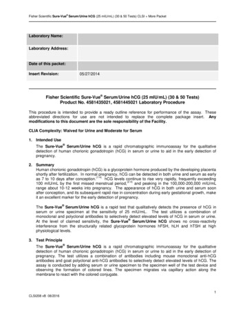
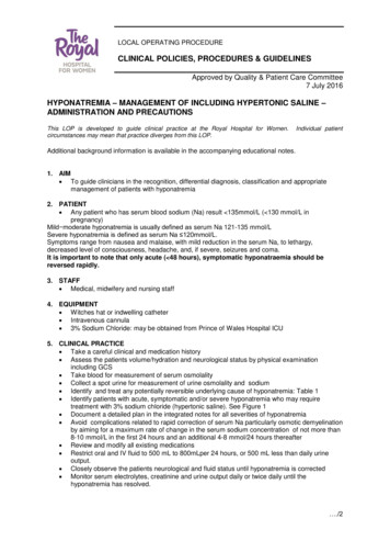
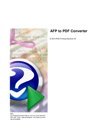
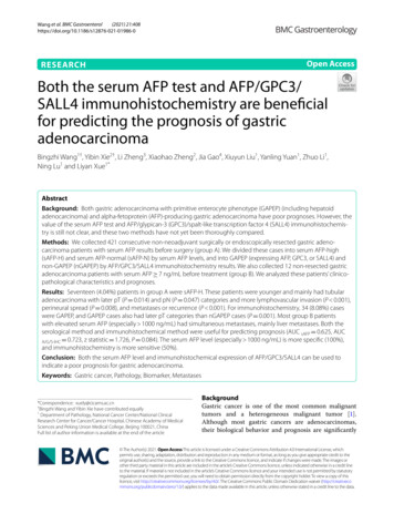
Wang et al. BMC Gastroenterol(2021) 21:408were reviewed jointly until a consensus was reached.We divided the group A cases by the AFP/GPC3/SALL4results into gastric cancer with primitive enterocyte phenotype (GAPEP, at least one of three was positive) andnon-GAPEP (nGAPEP, all three markers were negative).Statistical analysisThe clinicopathological characteristics and prognoses ingroups A and B were analyzed. We performed 1:1 propensity score matching (PSM) in group A (both sAFPN vs. sAFP-H and GAPEP vs. nGAPEP) to compare theProgression Free Survival (PFS), and the PSM predictorsincluded age, Lauren classification, and T and N categories (match tolerance 0.2, random seed to verify the reliability). Metastases in group B, including simultaneousmetastases and metachronous metastases, were analyzed.The heatmap of A/G/S-IHC of sAFP-H cases was drawnby Prism 8 software (GraphPad Software Ltd., San Diego,USA). The relationship between immunohistochemistry and a high risk of disease progression (simultaneous metastases or postoperative metastases within threeyears) was analyzed. The comparison of count data wasmade by the chi-square test with a significance level of0.05 on two-tailed P-values. SPSS (Statistical Product andService Solutions, IBM Corp., NY, United States) 25.0software was used for statistical analysis. Receiver operating characteristic (ROC) curves were plotted, and thearea under the curve (AUC) and z-test were calculated.ResultsClinicopathological characteristics in group AAccording to sAFP level, 404 (96.19%) cases wereassigned to the sAFP-N group, and 17 (3.81%) cases wereassigned to the sAFP-H group (Table 1). When the caseswere divided by a cutoff age of 50 years, significantlymore patients aged 50 years were noted in the sAFP-Hgroup than those in the sAFP-N group (P 0.023). Definite differences in both Lauren classification (P 0.030)and JCGA histological classification (P 0.021) wereidentified. Regarding the JGCA classification, the sAFPH cases mainly composed of tub2 type (8, 47.06%) andpor1 type (4, 23.53%). More por2 type were observed inthe sAFP-N cases. Notably, few cases of special subtypeswere noted in the sAFP-H group: only 2 cases were HAC,1 case was GAED, and the other cases were mostly moderately or poorly differentiated tubular adenocarcinomawith no special morphological features (Fig. 1). Therewere significant differences in T categories (P 0.014).More T4 cases were in the sAFP-H cases (10, 58.82%)than those in the sAFP-N cases (150, 37.13%). Significant difference in N categories was identified (P 0.047).The sAFP-H group had significantly more N3 cases thanthe sAFP-N group (52.94% vs 29.60%). LVI (76.47% vsPage 3 of 1035.15%, P 0.001) and PNS (64.71% vs 33.42%, P 0.008)were significantly higher in the sAFP-H group than thatin the sAFP-N group, respectively. The sAFP-H grouphad significantly more postoperative recurrence ormetastases than the sAFP-N group (23.53% vs 5.66%,P 0.001). Among the sAFP-H group, 3 patients had livermetastasis, and 1 patient had recurrence of the remnantstomach at one year after surgery. After 1:1 PSM, sAFPH patients also had worse PFS than sAFP-N patients(P 0.048) (Fig. 2A). There were no significant differences in sex (P 0.779), location (P 0.164), or macroscopic classification (P 0.064).By the A/G/S-IHC results, 34/421 (8.08%) cases wereGAPEP. The positive rate of AFP was 4.28% (18/421), thepositive rate of GPC3 was 4.51% (19/421), and the positive rate of SALL4 was 2.85% (12/421). The GAPEP casesalso showed similar characteristics as sAFP-H cases,especially in JGCA classification (P 0.009) and T category (P 0.001). The GAPEP group had significantlymore postoperative recurrence or metastases than thenGAPEP group (23.53% vs 2.07%, P 0.001) 1 year aftersurgery. After 1:1 PSM, GAPEP patients also had worsesurvival than nGAPEP patients (P 0.035) (Fig. 2B).Serum AFP was re-examined in 7 sAFP-H patientsafter surgery. The sAFP levels in these 7 patients weresignificantly decreased after surgery, and most fell in thenormal range within one year after surgery (Fig. 3A).Clinicopathological characteristics in group BIn group B, a total of 7 cases had simultaneous metastases(58.33%), of which 4 cases were liver metastases, 2 caseswere ovarian metastases, and 1 case was liver metastasesaccompanied by lung metastases at the same time. Therate of liver metastases reached 41.67%. Two patients hadmetachronous liver metastases during follow-up. Thetotal liver metastasis rate reached 58.33%. For the A/G/SIHC results, 11/12 cases were GAPEP.The clinicopathological characteristics of the 29sAFP-H cases in groups A and B are shown in Table 2.Interestingly, all patients with sAFP 1000 ng/mL ingroups A and B had liver metastases, most of whom hadliver metastases at diagnosis, while only one case hadmetachronous metastasis (Fig. 3B).Comparison and correlation of immunohistochemistryand sAFP testSerum AFP-H patients had significantly higher positiverates of AFP, GPC3, and SALL4 than sAFP-N patients(Table 3). Serum AFP-H was significantly related toGAPEP (P 0.001). Notably, more high-risk caseswere detected by immunohistochemistry, 17 surgicalcases of GAPEP with normal sAFP were identified, 4of which had postoperative recurrence or metastasis (2
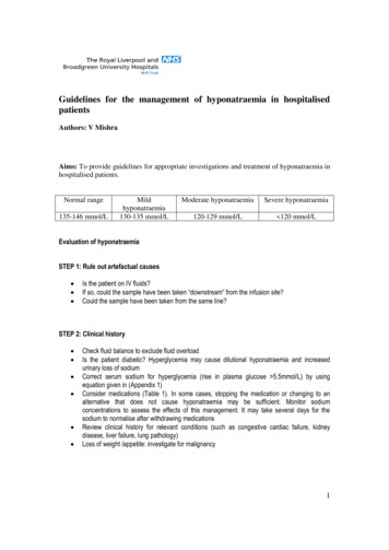
Wang et al. BMC Gastroenterol(2021) 21:408Page 4 of 10Table 1 Comparison of clinicopathological characteristics between sAFP-N vs. sAFP-H and between nGAPEP vs. GAPEP in the surgicalcases (group A)sAFP-NAgeSexLocation* 5093 (23.02%)8 (47.06%) 50311 (76.98%)9 (52.94%)Male272 (67.33%)12 (70.59%)Female132 (32.67%)5 (29.31%)ProximalDistalLauren classificationN categoryLVIPNSPostoperative recurrence/metastasis2 (11.76%)15 (88.24%)Overlapping60 (15.04%)0 (0.00%)88 (21.78%)8 (47.06%)262 (64.85%)6 (35.29%)Mixed54 (13.37%)3 (17.65%)Tub110 (2.48%)0 (0.00%)Tub272 (17.82%)8 (47.06%)PnGAPEP0.0230.7790.1640.0300.021GAPEP91 (23.51%)10 (29.41%)296 (76.49%)24 (70.59%)259 (66.93%)25 (73.53%)128 (33.07%)9 (26.47%)60 (15.67%)6 (18.18%)265 (69.19%)25 (75.76%)58 (15.14%)2 (6.06%)83 (21.45%)13 (38.24%)251 (64.86%)17 (50.00%)53 (13.69%)4 (11.76%)10 (2.58%)0 (0%)68 (17.57%)Muc6 (1.49%)0 (0.00%)5 (1.29%)1 (2.94%)139 (34.41%)0 (0.00%)137 (35.40%)2 (5.88%)Por162 (15.35%)4 (23.53%)58 (14.99%)8 (23.53%)Por261 (15.10%)2 (11.76%)56 (14.47%)7 (20.59%)54 (13.37%)3 (17.65%)T1142 (35.15%)0 (0.00%)T257 (14.11%)5 (29.41%)0.01453 (13.70%)4 (11.76%)140 (36.18%)2 (5.88%)52 (13.44%)10 (29.41%)T355 (13.61%)2 (11.76%)54 (13.95%)3 (8.82%)T4150 (37.13%)10 (58.82%)141 (36.43%)19 (55.88%)N0150 (40.00%)3 (17.65%)141 (39.39%)12 (35.29%)N163 (16.80%)1 (5.88%)62 (17.32%)2 (5.88%)0.047N251 (13.60%)4 (23.53%)47 (13.13%)8 (23.53%)N3111 (29.60%)9 (52.94%)108 (30.17%)12 (35.29%)No262 (64.85%)4 (23.53%)249 (64.34%)17 (50.00%)Yes142 (35.15%)13 (76.47%)138 (35.66%)17 (50.00%)No269 (66.58%)6 (35.29%)257 (66.41%)18 (52.94%)Yes135 (33.42%)11 (64.71%)130 (33.59%)16 (47.06%)No200 (94.34%)13 (76.47%)379 (97.93%)26 (76.47%)Yes12 (5.66%)4 (23.53%)8 (2.07%)8 (23.53%)0.0010.008 0.001P0.4400.4310.3590.0810.00912 (35.29%)SigOthersT category64 (16.04%)275 (68.92%)IntestinalDiffuseHistological classification**sAFP-H0.0010.1520.0960.114 0.001LVI, lymphovascular invasion; PNS, perineural spread*Proximal tumor in upper 1/2 with a proximal gastrectomy; dismal tumor in lower 1/2 with a dismal gastrectomy; overlapping large tumor or linitis plastica witha total gastrectomy**Based on the Japanese Gastric Cancer Association classification, tub1 well differentiated tubular type, tub2 moderately differentiated tubular type, por1 solidtype, por2 poorly cohesive type, sig signet-ring cell type, muc mucin type, and other typescases of recurrence in gastric remnants, 1 case of livermetastasis, and 1 case of lung metastasis).With a high risk of disease progression as the observation index, both methods had good efficacy (AUCsAFP 0.625, AUC A/G/S-IHC 0.723, z statistic 1.726,P 0.084) (Fig. 4). The serum test had higher specificity (100% vs 94.5%) while the immunohistochemicaltest had higher sensitivity (50% vs 25%). We also drewa heatmap of all sAFP-H cases (Fig. 5).DiscussionIn this study, we compared the serum AFP test andimmunohistochemical test of GAPEP to find the valueof the two methods in clinical diagnosis. The proportion of sAFP-H patients in group A was 1.82%, which wasslightly lower than the number reported in the literature[17]. The reason might be this study is a retrospectiveresearch and not every patient underwent the sAFP testin our hospital.
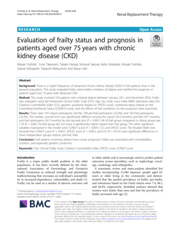
Wang et al. BMC Gastroenterol(2021) 21:408Page 5 of 10Fig. 1 A Hepatoid carcinoma area. B Adenocarcinoma with enteroblastic differentiation. C Moderately poorly differentiated adenocarcinomas withpositive AFP (D), GPC3 (E) and SALL4 (F). All X200Fig. 2 Progression-free survival (PFS) curves (months) after 1:1propensity score matching (matching predictors included the ages,Lauren classification, T and N categories). A sAFP-N versus sAFP-H(P 0.048). B GAPEP versus nGAPEP (P 0.035)Though some studies argued the predictive value ofserum AFP [18], most previous studies have confirmedthat the prognosis of even early gastric adenocarcinomawith elevated serum AFP is poor [19]. It is believed thatthe production of sAFP is more likely to be caused bygastric cancer itself. AFP produced by the tumor itselfthat leads to an increase in the serum, and sAFP loses itssource after tumor resection and then rapidly decreases,similar to our result (Fig. 3A). Some cases (3/8) withsAFP dropping after surgery had progression later, whichmight reflect the aggressive characteristics. However, thequantity of cases with both pre- and post-operation sAFPwas limited. Another phenomenon that must be noted isthat patients with extremely high sAFP values ( 1000 ng/mL) had liver metastases at diagnosis, which may reflecthigh tumor burden. It is suggested that the sAFP testcould be an important signal of liver metastasis. Thisstudy did not include biopsy cases with normal sAFP asa control for group B. Serum AFP-H patients often havesimultaneous metastases at diagnosis, which is sufficientto demonstrate the biological characteristics. Furthercontrolled studies of sAFP-H and sAFP-N in biopsy casesare necessary to better understand the implication ofsAFP-H.The mechanism of the special biological behavior ofliver metastasis has been explored in some studies [20,21]. One reason may be that this tumor produces many
Wang et al. BMC Gastroenterol(2021) 21:408Page 6 of 10Fig. 3 A The serum AFP value (ng/mL) varied from the preoperative period to the postoperative period in 8 cases in group A. B The serum AFPvalue (ng/mL) of 12 cases in group B. Each dot represents a case, and the red dot indicates liver metastasestumor thrombi, and the liver and lungs are vital forblood circulation, so the tumor thrombi easily spread tothe liver through the blood [22]. Another possible reason is that the tumor itself activates certain molecularpathways, so it easily colonizes and grows in the liver[23, 24]. From the perspective of embryonic development, the liver, gastrointestinal tract, and lung originatefrom the endoderm, and those organs develop closely in
talDistalDistalDistalDistalLocation**With enteroblastic 33.57.3645.564Diameter sAFP eousmetastasisLiverLiverStump ub2tub2por1*por2otherstub2por1por1JGCA N2N2N3N2N0N3N3N3N3N3N2N0pN LVI PNS AFP GPC3 SALL4(2021) 21:408*With hepatoid carcinoma ntgroupCaseTable 2 Clinicopathological characteristics of the sAFP-H cases in groups A and BWang et al. BMC GastroenterolPage 7 of 10
Wang et al. BMC Gastroenterol(2021) 21:408Table 3 Correlation betweenimmunohistochemical resultsAFPGPC3SALL4GAPEPserumPage 8 of 10AFPsAFP-NsAFP-H( )403 (99.75%)0( )1 (0.25%)17 (100.00%)( )392 (97.03%)10 (58.82%)( )12 (2.97%)7 (41.18%)( )399 (98.76%)10 (58.82%)( )5 (1.24%)7 (41.18%)( )387 (95.79%)0( )17 (4.21%)17 (100%)valueandP 0.001 0.001 0.001 0.001Fig. 5 Heatmap of all sAFP-H cases in groups A and B. Horizontal axis,AFP, GPC3 and SALL4 results. Vertical axis, sAFP-H cases in descendingorder of sAFP levelFig. 4 ROC curves of the two methods individually to predictprogression free survival in group A (AUC sAFP 0.625, AUC A/G/S-IHC 0.723, z statistic 1.726, P 0.084)a relatively long period [25, 26]. The lineage of primitiveepithelial differentiated tumors that produces AFP, suchas some gastric cancer, intestinal cancer and fetal adenocarcinoma of the lung [27]. The name "gastric cancer withprimitive enterocyte phenotype" is a possibly more scientific name.Consistent with the research by Yamazawa et al., theGAPEP tumors in this study were mostly adenocarcinomas of common morphology, and these tumors hadsimilar biological behaviors [28–30]. This suggests thatthe traditional pathological diagnosis of special morphology is insufficient. GPC3 is a glypican-related proteoglycan that is sensitive for diagnosing HCC, distinguishingit from benign hepatocellular lesions [10]. SALL4 is anovel sensitive and specific marker for both primary andmetastatic germ cell tumors [15]. Both GPC3 and SALL4have significance in gastric carcinoma [14]. This studyfurther confirms that the determination of GAPEP basedon the immunohistochemistry of AFP, GPC3, and SALL4is indeed of significance for judging prognosis. Molecular research on GAPEP and its related tumors has yieldedsome results [31, 32]. Noboru et al. found frequent lossof SMAD4 heterozygosity by immunohistochemistry ingastric adenocarcinoma with enteroblastic differentiation[33, 34]. Yoichi et al. found that GAED frequently harbors TP53 mutations and ERBB2 amplification [35]. Fujimoto et al. found that HER2 is frequently overexpressedin hepatoid adenocarcinoma and gastric carcinoma withenteroblastic differentiation [36]. According to our previous research, GAPEP accounted for 7.53% of surgicalspecimens [37]. Defining this particular type may havesome significance in selecting patients for more aggressive treatment [38]. However, several large-scale gastriccancer molecular typing studies, such as The CancerGenome Atlas (TCGA) and the Asian Cancer ResearchGroup (ACRG) [39–42], have not included GAPEP. Morestudies are needed on the molecular pathological featuresof GAPEP.There were still some weaknesses in this research. GAwith AFP-producing and GAPEP were rare tumor, and thequantity of those cases was limited. More evidences arein need to confirm the results in future. And we couldn’testablish the comparator arm in group B because the complex reasons for not getting operation. The two methodsboth have their own advantages, the serological methodis noninvasive and specific. However, the results may beaffected by other factors (liver disease, germ cell tumors,etc.). Although immunohistochemical examination isinvasive, it is sensible and can better reflect the nature ofthe tumor. In applying both methods to determine the
Wang et al. BMC Gastroenterol(2021) 21:408prognosis of a patient, the AUC was slightly better thanusing only one of the two. Therefore, we recommend usingsAFP and AFP/GPC3/SALL4 immunohistochemistry forall cases of gastric cancer.ConclusionsBoth the serum AFP level and immunohistochemicalexpression of can be used to indicate a poor prognosisfor gastric adenocarcinoma. The serum AFP level (especially 1000 ng/mL) is more specific, and immunohistochemistry is more sensitive.AcknowledgementsNone.Authors’ contributionsLX and YX performed study concept and design; BW, LZ and XZ performeddevelopment of methodology and writing, review and revision of the paper;NL, JG and ZL provided acquisition, analysis and interpretation of data, andstatistical analysis; YY and XL provided technical and material support. Allauthors read and approved the final paper.FundingThis work was supported by the Beijing Hope Run Special Fund (No.LC2018A12), the CAMS Initiative for Innovative Medicine (CIFMS) (No. 2016I2M-3-005), the Medical and Health Science and Technology InnovationProject of the Chinese Academy of Medical Sciences (2016-12M-1-007), andthe China International Medical Exchange Foundation Xiansheng Anti-TumorTherapy Special Research Fund (cimf-f-h001-314).Availability of data and materialsThe datasets used and/or analysed during the current study available from thecorresponding author on reasonable request.DeclarationsEthics approval and consent to participateEthical approval was approved by CICAMS ethics committee prior to commencing this study (No. 21/105-2776). All methods were carried out in accordance with relevant guidelines and regulations, and informed consent wasobtained from all participants.Consent for publicationNot applicable.Competing interestsThe authors declare that they have no competing interests.Author details1Department of Pathology, National Cancer Center/National Clinical ResearchCenter for Cancer/Cancer Hospital, Chinese Academy of Medical Sciencesand Peking Union Medical College, Beijing 100021, China. 2 Departmentof Abdominal Surgical Oncology, National Cancer Center/National ClinicalResearch Center for Cancer/Cancer Hospital, Chinese Academy of Medical Sciences and Peking Union Medical College, Beijing 100021, China.3Department of General Surgery, the First People’s Hospital of DongchengDistrict, Beijing 100075, China. 4 Department of Clinical Laboratory, NationalCancer Center/National Clinical Research Center for Cancer/Cancer Hospital,Chinese Academy of Medical Sciences and Peking Union Medical College,Beijing 100021, China.Received: 6 April 2021 Accepted: 18 October 2021Page 9 of 10References1. Torre LA, Siegel RL, Ward EM, Jemal A. Global cancer incidence andmortality rates and trends: an update. Cancer Epidemiol Biomarkers Prev.2016;25(1):16–27.2. He CZ, Zhang KH, Li Q, Liu XH, Hong Y, Lv NH. Combined use of AFP, CEA,CA125 and CAl9-9 improves the sensitivity for the diagnosis of gastriccancer. BMC Gastroenterol. 2013;13:87.3. Feng F, Tian Y, Xu G, Liu Z, Liu S, Zheng G, et al. Diagnostic and prognosticvalue of CEA, CA19-9, AFP and CA125 for early gastric cancer. BMC Cancer. 2017;17(1):737.4. Bourreille J, Metayer P, Sauger F, Matray F, Fondimare A. Existence of alphafeto protein during gastric-origin secondary cancer of the liver. PresseMed. 1970;78(28):1277–8.5. Ishikura H, Fukasawa Y, Ogasawara K, Natori T, Tsukada Y, Aizawa M. AnAFP-producing gastric carcinoma with features of hepatic differentiation:a case report. Cancer. 1985;56(4):840–8.6. Lakshmanan A, Kurian A, Subramanyan A, Srinivasan A. An alpha fetoprotein producing gastric tumor with yolk sac, hepatoid and papillaryadenocarcinoma components. J Clin Diagn Res. 2017;11(9):ED03–5.7. Murakami T, Yao T, Mitomi H, Morimoto T, Ueyama H, Matsumoto K, et al.Clinicopathologic and immunohistochemical characteristics of gastricadenocarcinoma with enteroblastic differentiation: a study of 29 cases.Gastric Cancer. 2016;19(2):498–507.8. Kinjo T, Taniguchi H, Kushima R, Sekine S, Oda I, Saka M, et al. Histologicand immunohistochemical analyses of alpha-fetoprotein–producingcancer of the stomach. Am J Surg Pathol. 2012;36(1):56–65.9. Xie Y, Zhao Z, Li P, Wang Y, Guo C, Wang X, et al. Hepatoid adenocarcinoma of the stomach is a special and easily misdiagnosed or misseddiagnosed subtype of gastric cancer with poor prognosis but curative forpatients of pN0/1: the experience of a single center. Int J Clin Exp Med.2015;8(5):6762–72.10. Yamazawa S, Ushiku T, Shinozaki-Ushiku A, Hayashi A, Iwasaki A, Abe H,et al. Gastric cancer with primitive enterocyte phenotype: an aggressive subgroup of intestinal-type adenocarcinoma. Am J Surg Pathol.2017;41(7):989–97.11. Chang YC, Nagasue N, Abe S, Taniura H, Kumar DD, Nakamura T. Comparison between the clinicopathologic features of AFP-positive andAFP-negative gastric cancers. Am J Gastroenterol. 1992;87(3):321–5.12.
determine the value of sAFP and A/G/S-IHC in the diag-nosis and treatment of gastric adenocarcinoma. Materials and methods Enrollment criteria For enrollment, patients must have met the following conditions: admission to the National Cancer Center/ National Clinical Research Center for Cancer/Can-cer Hospital, Chinese Academy of Medical Sciences