Transcription
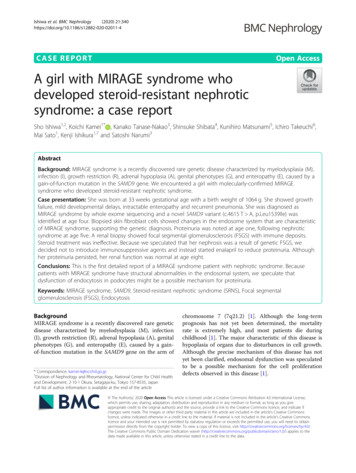
Ishiwa et al. BMC Nephrology(2020) SE REPORTOpen AccessA girl with MIRAGE syndrome whodeveloped steroid-resistant nephroticsyndrome: a case reportSho Ishiwa1,2, Koichi Kamei1* , Kanako Tanase-Nakao3, Shinsuke Shibata4, Kunihiro Matsunami5, Ichiro Takeuchi6,Mai Sato1, Kenji Ishikura1,7 and Satoshi Narumi3AbstractBackground: MIRAGE syndrome is a recently discovered rare genetic disease characterized by myelodysplasia (M),infection (I), growth restriction (R), adrenal hypoplasia (A), genital phenotypes (G), and enteropathy (E), caused by again-of-function mutation in the SAMD9 gene. We encountered a girl with molecularly-confirmed MIRAGEsyndrome who developed steroid-resistant nephrotic syndrome.Case presentation: She was born at 33 weeks gestational age with a birth weight of 1064 g. She showed growthfailure, mild developmental delays, intractable enteropathy and recurrent pneumonia. She was diagnosed asMIRAGE syndrome by whole exome sequencing and a novel SAMD9 variant (c.4615 T A, p.Leu1539Ile) wasidentified at age four. Biopsied skin fibroblast cells showed changes in the endosome system that are characteristicof MIRAGE syndrome, supporting the genetic diagnosis. Proteinuria was noted at age one, following nephroticsyndrome at age five. A renal biopsy showed focal segmental glomerulosclerosis (FSGS) with immune deposits.Steroid treatment was ineffective. Because we speculated that her nephrosis was a result of genetic FSGS, wedecided not to introduce immunosuppressive agents and instead started enalapril to reduce proteinuria. Althoughher proteinuria persisted, her renal function was normal at age eight.Conclusions: This is the first detailed report of a MIRAGE syndrome patient with nephrotic syndrome. Becausepatients with MIRAGE syndrome have structural abnormalities in the endosomal system, we speculate thatdysfunction of endocytosis in podocytes might be a possible mechanism for proteinuria.Keywords: MIRAGE syndrome, SAMD9, Steroid-resistant nephrotic syndrome (SRNS), Focal segmentalglomerulosclerosis (FSGS), EndocytosisBackgroundMIRAGE syndrome is a recently discovered rare geneticdisease characterized by myelodysplasia (M), infection(I), growth restriction (R), adrenal hypoplasia (A), genitalphenotypes (G), and enteropathy (E), caused by a gainof-function mutation in the SAMD9 gene on the arm of* Correspondence: kamei-k@ncchd.go.jp1Division of Nephrology and Rheumatology, National Center for Child Healthand Development, 2-10-1 Okura, Setagaya-ku, Tokyo 157-8535, JapanFull list of author information is available at the end of the articlechromosome 7 (7q21.2) [1]. Although the long-termprognosis has not yet been determined, the mortalityrate is extremely high, and most patients die duringchildhood [1]. The major characteristic of this disease ishypoplasia of organs due to disturbances in cell growth.Although the precise mechanism of this disease has notyet been clarified, endosomal dysfunction was speculatedto be a possible mechanism for the cell proliferationdefects observed in this disease [1]. The Author(s). 2020 Open Access This article is licensed under a Creative Commons Attribution 4.0 International License,which permits use, sharing, adaptation, distribution and reproduction in any medium or format, as long as you giveappropriate credit to the original author(s) and the source, provide a link to the Creative Commons licence, and indicate ifchanges were made. The images or other third party material in this article are included in the article's Creative Commonslicence, unless indicated otherwise in a credit line to the material. If material is not included in the article's Creative Commonslicence and your intended use is not permitted by statutory regulation or exceeds the permitted use, you will need to obtainpermission directly from the copyright holder. To view a copy of this licence, visit http://creativecommons.org/licenses/by/4.0/.The Creative Commons Public Domain Dedication waiver ) applies to thedata made available in this article, unless otherwise stated in a credit line to the data.
Ishiwa et al. BMC Nephrology(2020) 21:340To date, renal complications have been reported inseven patients with this disease [2–8]. Focal segmentalglomerulosclerosis (FSGS) was diagnosed in two patients[2, 3], renal tubular acidosis in one [4], interstitial nephritis in one [4], renal hypoplasia in one [5], C1q nephropathy after bone marrow transplantation (BMT) inone [6, 7], and renal injury after BMT in one [8]. Theselines of evidence indicate the importance of renalcomplications, although detailed clinical descriptions arelacking. Here, we report a girl with molecularlyconfirmed MIRAGE syndrome with a particular emphasis on the detailed clinical information of hersteroid-resistant nephrotic syndrome (SRNS). Consentfor publication was obtained from her family.Case presentationThis case was a female born at 33 weeks gestational agewith a birth weight of 1064 g, no asphyxia, and no familyhistory. At birth, she showed transient low plateletcounts (28,000/mm3), although it improved spontaneously. She had mild global developmental delays (rolling over, 8 months; walking with assistance, 16 months;standing alone, 19 months; walking without assistance,27 months of corrected age). Her postnatal growth waspoor. She had esophageal dysmotility, and she requiredtube-feeding for disturbances in oral intake. She was alsosuffering from clumpy abdominal pain and waterydiarrhea. She had an episode of “autoimmune encephalitis” at age four. She developed recurrent aspirationpneumonia, and a gastrostomy was performed at agefive. Whole exome sequencing was performed at agefour, and a novel SAMD9 variant (c.4615 T A,p.Leu1539Ile) was identified. Biopsied skin fibroblastcells showed changes in the endosome system that arecharacteristic of MIRAGE syndrome, supporting thegenetic diagnosis (Fig. 1). She did not show monosomy 7nor myelodysplastic syndrome.She showed recurrent infections (I), growth restriction(R), and intractable enteropathy (esophageal dysmotility,episodic vomiting, clumpy abdominal pain, and waterydiarrhea) (E), although she did not suffer from myelosuppression (M) except for the transient low plateletcount during the neonatal period. She also did not sufferfrom episodes of adrenal insufficiency (A) (basic cortisollevel, 6.7 μg/dL; basic adrenocorticotropic hormone(ACTH) level, 12.8 pg/mL; peak cortisol level aftercorticotropin-releasing hormone (CRH) stimulation test,13.2 μg/dL; peak ACTH level after CRH stimulation test,58.7 pg/mL). She showed normal female externalgenitalia and a normal uterus (G). Her ovaries could notbe detected by abdominal ultrasonography, which is notspecific for her age. She showed insensitivity to painwith anhidrosis. The somatosensory-evoked potentialsshowed poorly depicted waveforms, and a sympatheticPage 2 of 6skin response was absent. These examinations uropathy.Proteinuria was first noted at age one. Although heavyproteinuria persisted, her serum albumin levels remainednormal until age three. Her serum albumin levelsdecreased to less than 3.0 g/dL at 3 years and 11 months,showing nephrotic syndrome. At this time, she wasobserved by outpatient clinic as she showed no edema.She developed prominent edema due to severe hypoalbuminemia of less than 2.0 g/dL at 5 years and 7 months.Renal biopsy revealed collapse in one glomerulus,segmental sclerosis in two glomeruli, and mild mesangialcell proliferation in 3 of 21 glomeruli (Fig. 2). Immunostaining showed codominant deposition of IgG, C3, andC1q in the mesangial area. Electron dense deposits wereobserved in mesangial matrix, subepithelial, and subendothelial space in electron microscopy. Mesangial interposition was also revealed. Mesangial matrix was slightlyenlarged, although mesangial cells were not proliferated.Hyalinosis is found in some glomerular loops. Abnormalfindings of epithelial cells were not detected except forpartial disappearance of the foot process. The pathological diagnosis was FSGS. She received steroidtreatment (60 mg/m2/day of oral prednisolone), althoughit did not lead to remission at 4 weeks. She wasdiagnosed with SRNS. Her treatment was switched fromoral prednisolone to an angiotensin converting enzymeinhibitor (enalapril).A physical examination at age six showed no abnormalities, except for extreme growth failure (height, 93.3cm, 4.4 SD; body weight, 10.0 kg, 3.2 SD). Her whiteblood cell count (8070/μL), hemoglobin level (10.9 g/dL),and platelet count (419,000/μL) were within normalranges. She displayed hypoalbuminemia (2.0 g/dL) withnormal renal function (blood urea nitrogen level of 13.3mg/dL and serum creatinine level of 0.26 mg/dL). Herserum immunoglobulin G level was low (228 mg/dL).Her complement activity, antinuclear antibody, antineutrophil cytoplasmic antibody, and anti-glomerularbasement membrane antibody levels were normal. Theurinalysis demonstrated heavy proteinuria (5 g/g Cr)without hematuria. Her serum sodium (138 mEq/L),serum potassium (4.0 mEq/L), serum calcium (9.6 mg/dL), and serum phosphate (4.9 mg/dL) levels were normal, and the parameters of renal tubular functions werewithin normal ranges (fraction excretion of sodium,0.2%; fraction excretion of potassium, 5.7%; calcium/creatinine ratio, 0.09; tubular reabsorption of phosphate,96.2%; urinary β2 microglobulin, 0.32 μg/L), in which nosign of Fanconi syndrome was observed. The immunological examinations reported no abnormalities in the Tcells and B cells, indicating normal cellular and humoralimmunity. According to the abdominal ultrasound, her
Ishiwa et al. BMC Nephrology(2020) 21:340Page 3 of 6Fig. 1 Ultrastructural findings of skin fibroblasts. Electron microscopic images (magnification, 1,000) of biopsied skin fibroblast cells derived froma healthy child (a) and the MIRAGE syndrome patient with the p.L1539I SAMD9 mutation (b). Giant vesicles without a specific inner structurewere frequently observed (arrows). Bars, 5 μmkidney size was normal for her body size (right kidney,62 35 mm; left kidney, 61 30 mm).Despite her heavy proteinuria, we decided not to initiate further immunosuppressive treatment such as cyclosporin or steroid-pulse therapy because we thought hernephrotic syndrome was genetic FSGS due to MIRAGEsyndrome. At the last observation when she was 8 yearsold, her renal function was normal (serum creatininelevel of 0.34 mg/dL and estimated glomerular filtrationrate of 104 mL/min/1.73 m2) with mildly decreasedserum albumin levels (2.9 g/dL) and persisting proteinuria (5.4 g/g Cr) under enalapril treatment.Discussion and conclusionsThis case report describes a girl with MIRAGE syndrome who developed SRNS. A renal biopsy showedFSGS with immune deposits. This is the first detailedreport of a MIRAGE syndrome patient with nephroticsyndrome.Previously, there have been two case reports of FSGSwith MIRAGE syndrome [2, 3]. We speculate thatdysfunction of endocytosis in podocytes might play apossible role in proteinuria, leading to FSGS, althoughthis was not demonstrated at the molecular level.Patients with MIRAGE syndrome have structural alterations in the endosomal system, including enlargedendosomes and the presence of structureless giantvesicles [1]. These findings were also observed in ourpatient. In recent years, animal studies have shown thatendocytosis plays an important role in podocytes, andgenetic defects involving the endosome system causeprotein leakage [9–11]. Lysosomes are involved inprocessing endocytosed albumin in podocytes, andlysosomal dysfunction may contribute to podocyte injuryand glomerulosclerosis [10].A previous case report described a patient with renaltubular acidosis and a patient with interstitial nephritis[4]. Because immune abnormality is one of the majorsymptoms of MIRAGE syndrome, interstitial nephritismight be caused by autoimmune mechanisms of this disease. SAMD9 is shown to be expressed not only inglomeruli but also in renal tubules [12]. The proximaltubule plays a role in endocytosis via megalin or cubilin.However, the patient’s renal tubular function was normal, and she did not suffer from Fanconi syndrome. Furthermore, the renal biopsy did not show interstitialnephritis.Because this patient showed the codominant deposition of IgG, C3, and C1q on the glomeruli, nephroticsyndrome might have resulted from the immune abnormalities of this syndrome. C1q nephropathy was previously reported in a patient with MIRAGE syndrome [6,7], although this case developed nephrosis after BMT.C1q deposition usually indicates immune-complexmediated glomerulopathy and is typically observed insystemic lupus erythematosus. However, the clinicalsignificance of C1q nephropathy remains obscure [13].Light microscopic features of C1q nephropathy are
Ishiwa et al. BMC Nephrology(2020) 21:340Page 4 of 6Fig. 2 Kidney biopsy findings. a, b: Light microscopy shows FSGS (A, periodic acid-Schiff stain, 200; B, periodic acid methenamine silver stain, 200). c-h: Immunofluorescent microscopy (c, IgG; d, IgA; e, IgM; f, C3; g, C4; h, C1q). IgG ( ), IgA ( ), IgM ( ), C3 ( ), C4 ( ), C1q ( ). i-k: Electronmicroscopy (i, mesangial deposits; j, subepithelial deposits; k, subendothelial deposits)variable and heterogeneous, such as minor glomerularabnormalities, FSGS, and sometimes membranousnephropathy [14]. Its deposition is sometimes nonspecifically observed. There has even been a case reportof a girl with congenital nephrotic syndrome with granular mesangial C1q deposition on her glomeruli, althoughcongenital nephrotic syndrome is typically a geneticdisease [15].Hyperfiltration of glomeruli due to reduced nephronby premature birth might be one of the mechanismsof proteinuria as she was born at 33 weeks gestationalage with a birth weight of 1064 g. However, nephroticrange proteinuria is relatively rare on this situation.Moreover, her glomeruli were not large, which wasnot typical for FSGS due to hyperfiltration by nephron reduction.The patient’s kidney size was normal for her bodyweight, and her renal function was normal. Although themajor characteristics of this disease include hypoplasiaof organs due to disturbances of cell growth and renal
Ishiwa et al. BMC Nephrology(2020) 21:340hypoplasia is also reported in a patient with this disease[5], hypoplastic kidney was not observed in this case.Other specific symptoms of this case were insensitivityand anhidrosis. To date, three cases of hypolacrima withcorneal ulcer [4, 16, 17], one case of anhidrosis [8] andtwo cases of hyperhidrosis [4, 17] have been reported inMIRAGE syndrome. There have been no reports ofinsensitivity. We believe dysautonomia, such as insensitivity and anhidrosis, might be important clinical symptoms of this syndrome. More clinical information shouldbe accumulated in the future.We decided not to introduce immunosuppressiveagents. We only continued the angiotensin convertingenzyme inhibitor as the conservative treatmentbecause we thought this case was genetic FSGS,although recent review article did not introduce“SAMD9” as a causative gene of SRNS [18]. Untilnow, her renal function has remained normal,although proteinuria persisted under enalapril treatment for three years after the onset of nephroticsyndrome. The long-term prognosis of renal functionis obscure in this disease; thus, it is necessary for usto follow-up and observe the renal function of thispatient for a long time. We also have to obtain moreinformation about the renal complications of this disease by evaluating more cases to determine the mechanism of this rare disease in the future.AbbreviationsFSGS: Focal segmental glomerulosclerosis; BMT: Bone marrowtransplantation; ACTH: Adrenocorticotropic hormone; CRH: Corticotropinreleasing hormone; SRNS: Steroid-resistant nephrotic syndromeAcknowledgementsWe would like to thank Drs K. Ogata and T. Yoshioka for the pathologicaldiagnosis; Drs E. Tamura, T. Kawai, and S. Nonoyama for the immunologicalanalysis; and Drs M. Ogura, T. Kanamori, K. Nishi, M. Okutsu, M. Sako, T.Ogawa, I. Hayakawa, and K. Arai for their contributions to this report. We alsothank the patient and her family for providing permission to share herinformation.Authors’ contributionsSI and KK prepared the manuscript. KT-N examined the genetic analysis. SSexamined the microscopic analysis of the fibroblast. KM and IT are physiciansin charge of this case and collected the clinical data. MS and KI revised themanuscript. SN examined the genetic analysis, revised the manuscript andoversaw the work. All authors participated in the discussions about themanuscript and approved the final version.FundingThere was no external funding for this manuscript.Availability of data and materialsThe datasets used and/or analyzed during the current study are availablefrom the corresponding author upon reasonable request.Ethics approval and consent to participateEthics approval was not applicable. Consent for publication was obtainedfrom her family.Consent for publicationWritten informed consent for publication was obtained from her family asparticipants were under 16 years old.Page 5 of 6Competing interestsKamei received grants from the Terumo Foundation for Life Sciences andArts, Chugai Pharmaceutical Co. Ltd., Astellas Pharma Inc., OnoPharmaceutical Co., and Pfizer Japan, Inc., and lecture fees from TanabeMitsubishi Pharma, Novartis Pharma K.K., Chugai Pharmaceutical Co. Ltd. andAbbVie Inc. Ishikura received grants from Asahi Kasei Pharma, ChugaiPharmaceutical Co., Ltd., Novartis Pharma, and Zenyaku Kogyo Co. Ltd. andlecture fees from Asahi Kasei Pharma, Chugai Pharmaceutical Co., Ltd.,Zenyaku Kogyo Co., Ltd., and Novartis Pharma K.K.Author details1Division of Nephrology and Rheumatology, National Center for Child Healthand Development, 2-10-1 Okura, Setagaya-ku, Tokyo 157-8535, Japan.2Department of Pediatric Nephrology, Tokyo Women’s Medical University,Tokyo, Japan. 3Department of Molecular Endocrinology, National ResearchInstitute for Child Health and Development, Tokyo, Japan. 4ElectronMicroscope Laboratory, Keio University School of Medicine, Tokyo, Japan.5Department of Pediatrics, Gifu Prefectural General Medical Center, Gifu,Japan. 6Center for Pediatric Inflammatory Bowel Disease, Division ofGastroenterology, National Center for Child Health and Development, Tokyo,Japan. 7Department of Pediatrics, Kitasato University School of Medicine,Sagamihara, Kanagawa, Japan.Received: 9 June 2020 Accepted: 5 August 2020References1. Narumi S, Amano N, Ishii T, Katsumata N, Muroya K, Adachi M, et al. SAMD9mutations cause a novel multisystem disorder, MIRAGE syndrome, and areassociated with loss of chromosome 7. Nat Genet. 2016;48:792–7.2. Perisa MP, Rose MJ, Varga E, Kamboj MK, Spencer JD, Bajwa RPS. A novelSAMD9 variant identified in patient with MIRAGE syndrome: further definingsyndromic phenotype and review of previous cases. Pediatr Blood Cancer.2019;66:e27726.3. Ahmed IA, Farooqi MS, Vander Lugt MT, Boklan J, Rose M, Friehling ED,et al. Outcomes of hematopoietic cell transplantation in patients withGermline SAMD9/SAMD9L mutations. Biol Blood Marrow Transplant. 2019;25:2186–96.4. Shima H, Koehler K, Nomura Y, Sugimoto K, Satoh A, Ogata T, et al. Twopatients with MIRAGE syndrome lacking haematological features: role ofsomatic second-site reversion SAMD9 mutations. J Med Genet. 2018;55:81–5.5. Bluteau O, Sebert M, Leblanc T, Peffault de Latour R, Quentin S, Lainey E,et al. A landscape of germ line mutations in a cohort of inherited bonemarrow failure patients. Blood. 2018;131:717–32.6. McDonald S, Wilson DB, Pumbo E, Kulkarni S, Mason PJ, Else T, et al.Acquired monosomy 7 myelodysplastic syndrome in a child with clinicalfeatures suggestive of dyskeratosis congenita and IMAGe association.Pediatr Blood Cancer. 2010;54:154–7.7. Wilson DB, Bessler M, Ferkol TW, Shenoy S, Amano N, Ishii T, et al. Commenton: acquired monosomy 7 myelodysplastic syndrome in a child with clinicalfeatures of dyskeratosis congenita and IMAGe association. Pediatr BloodCancer. 2018;65. https://doi.org/10.1002/pbc.26747.8. Sarthy J, Zha J, Babushok D, Shenoy A, Fan JM, Wertheim G, et al. Pooroutcome with hematopoietic stem cell transplantation for bone marrowfailure and MDS with severe MIRAGE syndrome phenotype. Blood Adv.2018;2:120–5.9. Inoue K, Ishibe S. Podocyte endocytosis in the regulation of the glomerularfiltration barrier. Am J Physiol Renal Physiol. 2015;309:F398–405.10. Carson JM, Okamura K, Wakashin H, McFann K, Dobrinskikh E, Kopp JB, et al.Podocytes degrade endocytosed albumin primarily in lysosomes. PLoS One.2014;9:e99771.11. Dobrinskikh E, Okamura K, Kopp JB, Doctor RB, Blaine J. Human podocytesperform polarized, caveolae-dependent albumin endocytosis. Am J PhysiolRenal Physiol. 2014;306:F941–51.12. The Human Protein Atlas. Kidney. SAMD9. /tissue/kidney#. Accessed 28 Apr 2020.13. Gunasekara VN, Sebire NJ, Tullus K. C1q nephropathy in children: clinicalcharacteristics and outcome. Pediatr Nephrol. 2014;29:407–13.14. Zhang MF, Cui Z, Zhang YM, Qu Z, Wang X, Wang F, et al. Clinical andprognostic significance of glomerular C1q deposits in primary MN. ClinChim Acta. 2018;485:152–7.
Ishiwa et al. BMC Nephrology(2020) 21:34015. Kuwano M, Ito Y, Amamoto Y, Aida K. A case of congenital nephroticsyndrome associated with positive C1q immunofluorescence. PediatrNephrol. 1993;7:452–4.16. Shima H, Hayashi M, Tachibana T, Oshiro M, Amano N, Ishii T, et al. MIRAGEsyndrome is a rare cause of 46,XY DSD born SGA without adrenalinsufficiency. PLoS One. 2018;13:e0206184.17. Jeffries L, Shima H, Ji W, Panisello-Manterola D, McGrath J, Bird LM, et al. Anovel SAMD9 mutation causing MIRAGE syndrome: an expansion andreview of phenotype, dysmorphology, and natural history. Am J Med GenetA. 2018;176:415–20.18. Trautmann A, Vivarelli M, Samuel S, Gipson D, Sinha A, Schaefer F, et al.IPNA clinical practice recommendations for the diagnosis and managementof children with steroid-resistant nephrotic syndrome. Pediatr Nephrol.2020;35:1529–61.Publisher’s NoteSpringer Nature remains neutral with regard to jurisdictional claims inpublished maps and institutional affiliations.Page 6 of 6
lacking. Here, we report a girl with molecularly-confirmed MIRAGE syndrome with a particular em-phasis on the detailed clinical information of her steroid-resistant nephrotic syndrome (SRNS). Consent for publication was obtained from her family. Case presentation This case was a female born at 33weeks gestational age