Transcription
Stem Cell Reviews and dipose Mesenchymal Stem Cell‑Derived Exosomes Promote WoundHealing Through the WNT/β‑catenin Signaling Pathway in DermalFibroblastsCong Li1· Yu An1· Yu Sun1· Fan Yang1· Quanchen Xu2· Zhiguo Wang3Accepted: 13 April 2022 The Author(s) 2022AbstractThe differentiation, migration, and proliferation of skin fibroblasts are identified as key factors in cutaneous wound healing.Adipose-derived mesenchymal stem cells (ADMSCs) and their exosomes (ADMSC-Exos) have been considered as potential therapeutic tools for tissue regeneration; however, the underlying mechanisms on cutaneous wound healing are still notwell understood. In this study, we successfully obtained ADMSC-Exos and found ADMSC-Exos significantly promoted themigration and proliferation of fibroblasts in a dose-dependent manner in vitro. The expression levels of COL-I and COL-IIIin fibroblasts treated with ADMSC-Exos were significantly increased, while the expression level of α-SMA was decreased. Inaddition, the enhanced protein expression of WNT2b and β-catenin confirmed the activation of the WNT/β-catenin signalingpathway and the WNT/β-catenin inhibitor (XAV939) reversed the promoting effect of ADMSC-Exos on wound healing andthe β-catenin expression. Taken together, our study partially elucidates the mechanism of ADMSC-Exos in wound healing,illustrating the potential of ADMSC-Exos as a new therapeutic approach to promote skin wound healing.Keywords Wound healing · Exosomes · Adipose mesenchymal stem cells · Fibroblasts · WNT/β-catenin signaling pathwayIntroductionCutaneous wound healing is a complex and continuous process that includes haemostatic, inflammatory, proliferative,and remodelling stages. Various factors, such as high woundtension, infection, radiation damage, and metabolic disease,Cong Li and Yu An contributed equally to this work.lead to prolonged wound healing time, causing physical ormental pain and imposing a severe treatment burden onpatients [1, 2]. Fibroblasts are the most abundant cells inthe dermis and have the ability to synthesize and remodel theextracellular matrix (ECM). During different wound healingstages, integration of the proliferation, differentiation, andmigration of skin fibroblasts plays an important role [3–5].Therefore, regulation of fibroblasts is a very important targetin scar treatment.Cong Li and Yu An are co-first authors.* Quanchen Xuqyfyxqc@126.com* Zhiguo Wangqyfywangzhiguo@qdu.edu.cnCong Lilclicong1989@163.comYu Ananyu1015@outlook.comYu Sun978141467@qq.com1The Affiliated Hospital of Qingdao University, QingdaoUniversity, Qingdao, Shandong Province, 266021,People’s Republic of China2Department of Stomatology, the Affiliated Hospitalof Qingdao University, Qingdao University, Qingdao,Shandong Province, 266021, People’s Republic of China3Department of Burn and Plastic Surgery, theAffiliated Hospital of Qingdao University, QingdaoUniversity, Qingdao, Shandong Province, 266021,People’s Republic of ChinaFan Yangyangfan980421@163.com13Vol.:(0123456789)
Stem Cell Reviews and ReportsRecently, cell therapy has received much attention forits applications in skin tissue repair [6]. Adipose-derivedmesenchymal stem cells (ADMSCs) have become oneof the most promising stem cell populations identified todate because they can be harvested in larger quantities easily with minimal damage to the donor site [7–9]. Numerousstudies have reported that ADMSCs can promote the proliferation and migration of fibroblasts and epidermal cellsduring tissue injury, promote the formation of new bloodvessels during ischaemic tissue injury, and finally acceleratetissue regeneration and wound healing [10–12]. However, asthe high clearance rate of stem cells in vivo and their potential carcinogenic risk, the clinical application of stem cellsis limited [13]. Recent studies have shown that one of themechanisms of action of mesenchymal stem cells (MSCs) ismediated through the paracrine pathway, in which the discovery of exosomes is highly important [14, 15].Exosomes enclosed by lipid membranes are a type ofextracellular vesicle secreted by cells that contain a variety of microRNAs, proteins, cytokines, lipids, and uneditedRNAs [16]; thus, they have information transmission functions similar to those of the source cell and can transport avariety of cellular components to target cells. Many cells,such as lymphocytes, tumour cells, MSCs, oligodendrocytes,epithelial cells, etc., have been indicated to secrete exosomes[16]. Exosomes are formed in cells via endocytosis [17].Exosomes play an essential role in regulating differentphysiological and pathological processes, such as substancetransport, cell survival, apoptosis, and cell proliferation. Furthermore, exosomes can regulate angiogenesis and immuneregulation and can reduce ischaemia–reperfusion injury[18]. In addition, exosomes have the following characteristics: long-term activity, easy transport, low immunogenicity, easy control of the concentration and modification oftheir contents with the microenvironment [19]. Researchersbelieve that exosomes are the paracrine effectors of MSCs,which participate in a wide range of biological processes byaffecting tissue responses to injury, infection, and disease[16], while the mechanism is poorly understood [17].The WNT/β-catenin signaling pathway is a classical WNT signaling pathway that plays an important rolein wound healing [20]. Our previous studies showed thatWNT-responsive stem cells residing in the oral mucosa andbone are responsible for oral wound healing[21, 22]. There isalso evidence that MSCs-derived exosomes may play a rolein promoting cardiovascular disease healing by activatingthe WNT/β-catenin signaling pathway [23]. However, therelationship between ADMSC-Exos and the WNT/β-cateninsignaling pathway in wound healing remains unclear.In this study, we isolated ADMSC-Exos by an ultracentrifugation method and investigated their effect on normalskin fibroblasts to explore their role in the wound healingprocess. We also used an SD rat model of skin defects to13investigate the potential relationship between ADMSCExos and the WNT/β-catenin signaling pathway in woundhealing. Therefore, this study aimed to clarify the regenerative effects of ADMSC-Exos in cutaneous wound repair.Materials and MethodsIsolation and Characterization of ADMSCsHuman adipose tissue, which was obtained from patientsundergoing liposuction at the Affiliated Hospital of Qingdao University, was digested with 0.075% collagenase typeI (Invitrogen, USA). The digestion reaction was stoppedusing complete DMEM (HyClone, China), and the solution was then centrifuged at 1300 g (Beijing Era Beili,China) for 10 min. After resuspension in medium containing 15% FBS (Procell, China), the ADMSC solution wastransferred to culture dishes and cultured at 37 C in 5% CO2. Images of typical areas were acquired with a microscope (Olympus Corporation, Japan).After passage 3 ADMSCs were prepared into a cell suspension (1 103 cells/ml), they were divided into 6 partsand transferred to centrifuge tubes. Then, PE-anti-humanCD29 (# FAB17781P; R&D Systems, USA), 488-antihuman CD44 (# FAB3660G; R&D Systems, USA),PE-anti-human CD45 (# FAB1430P-100; R&D Systems,USA), PE-anti-human CD90 (# FAB2067P; R&D Systems, USA), and 488-anti-human CD105 (# FAB10971G100UG; R&D Systems, USA) antibodies and PBS wereadded to centrifuge tubes (5 μl each) and incubated atroom temperature in the dark for 30 min. After centrifugation at 900 g for 5 min, the samples resuspended in200 μl of PBS for flow cytometry. The data were analyzedwith FlowJo 7.6.1 software.Induction of Osteoblastic and AdipogenicDifferentiation of ADMSCsAfter passage 3 ADMSCs were grown to a confluence of80% to 90%, the medium was replaced with osteoblastinduction medium (ScienCell, USA) or adipogenic induction medium (ScienCell, USA). Alkaline phosphatase(Beyotime, China) staining was performed on the 9th dayof induction, and Alizarin red S (Solarbio, China) stainingwas performed on the 21st day of induction to evaluateosteogenesis. Oil red O (Solarbio, China) staining was performed on the 14th day of induction to evaluate lipid droplet formation. Staining was observed and imaged under amicroscope (Olympus Corporation, Japan).
Stem Cell Reviews and ReportsIsolation and Characterization of ADMSC‑ExosThe exosome isolation procedure was based on our previousstudy and modified appropriately [24]. In brief, cells werecultured in serum-free medium for 36 h to a confluence of70% 80%. After collection, the ADMSC media were centrifuged (Beijing Era Beili, China) for 10 min at 300 g,10 min at 2000 g, and 30 min at 10 000 g to removedead cells and large pieces of cell debris. The supernatantwas filtered through a 0.22 µm-pore syringe filter (MilliporeMillex-GP, USA) to remove cell debris and large vesicles.The supernatant was then ultracentrifuged twice at 100000 g for 70 min at 4 C using a 70Ti rotor (BeckmanCoulter, USA). The obtained exosomes were resuspendedin 200 µl of PBS, stored at -80 C and used for experimentsas soon as possible. The exosome suspension concentrationwas determined according to the instructions of a BCA protein concentration assay kit (Elabscience, China). Exosomemorphology was observed by transmission electron microscopy. The particle diameter distribution was measured bynanoparticle tracking analysis (NKT, China).Wound Healing Experiments in RatsTo evaluate the effects of ADMSC-Exos on wound healing,a total of 9 male SD rats (270 g 290 g) (Pengyue Laboratory Animal Breeding Co. Ltd, China) were randomlydivided into a blank control group, PBS treatment groupand ADMSC-Exos treatment group. Full-thickness roundwounds of equal sizes (0.8 0.8 cm2) were aseptically generated in the middle of the dorsal skin, and a ring-shapedrubber ring was sewn around the wound and left in placefor 10 days to apply the proper tension to the wound. On the1st, 5th and 10th days after injury, 200 μl of PBS containing 100 μg of ADMSC-Exos was injected subcutaneouslyand intradermally with a 1 ml disposable syringe. Rats inthe PBS group were injected with the same amount of PBSbuffer solution, while rats in the control group were not subjected to any treatment. On days 0, 3, 7 and 14 after injury,the wounds were imaged with a camera (Canon, China) andmeasured with Image-Pro Plus 6.0 software. The rats weresacrificed 14 days after the operation for immunohistochemical analysis.Immunohistochemical AnalysisAfter fixation with formaldehyde, the skin samples excisedfrom the wound sites were dehydrated in a low to high concentration alcohol gradient and immersed in xylene to obtaina transparent tissue mass. After paraffin embedding, the tissue blocks were cut into 4 µm sections. After the paraffinsections were dewaxed with xylene and rehydrated withanhydrous ethanol, antigen repair was performed with citricacid antigenic repair buffer (pH 6.0). Endogenous peroxidase activity was blocked with H 2O2, and the tissues wereblocked with 3% BSA (Solarbio, China). After removing theblocking solution, the prepared primary antibodies againstWNT2b (1:200; # ab203225; Abcam, UK), β-catenin (1:500;# ab32572; Abcam, UK), COL-I (1:100; # E-AB-62684;Elabscience, China), COL-III (1:100; # ab184993; Abcam,UK), and α-SMA (1:200; # ab5694; Abcam, UK) wereadded to the tissue sections, and the sections were incubatedovernight at 4 C. The tissue sections were washed with PBS(pH 7.4) 3 times for 5 min each. After the sections weredried slightly, the secondary antibody (HRP-conjugated)(Elabscience, China) of the species corresponding to thatof the primary antibody was added to cover the tissues, andthe tissues were incubated at room temperature for 50 min.The tissue sections were washed with PBS (pH 7.4) 3 timesfor 5 min each, and freshly prepared DAB (Solarbio, China)solution was added for colour development. The colourdevelopment time was controlled by observation under amicroscope. After restaining nuclei with haematoxylin(Solarbio, China), the tissue sections were dehydrated andsealed. Micrographs were acquired using an FSX100 microscope (Olympus, Japan) for further analysis.Effects of WNT signaling pathway inhibitor (XAV939)on wound healing in ratsTo verify that WNT/β-catenin pathway inhibitor canblock the activation of ADMSC-Exos on WNT pathwayin wound healing, a total of 9 male SD rats (270 290 g)were randomly divided into blank control group, XAV939group, ADMSC-Exos treatment group and ADMSCExos XAV939 (ADMSC-Exos X) treatment group.The wound model was made in the same way as above. Onday 1, day 5, and day 10 after injury, the ADMSC-Exostreatment group was injected with 100 μg exosome locallyaround the wound, and the ADMSC-Exos XAV939 treatment group was injected with the same amount of exosomelocally around the wound. XAV939 treatment group andADMSC-Exos X treatment group were intraperitoneallyinjected XAV939 (# M1796, AbMole, USA) four times onday 1, each time 1.25 mg/kg. XAV939 selectively inhibitedownstream β-catenin in the WNT pathway. The controlgroup was not treated. The wound healing of each groupwas recorded by camera on day 0, day 3, day 7, and day14, and the wound area was calculated by Image-Pro Plus6.0 software. The rats were sacrificed on the 14th day aftersurgery for H&E staining, Masson staining and immunohistochemical analysis.13
Stem Cell Reviews and ReportsIsolation of Dermal FibroblastsSkin fibroblasts were isolated from male foreskin tissue. Theouter epidermis and subcutaneous tissue were removed withsterile surgical scissors, and only the dermis was kept. Theremaining white tissue was cut into square tissue blocks ofapproximately 0.5 0.5 cm2 in size and evenly spread onthe bottom of a Petri dish (1 cm interval). An appropriateamount of complete medium supplemented with 15% FBS(Procell, China) was added to ensure that the medium completely covered the tissue blocks and that the bottom surfaceof each tissue block was close to the bottom of the Petri dish.The Petri dishes were transferred to a constant temperatureincubator (SANYO, Japan) for culture at 37 C in 5% C O 2.Images of typical areas were acquired with a microscope(Olympus Corporation, Japan).Migration AssayThe effects of ADMSC-Exos on fibroblast migration wereevaluated by a scratch assay. Briefly, fibroblasts wereseeded in 6-well plates at 5 105 cells/well and cultured ina humidified incubator containing 5% C O2 at 37 C. Whenthe cells were 90% confluent, the medium was replaced withfresh FBS-free medium after two washes with PBS, and theconfluent cell monolayer was then scratched using a sterile200 μl pipette tip. Different concentrations of ADMSC-Exos(0 μg/ml, 25 μg/ml, 50 μg/ml, and 100 μg/ml) were added tothe wells. Images were acquired at 0 h, 12 h, and 24 h afterthe monolayer was scratched. The migration area was measured by using ImageJ software (Medical Cybernetics, USA)and quantified as follows: migration area (%) (A0 – A n)/00A 100%, where A is the initial wound area (t 0 h) and An is the residual area of the wound at the time of measurement (t n h).Cell Growth AssayFibroblasts were seeded into 96-well plates at a density of2 103 cells/well for 6 h. The cells were then divided into4 groups and treated with ADMSC-Exos in a concentrationgradient of 0 μg/ml, 25 μg/ml, 50 μg/ml and 100 μg/ml.Each group had 6 replicate wells. 10 μl CCK-8 (MedChemExpress, China) reagent was added into the 96-well plates at12 h, 24 h and 48 h, respectively. The plates were incubatedin an incubator at 37 C for 2 h in the dark. The absorbanceat 450 nm was measured with a microplate reader, and theproliferation of cells in each well was compared.Real‑Time PCR AnalysisThe expression of each gene was evaluated by qRT-PCR.Fibroblasts in logarithmic growth phase were inoculated13into 6-well culture plates at 5 105 cells/well. After the cellsgrew to a confluence of approximately 70%, the medium wasremoved by aspiration, and the cells were washed twice withPBS. Then, the cells were divided into 4 groups, which weretreated with different concentrations of exosomes (0 μg/ml,25 μg/ml, 50 μg/ml and 100 μg/ml) and cultured for 36 h forsubsequent experiments.RNA extraction from fibroblasts was performed usingTRIzol reagent (Takara, China) at 1 ml/well followingthe manufacturer’s instructions. Then, 300 ng of RNAwas reverse transcribed into cDNA using a Prime ScriptRT Reagent Kit (Takara, China). Quantitative PCR wasperformed using an RT-PCR system (Takara, China) withSYBR Premix Ex Taq II (Takara, China) in a 10 μl reactionvolume. After an initial denaturation step at 95 C for 90 s,amplification was performed under the following thermalcycling conditions: 40 cycles of denaturation at 95 C for10 s, annealing at 55 C for 10 s, and extension at 72 C for30 s. GAPDH was used as the reference gene for calculations. Expression levels were expressed relative to that ofGAPDH via the comparative CT method. Oligonucleotideswere synthesized by Integrated DNA Technologies (Takara,China). COL-I oligonucleotide primer sequences are as follows: forward, 5’-TAG GGT CTA GAC ATG TTC AGC TTT G-3’; reverse, 5’-CGTT CTG TAC GCA GGT GAT TG-3’. Thesequences of COL-III are as follows: forward, 5’-TCA GGC CAG TGG AAA TGT AAAGA-3’; reverse, 5’-CAC AGC CTT GCG TGT TCG ATA-3’. The sequences of α-SMA are as follows: forward, 5’- CTC TGG ACG CAC AAC TGG CATC-3’;reverse, 5’-GGC ATG GGG CAA GGC ATA GC-3’.Western Blot AnalysisThe exosome-specific protein markers CD9 and CD63 wereanalyzed by Western blotting. After protein sampling, electrophoresis and transfer to a PVDF membrane (Millipore,USA), protein bands were successfully obtained. The PVDFmembrane was placed face up in an antibody incubator, and3% skim milk powder (Yili, China) was added for blocking for 1.5 h. After blocking, the membrane was washedwith TBST (Solarbio, China) 3 times for 10 min each. Forprimary antibody incubation, the PVDF membrane wasimmersed in a preprepared primary antibody solution (CD91:1500; CD63 1:1500) (# PA5-11,559, # 10628D; Invitrogen, USA) and placed in a refrigerator at 4 C overnight.After primary antibody incubation, the membrane waswashed with TBST 3 times for 10 min each, and the residualprimary antibody solution was removed by washing. Forsecondary antibody incubation, the PVDF membrane wasimmersed in a goat anti-rabbit secondary antibody solution(1:1500) (Elabscience, China) prepared with 3% skim milkpowder and incubated for 1.5 h. After secondary antibody
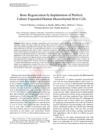
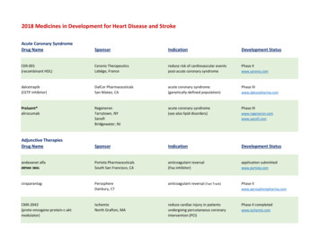
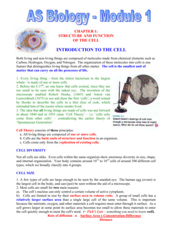
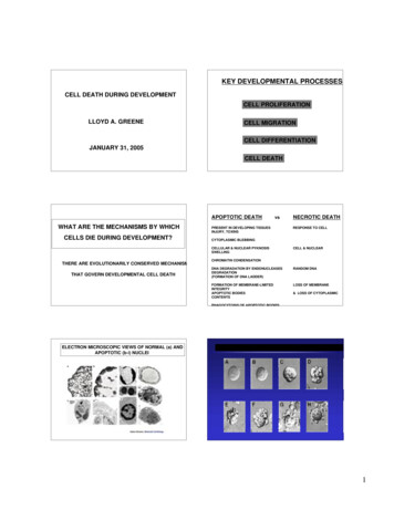
Stem Cell Reviews and Reportsincubation, the membrane was washed with TBST 3 timesfor 10 min each, and the residual secondary antibody solution was removed by washing. Finally, the PVDF membranewas developed and imaged, and the image analysis softwareImageJ was used for analysis.Western blotting assay was used to detect the expressionof COL-I, COL-III, α-SMA and WNT2b/β-catenin in fibroblasts treated with ADMSC-Exos. Fibroblasts were seededin a 6-well plate at a density of 5 106 cells/well. After thecells grew to a confluence of approximately 70%, the cellswere treated with different concentrations of exosomes(0 μg/ml, 25 μg/ml, 50 μg/ml and 100 μg/ml) and incubatedat 37 C in 5% C O2 for 36 h to detect collagen synthesis andthe expression of α-SMA. As for the western blotting assayof WNT2b/β-catenin expression, the cells were treated with2 ml of culture medium containing 50 μg/ml exosomes, PBSbuffer or serum-free DMEM medium.Then, the cells were treated with lysis buffer (RIPA protein lysis buffer: protease inhibitor 100:1), and the concentration of the protein sample was determined with the BCAkit (Elabscience, China) according to the kit instructions.After adjusting the protein concentration, a 1/4 sample volume of loading buffer was added and mixed by vortexing.Proteins were denatured in a dry bath at 95 C for 5 min.Primary antibodies against WNT2b (1:3000; # ab203225;Abcam, UK), β-catenin (1:5000; # ab32572; Abcam, UK),COL-I (1:2500; # E-AB-62684; Elabscience, China), COLIII (1:2500; # ab184993; Abcam, UK), α-SMA (1:2000; #ab5694; Abcam, UK), and GAPDH (1:2000; # E-AB-20079;Elabscience, China) were used. The corresponding goat antirabbit secondary antibody was also used. The remainingsteps were the same as those used for Western blot analysisfor the identification of exosome-specific protein markers.Signals were monitored with an Enhanced Chemiluminescence Detection System (Millipore, USA).Statistical AnalysisAll experimental data were collected and analyzed withGraphPad 7. A t-test was used for comparisons betweensingle groups, and one-way analysis of variance was usedfor comparisons among multiple ( 2) groups. The experimental data are expressed as the mean standard deviation(X SD) values. Differences were considered statisticallysignificant when P 0.05.Resultswere found to have a spindle‐like shape during cell culture.After the third passage, the morphology of ADMSCs gradually stabilized to an elongated spindle shape, similar to thatof fibroblasts, with larger nuclei (Fig. 1A). Flow cytometric analysis of ADMSCs showed positive expression of thestem cell-specific markers CD29, CD44, CD90, and CD105and negative expression of the haematopoietic marker CD45(Fig. 1E-I). Osteogenic induction of ADMSCs showedbluish-purple alkaline phosphatase staining on the 9th day(Fig. 1B). On the 21st day of induction, the cells showedlamellar growth with an indistinct structure, and red calciumnodules were observed in the extracellular matrix by Alizarinred S staining (Fig. 1C). Adipogenic induction of ADMSCsshowed that small vacuoles with good light transmissibilitybegan to appear in the cytoplasm of adipogenic cells on the4th day of adipogenic induction and that a large number ofvacuoles of different sizes with good light transmissibilityappeared on the 14th day of adipogenic induction. Oil redO staining appeared orange-red (Fig. 1D). The above resultsproved that ADMSCs were successfully isolated.Isolation and Characterization of ADMSC‐ExosThe cup-shaped morphology of ADMSC‐Exos was observedby transmission electron microscopy (TEM) (100 000 magnification) (Fig. 2A). Exosomes purified from ADMSC culture supernatants were characterized by particle size analysis, and the results showed that the exosomes were circularmembrane-bound vesicles with a diameter of 40 to 100 nm(Fig. 2C), which was consistent with the previously reportedexosome size distribution [25]. Western blotting also showedthat the exosomal markers CD9 and CD63 were expressedin ADMSC‐Exos (Fig. 2B). These results indicated thatexosomes from human ADMSCs were successfully isolated.ADMSC‑Exos Accelerate Cutaneous Wound Healingin RatsWe locally injected 100 μg of ADMSC-Exos into full-thickness skin defects in rats at different time points (1, 5, 10 days)and photographed wound healing at different time points (0,3, 7, 14 days). The wound healing rate was statistically analyzed using Image-Pro Plus 6.0, and the results showed thatthe wound healing rate in the ADMSC-Exos injection groupwas significantly faster than that in the PBS injection groupor the blank control group, indicating the positive effect ofADMSC-Exos on wound healing (Fig. 2D, E, F).Isolation and Characterization of ADMSCsADMSC‑Exos can Promote Skin FibroblastProliferation and MigrationAfter initial isolation and culture for 36 h, ADMSCs wereobserved via a microscope, and most of the adherent cellsTo study the effect of ADMSC-Exos on the proliferation andmigration of fibroblasts, we treated fibroblasts with different13
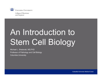
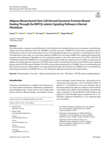
Stem Cell Reviews and ReportsFig. 1 Characterization of ADMSCs. A Results of passage 3 ADMSCs observed under light microscope. B The alkaline phosphatasestaining of ADMSCs on day 9 of osteogenesis is blue-purple, asshown by arrow. C On day 21 of osteogenic induction, the cellswere lamellar with unclear structure, and red calcium nodules wereobserved in the extracellular matrix stained with Alizarin red S, asshown by arrow. D On day 14 of adipogenesis, fat vacuoles werestained orange by oil red O, as shown by the arrow. E-I Flow cytometry showed positive expression of CD29, CD44, CD90 and CD105,and negative expression of CD45concentrations of exosomes (0 μg/ml, 25 μg/ml, 50 μg/ml,and 100 μg/ml), and the results of the CCK-8 assay showedthat as the exosome concentration increased, the proliferation ability of fibroblasts was significantly increased compared with that of blank control cells (Fig. 3C) and that thedifferences in the proliferation ability among the groupstreated with different concentrations were statistically significant (P 0.05). In addition, the scratch assay resultsshowed that the migration rate of fibroblasts increased withincreasing exosome concentration (Fig. 3A, B) and that thedifferences in the migration rate among the groups treatedwith different concentrations were statistically significant(P 0.05). ADMSC-Exos showed concentration-dependent effects on the proliferation and migration of fibroblasts;100 μg/ml ADMSC-Exos had the most significant effect.These results suggest that ADMSC-Exos can promote theproliferation and migration of fibroblasts.ADMSC‑Exos Promoted Collagen Synthesisand Decreased α‑SMA Synthesis in Fibroblasts13The mRNA and protein expression levels of COL-I, COL-IIIand α-SMA in fibroblasts treated with ADMSC-Exos wereanalyzed by Western blotting and qRT-PCR. The resultsshowed that ADMSC-Exos could promote collagen synthesis by stimulating the synthesis of COL-I and COL-III infibroblasts and that this promotive effect was enhanced asthe concentration of ADMSC-Exos increased. At the sametime, the expression of α-SMA was decreased, indicatingthat myofibroblast differentiation was inhibited by ADMSCExos (Fig. 4A, B, C). Interestingly, at the high concentrationof ADMSC-Exos (100 μg/ml), the collagen synthesis abilityof fibroblasts was decreased compared with that at the lowconcentration, but it was still higher than that at 0 μg/ml.Immunohistochemical staining of rat wounds also showed
Stem Cell Reviews and ReportsFig. 2 Exosomes were successfully isolated from ADMSCs medium;ADMSC-Exos promotes wound healing. A Exosomes were observedby transmission electron microscopy (TEM). B The expression of theexosome protein markers CD9 and CD63 was confirmed by Westernblotting. C Nanoparticle tracking analysis (NTA) showed that theADMSC-Exos distribution diameter ranged from 40 to 100 nm. D, EAs can be seen from the wound images at different time points, thewound shrinkage rate of the AMDMSC-Exos group was significantlyfaster than that of the PBS group and the blank control group. At the14th day, the remaining wound area was significantly reduced compared with the other two groups. F Traces of wound-bed closure during 14 days in vivo for each treatment category. The data were statistically analyzed using GraphPad. Results are presented as mean SD;n 3; *p 0.05, compared with PBS and blank control groupthat the expression of COL-I and COL-III was increased andthat of α-SMA was decreased in the ADMSC-Exos group(Fig. 4D, E, F).ADMSC-Exos may promote wound healing by activatingthe WNT/β-catenin signaling pathway.ADMSC‑Exos may Promote Wound Healingby Activating WNT/β‑catenin SignalingTo investigate the potential molecular mechanism ofADMSC‐Exos in wound healing, we performed two experimental protocols in vivo and in vitro. First, ADMSC-Exoswere applied to normal skin fibroblasts, and the proteinexpression levels of WNT2b and β-catenin were determinedby Western blotting. Second, we performed immunohistochemical analysis to detect the protein expression of WNT2band β-catenin in the scar tissue of rats after treatment withADMSC-Exos. We found that in both the in vivo and in vitroexperiments, the expression of WNT2b and β‐catenin wasnoticeably enhanced in the ADMSC-Exos group compared with the control group (Fig. 5A-D), suggesting thatActivation of WNT Pathway by ADMSC‑Exoswas Inhibited by XAV939We applied WNT2b/β-catenin pathway inhibitor(XAV939) to further observe the changes of ADMSCExos on wound healing. After treatment with WNTpathway inhibitor, the wound closure rate of ADMSCExos X treatment group decreased compared with thatof ADMSC-Exos treatment group, and the difference wasstatistically significant on day 7 and 14, suggesting thatblocking the WNT2b/β-catenin pathway can inhibit thewound healing promoted by ADMSC-Exos (Fig. 6A, B).Although there was no statistical difference in the healing rate between the XAV939 treatment group and theblank control group on day 3 and 14, the healing ratein the XAV939 treatment group was significantly lower13
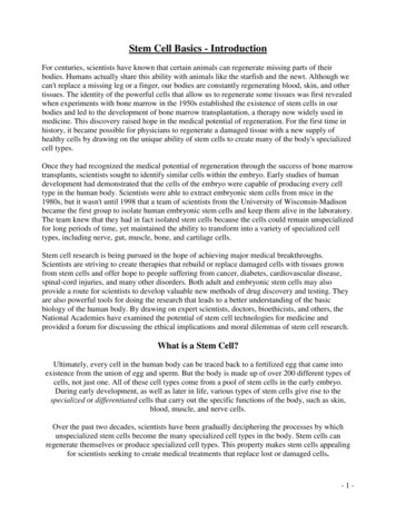
Stem Cell Reviews and ReportsFig. 3 ADMSC-Exos promote fibroblasts cell proliferation andmigration. A, B Representative images from the scratch wound assayand quantitative analysis of cell migration in each group at 12 h and24 h. C CCK-8 analysis shows the proliferation results of fibroblastsat different concentrations of ADMSC-Exos. The data were statistically analyzed using GraphPad. Results are presented as mean SD;n 3; *p 0.05, comparison between the two groups. Scale bars,200 μmthan that in the blank control group on day 7, suggesting that the activation of the WNT pathway has a positive effect on wound healing. H&E analysis showed thatthere was no obvious inflammatory cell infiltration inthe three groups on the 14th day, and the fibrous tissuebecame dense, orderly and continuous with epithelialization, especially in the ADMSC-Exos group, followed bythe ADMSC-Exos X group. Masson analysis showedthat ADMSC-Exos group had more compact collagenand the most orderly and continuous arrangement. In theXAV939 treatment group, the collagen arrangement wasdisordered, coarser and sparse and the degree of the uppercortex was minimal (Fig. 6C ).After XAV939 treatment, immunohistochemicalaverage optical density (AOD) analysis showed thatthe expression
Healing Through the WNT/β‑catenin Signaling Pathway in Dermal . lead to prolonged wound healing time, causing physical or mental pain and imposing a severe treatment burden on patients [1, 2]. . pension (1 10 3 cells/ml), they were divided into 6 parts and transferred to centrifuge tubes. Then, PE-anti-human CD29 (# FAB17781P; R&D .