Transcription
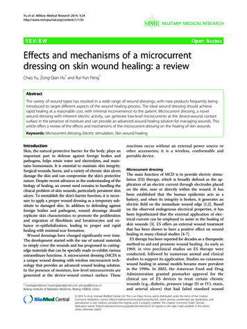
Yu et al. Military Medical Research 2014, Open AccessEffects and mechanisms of a microcurrentdressing on skin wound healing: a reviewChao Yu, Zong-Qian Hu* and Rui-Yun Peng*AbstractThe variety of wound types has resulted in a wide range of wound dressings, with new products frequently beingintroduced to target different aspects of the wound healing process. The ideal wound dressing should achieverapid healing at a reasonable cost, with minimal inconvenience to the patient. Microcurrent dressing, a novelwound dressing with inherent electric activity, can generate low-level microcurrents at the device-wound contactsurface in the presence of moisture and can provide an advanced wound healing solution for managing wounds. Thisarticle offers a review of the effects and mechanisms of the microcurrent dressing on the healing of skin wounds.Keywords: Microcurrent dressing, Electric stimulation, Skin wound healingIntroductionSkin, the natural protective barrier for the body, plays animportant part in defense against foreign bodies andpathogens, helps retain water and electrolytes, and maintains homeostasis. It is essential to maintain skin integrity.Surgical wounds, burns, and a variety of chronic skin ulcersdamage the skin and can compromise the skin’s protectivenature. Despite recent advances in the understanding of thebiology of healing, an unmet need remains in handling theclinical problem of skin wounds, particularly persistent skinulcers. To reestablish the skin’s barrier function, it is necessary to apply a proper wound dressing as a temporary substitute to damaged skin. In addition to defending againstforeign bodies and pathogens, wound dressings shouldreplicate skin characteristics to promote the proliferationand migration of fibroblasts and keratinocytes and enhance re-epithelialization, leading to proper and rapidhealing with minimal scar formation.Wound dressings have changed significantly over time.The development started with the use of natural materialsto simply cover the wounds and has progressed to cuttingedge materials that can be specially made to exhibit variousextraordinary functions. A microcurrent dressing (MCD) isa unique wound dressing with wireless microcurrent technology that provides an advanced wound healing solution.In the presence of moisture, low-level microcurrents aregenerated at the device-wound contact surface. These* Correspondence: huzongqian@gmail.com; pengry@bmi.ac.cnBeijing Institute of Radiation Medicine, Beijing 100850, Chinareactions occur without an external power source orother accessories; it is a wireless, conformable andportable device.Microcurrent dressingThe main function of MCD is to provide electric stimulation (ES) therapy, which is broadly defined as the application of an electric current through electrodes placedon the skin, near or directly within the wound. It hasbeen established that the human epidermis acts as abattery, and when its integrity is broken, it generates anelectric field on the immediate wound edge [1,2]. Basedon the observed endogenous electrical properties, it hasbeen hypothesized that the external application of electrical current can be employed to assist in the healing ofskin wounds [3]. ES offers an external wound treatmentthat has been shown to have a positive effect on woundhealing in many clinical studies [4-7].ES therapy has been reported for decades as a therapeuticmethod to aid and promote wound healing. As early as1969, in vivo preclinical studies on ES therapy wereconducted, followed by numerous animal and clinicalstudies to support its application. Studies on cutaneouswound healing in animal models became more prevalentin the 1990s. In 2002, the American Food and DrugAdministration granted premarket approval for theclinical use of ES devices to treat certain chronicwounds (e.g., diabetic, pressure (stage III or IV), stasis,and arterial ulcers) that had failed standard wound 2014 Yu et al.; licensee BioMed Central Ltd. This is an Open Access article distributed under the terms of the CreativeCommons Attribution License (http://creativecommons.org/licenses/by/4.0), which permits unrestricted use, distribution, andreproduction in any medium, provided the original work is properly credited. The Creative Commons Public DomainDedication waiver ) applies to the data made available in this article,unless otherwise stated.
Yu et al. Military Medical Research 2014, ies [8]. Recent advances in research of the electrical phenomena in the skin have aroused an interestin this modality [2,9].There are several types of ES devices have been used totreat wounds. One example is a portable electric dressingdevice that integrates low-level ES into wound dressings[10]. Generally, electric dressing devices can be divided intotwo types: the wired electric dressing and the latest wirelessMCD (Figure 1). The former delivers microcurrent stimulation by connecting to an exogenous power source [11], andthe wireless MCD does not need any accessories andjust looks like a common woven dressing. The inherentelectrical properties of the wireless MCD are activated bymoisture from the wound through a pattern of alternatelyprinted metallic dots, which employ the endogenous electric potential. The wireless MCD is commercially referredto as a bioelectric dressing (BED).Effects of microcurrent stimulation on wound healingThere have been many studies on the effects of microcurrent in both human and animal wound healing models[12-14] and on the cells involved in wound healing [15-17].The application of the MCD, which transmits a low-levelelectric current to wounds, has been shown to facilitatedifferent stages of wound repair process.Anti-inflammation effectsThe three phases of healing are inflammation, proliferation,and remodeling; they determine physiological wound healing. The normal process of healing in chronic wounds isinterrupted by a prolonged inflammation phase [18,19].Both animal and human studies suggest that ES may aid indecreasing the duration of the inflammatory phase, therebyincreasing the rate of healing, by reducing edema formation[20,21] or reducing the pathogens in the wound area anddecreasing their motility [22].Using a rat ear skin model, Kaur et al. [20] studied theeffects of microcurrent stimulation on the inflammationPage 2 of 8responses in intact skin. Tetradecanoyl phorbol acetate(TPA)-induced ear edema was performed, and the electriccurrent was provided by galvanic zinc-copper (Zn-Cu)particles producing an electric current in the range of70-90 μА (a current similar in magnitude to the endogenous wound current). By comparing the differencein ear weight and the levels of macrophage inflammatoryprotein 2 (MIP-2) between the control and Zn-Cu-treatedmice, they found that Zn-Cu significantly reduced theTPA-induced edema response, thus inhibiting excessiveinflammatory response.In a rat hind limb model, Cook and colleagues [21]found that an applied microcurrent stimulated themovement of blue-dye-labeled albumin into the lymphaticvessels, increased oncotic pressure, and drew fluid intothe vessels, thus reducing edema formation in the limb.Additionally, another study in rats that compared thetherapeutic effects of microcurrent therapy and lasertherapy in the process of wound healing reported that amicrocurrent was effective in reducing the inflammatory reaction [23].Similar to animal studies, anti-inflammation effectshave been observed for electrical stimulation applied tohuman wounds. Lee et al. [24] used ultraviolet irradiationto induce inflammatory reactions in two areas in thelumbar region of 22 subjects. A microcurrent was appliedto one region at an intensity of 50 μA. Measurements ofthe changes in chromatic red and luminance were takenover time, and a comparison of wound contraction inthe two regions indicated that the applied-microcurrentregion healed faster.The antibacterial effects of ES also assist in reducinginflammation. All Gram-negative and Gram-positivemicrobes in the wound area carry a negative charge [25].The positive polarity of the electric field attracts microbes and decreases their motility, thereby reducingthe bacteria-caused inflammation response. In a study byDaeschlein et al. [22], the application of positive polarityFigure 1 The sketch of a microcurrent dressing. (A) The dots of different color stand for dissimilar reservoirs. One type of reservoir includesoxidizing agents, and the other includes reducing agents. (B) One of the coupled dissimilar reservoirs. In the presence of moisture, redoxreactions will occur, and currents will thus be produced. Accordingly, a field of multiple currents will be produced across a surface of a substrate.
Yu et al. Military Medical Research 2014, 1:24http://www.mmrjournal.org/content/1/1/24had greater antibacterial effects than did negative polarity,regardless of whether the bacteria were Gram-negativeor Gram-positive. In general, an applied microcurrentfosters an anti-inflammatory response and enhanceswound recovery.Effects of electrical stimulation on angiogenesis andblood circulationThe wound healing process consists of complex phasesthat begin right after injury, and there is an interactionbetween several tissues and cells [26]. The most crucialsteps occur during the proliferative phase, ensuring thesuccessful closure of the wound. In this phase, the formation of new blood vessels occurs through the bifurcationand extension of existing capillaries, an indispensableprocess for successful wound healing [27,28]. ES has beendemonstrated to induce important pre-angiogenic responsesin in vitro mature endothelial cells.Using an image analyzer, Zhao et al. [29] quantifiedthe behavior of in vitro vascular endothelial cells beforeelectric field (EF) exposure and after 8, 12 and 24 hoursof EF (100 mV/mm) exposure. It showed that cells cultured without exposure to an EF had typical cobblestonemorphology, with the long axis of each cell body orientedrandomly. By contrast, endothelial cells cultured in anEF performed a dramatic reorientation, with their longaxis lying perpendicular to the vector of the appliedEF. This remarkable elongation and alignment in anapplied EF resembles the angiogenic responses of endothelial cells. Additionally, angiogenesis is governed by endothelial migration. Zhao and colleagues [17] found that a smallEF could also induce angiogenic responses in endothelialprogenitor cells by causing significant directional migrationand orientation. This was solely a response of the endothelial progenitor cells and did not require any other cell types.In addition to promoting angiogenesis, microcurrentstimulation has been proven to increase the blood flowrate and promote local blood circulation. Park et al. [30]investigated the effect of microcurrent electrical stimulation,which was provided through a shoe, on blood circulationand pain in the feet of diabetes patients. The microcurrentdelivered by the shoes was a pulsed microcurrent of nomore than 300 μА. The subjects assumed a supine positionbefore the measurement. The stable blood flow rate wasthen measured in that position, and a second measurementwas taken after 1 h of walking exercise. The results showedthat the increased blood flow rate of the experimental groupwas 1.19 mv/V, whereas that of the control group was 0.52mv/V (P 0.05). In this study, blood circulation in the feetof diabetes patients was significantly improved by applyingmicrocurrent stimulation therapy. Similarly, Clarke et al.[31] reported that the lower extremity blood flow rate increased with applied of microcurrent stimulation in patientswith chronic venous insufficiency.Page 3 of 8Effects of electrical stimulation on granulationDuring the proliferation phase of wound healing, granulation begins through increased collagen production [32].Fibroblasts, which synthesize and secrete collagen protein,are the major component of granulation tissue and playan important role in the wound healing process. Duringthe course of normal wound healing, fibroblasts at thewound edge are exposed to an electric field ranging from40 to 200 mV/mm [2]. Various electric field conditionsinfluence fibroblast migration, proliferation, and proteinsynthesis. Jennings et al. [33] explored the role of electricfields during the normal progression of healing, comparinggene expression in normal adult dermal fibroblasts exposedto a 100 mV/mm electric field for 1 h to non-stimulatedcontrols. Significantly increased expression of 162 transcriptsand decreased expression of 302 transcripts were detectedusing microarrays. Further, in a porcine model with 0.3 mmexcisional wounds, direct current (50-300 μА) led to anincrease in collagen synthesis from days 5 to 7, whichwas attributed to an augmentation of the number ofcollagen-producing cells. This increased number ofcells could be due to proliferation or chemoattractionwithin the wound [34]. Electric fields appear to play animportant role in controlling fibroblast activity in theprocess of wound healing.Fibroblasts, the major cells of the dermis, show significant migration in the electric field, which is knownas galvanotaxis. Sugimoto and colleagues [35] developedmethods to measure galvanotaxis of fibroblasts and determined the optimal conditions for electrical stimulation.The results suggested that a low-intensity direct currentpromoted migration to the negative pole of human dermalfibroblasts. Tandon et al. [36] designed a wound-healingmodel in vitro for studying the effects of microcurrentsgenerated by galvanic microparticles on cultured dermal fibroblasts. Single vertical scratches were made through thecenter of monolayers of human adult dermal fibroblasts.The imaging results suggested that the presence of galvanicmicroparticles significantly increased the speed of woundclosing compared with the control. Taken together, thegranulation phase is promoted by electrical stimulationthrough enhanced activity and migration of fibroblasts.Effects of electrical stimulation on re-epithelializationOne key aspect of wound closure is re-epithelialization,namely the healing of the epidermis. Keratinocytes arethe main cell population of the epidermis, and the migration and proliferation of keratinocytes are critical tore-epithelialization. Studies have found that keratinocytesmigrated toward the negative pole and that applyingdirect-current electric field of as low as 10 mV/mm wasenough to induce directional keratinocytes [37]. Banerjeeet al. [38] studied the influence of BEDs on humankeratinocyte cell migration. A scratch assay was performed,
Yu et al. Military Medical Research 2014, 1:24http://www.mmrjournal.org/content/1/1/24and cell migration was observed at 6 h and 9 h followingthe scratches. The results demonstrated with statisticalsignificance that the gap closed faster in the presenceof the BED than when treated with the placebo.In addition, more findings from human and animalstudies indicate accelerated wound healing rates andenhanced healing outcomes with the use of the BED[25,39,40]. In vivo porcine studies showed partial andfull-thickness wounds treated with BED epithelializedsignificantly faster (up to 3 times at Day 5) comparedto controls [40]. The interleukin-1α (IL-1α) responsewas reduced at Day 8, and the collagen markers, type-1collagen (COL-1) and COL-3, evolved to perform abetter long-term recovery in terms of remodeling andwound strength. The findings from a prospective caseseries of skin graft harvest sites demonstrated the sameresults [25]. Thirteen patients who underwent skingrafting were enrolled. Half of the skin graft donorsites were treated with the BED and a semi-occlusivedressing (SOD), and the other half used only an SOD. Thefinal results showed promise of faster healing, improvedscarring, and improved patient subjective outcome withthe use of the BED on acute wounds.Mechanisms of accelerated wound healing by ESThe exogenous application of microcurrent stimulationcan direct cell migration and proliferation, stimulateangiogenesis, reduce the inflammatory response and improve wound healing. However, clinical application ofthe therapy remains elusive due to lack of a suitable device; hence, limitations in understanding the definitemolecular mechanisms exist despite numerous completed studies. The main potential mechanisms to dateare summarized below.Increased expression of the BMP/SMAD signaling pathwayThe expression of bone morphogenetic protein 6 (BMP6)has been proven in various tissues, including bone, skinand liver [41]. In skin, BMP6 signaling has been found to(1) regulate the developing and postnatal skin; (2) controlcellular proliferation, differentiation, and tissue remodeling; and (3) govern a variety of pathological processes,including wound healing [42]. BMP6 is mainly producedby fibroblasts during wound healing, and it is found in theregenerating epidermis at the edge of the wound, as wellas in fibroblasts of the granulation tissue. It is possible thatthe BMP6 is involved in the process of skin wound healinginduced by microcurrent stimulation [36].Tandon et al. [36] analyzed the response of fibroblastgene expression of BMP6 in response to the continuousapplication of microcurrent generated by galvanic microparticles. On days 1 and 3, significant increases inexpression of the genes for BMP6, drosophila mothersagainst decapentaplegic protein 7 (SMAD7), and inhibitorPage 4 of 8of differentiation-1 (ID1) mRNA were observed. Theup-regulation of BMP6, SMAD7, and ID1 is in accordancewith the typical BMP signaling pathway (Figure 2). In theBMP/SMAD pathway, the interaction of BMP6 dimers withtheir receptors leads to the activation of the receptor kinase,followed by the phosphorylation of SMAD1/5/8 and theformation of the SMAD1/5/8-SMAD4 complex. The complex translocates to the nucleus, activating the promoters ofthe target genes [43] and then regulating SMAD7 and ID1[44,45]. A proposed representation of the BMP6 signalingpathway could be either via ID1, stimulating proliferationand cell motility after skin injury [46], or via SMAD7,which stimulates wound healing [47]. The potential mechanism to stimulate BMP6 into action may relate to the electrons produced by microcurrent stimulation. Studies haveshown that the electrons from iron in the liver may influence the BMP6 signaling pathway [43], although this effecthas not been reported in the skin. Thus, it is possiblethat microcurrent stimulation mimics these effects inskin by producing free electrons, thereby activating theBMP/SMAD signaling pathway.Suppression of NF-κB activityNuclear factor-κB (NF-κB) is a nuclear transcriptionfactor and a key molecular target because it modulatesmany types of genes governing immune and inflammatory responses [48]. In most cells, NF-κB proteins areseparated in the cytoplasm, bound by a member of theNF-κB (IκB)-inhibitor family, which include IκBα, IκBβ,and IκBε [49]. The exposure of cells to extracellular stimuli, such as tumor necrosis factors (TNF), leads to the activation of the IκB kinase (IKK) complex, which includestwo catalytic subunits, IKKα and IKKβ, and results inthe phosphorylation and ubiquitination of the IκB proteins and their proteasome mediated degradation [50].When IκB is degraded, NF-κB subunits, including P65,P50 and relA, are free to transfer to the nucleus and turnon genetic transcription of downstream targets. Expression of the pro-inflammatory cytokines, such as IL-1α,IL-2, nitric oxide (NO) and TNF-α, is transcriptionallyregulated by NF-κB [51].In an attempt to clarify the potential mechanism of antiinflammation reaction by low-level ES, Kaur and colleagues[20] treated the cultured primary keratinocytes with Zn-Cugalvanic particles, followed by treatment with TNF-α for24 h, and then examined the activity of NF-κB. The activityof TNF-α induced NF-κB was almost entirely inhibited.They followed the degradation of IκBα in the untreated andZn-Cu treated keratinocytes. TNF-α treatment caused thedegradation of IκBα and P65 phosphorylation [52], whileZn-Cu treatment inhibited this effect. And further, the result was confirmed by visualizing the nuclear localizationof the NF-κB P65 subunit. The P65 subunit translocatedto the nucleus with the treatment of TNF-α, whereas cells
Yu et al. Military Medical Research 2014, 1:24http://www.mmrjournal.org/content/1/1/24Page 5 of 8Figure 2 The BMP/SMAD signaling pathway. In the pathway, the interaction of BMP6 dimers with their receptors leads to the activation of thereceptor kinase, followed by the phosphorylation of SMAD1/5/8 and the formation of the SMAD1/5/8-SMAD4 complex, which, when translocatedto the nucleus, activates the promoters of the target genes and then regulates SMAD7 and ID1.pretreated with Zn-Cu for 2 h followed by TNF-α stoppedNF-κB from translocating. In summary, suppression ofNF-κB activity is one of the potential pathways throughwhich microcurrent exerts its anti-inflammatory effects.Activation of PI3K signaling pathwayUnder the effect of chemoattraction, cells polarize and migrate directionally, and the activation of phosphoinositide3kinases (PI3K) is evident on the leading edge; meanwhile,phosphatase and tensin homolog (PTEN), the negativelyregulating PI3K factor, collect at the opposite pole of thecells [53]. Thus PI3 kinase and PTEN are called ‘compassmolecules’ for directional sensing and polarization inchemotactic cells [54].Studies have found that the physiological electric fieldactivated the PI3K-signaling pathway in neutrophils andkeratinocytes cultured in a serum-free medium [55]. Whenthe cells that expressed green fluorescent protein-markedAkt (Akt-GFP) were exposed to an electric field, Akt-GFPdispersed and polarized in the cell migration direction; thisindicated that PI3K was activated in a polarized manner atthe cathode-facing side and that cells extended to form theleading edge [56]. Once the polarity of the electric fieldchanged, Akt-GFP oriented to the new cathode-facing side;this outcome shows that the electric field can initiate thedirectional activation of PI3K in the cells. At the side of thecell where PI3K was activated, the cell membrane stretchedout, and the cell showed migration in that direction [55,57].Moreover, using mutant mice whose PI3K was genetically knocked down, the important function of PI3K inmodulating the electric field-induced directional cellmigration was proven [55]. This characteristic abatedelectrotactic migration in wound healing in cell andtissue cultures. In cellular experiments, tissue-specificremoval of the PTEN gene in keratinocytes intensifiedAkt phosphorylation; this mutation in turn enhancedelectrotaxis. Thus, PI3K and PTEN represent a class ofmolecules modulating electrotaxis that have been confirmed at gene levels.In addition, electric fields of physiological magnitude alsoactivate other signaling pathways, such as the epidermalgrowth factor receptor (EGFR) and mitogen-activated protein kinases (MAPK) [58]; the activation of these signalingpathways is associated with a direction of cell migration.Enhancement of VEGF releaseAngiogenesis is an important event that is involved inthe healing of various types of wounds and is primarilymodulated by the release of vascular endothelial growthfactor (VEGF) from endothelial cells, platelets, keratinocytes, and fibroblasts [59]. VEGF regulates the multiple biological functions of endothelial cells, therebyenhancing the production of vasodilatory mediators,increasing vascular permeability, and stimulating theirmigration, proliferation, and formation [27]. As the basicangiogenesis regulator, the VEGF improves angiogenesisand plays a crucial role in the healing of wounds. Microcurrent stimulation has been demonstrated to promoteangiogenesis, a mechanism related to the enhancement ofVEGF release.
Yu et al. Military Medical Research 2014, mental studies on endothelial cells have shownthat ES significantly increases the levels of VEGF release.Bai and colleagues [60] cultured endothelial cells of ahuman umbilical vein in a 200 mV/mm direct-currentfield and then quantified the release of VEGF. A markedincrease of VEGF in the culture medium was observedas early as 30 min after the exposure to the electric field.The level decreased between 1 and 2 h, and rose againat 4 h, reaching the highest level by 24 h. Additionally,the expression of VEGF mRNA demonstrated significantup-regulation at 4-24 h. In rat models, Asadi et al. [61]proved that a direct current of 600 μА was more effective than a monophasic pulsed current of 2.5-3.0 mA inpromoting wound healing of a full-thickness skin incision due to higher skin VEGF levels on the 7th day.Although it is known that the application of microcurrent stimulation promotes the release of VEGF bothin vitro and in animal models, it is encouraging that thesame results are observed in clinical practice. Ferroniet al. [62] conducted a pilot study to verify the effect ofES on circulating VEGF levels in patients with peripheralarterial ulcers. Nine patients were recruited and receivedlocal pulse ES (100 μА max). Samples of peripheral venous blood were withdrawn, and the VEGF levels weredetected in samples obtained before, during, and aftertreatment. An immediate increase in the mean VEGFlevel was observed during the treatment, and a peak appeared at 7 min. Sebastian et al. [63] compared cutaneouswound healing in healthy human volunteers with or without an electric ES. A punch biopsy of full-thickness skinwas obtained and then treated with ES. VEGF was assessedand showed significant expression in the treatment group(66%) and the non-treatment group (38%), compared with24% in normal skin at the 14th day. These data indicate thepromising effect of microcurrent for enhancing the releaseof VEGF and promote wound healing.Improvement of mitochondrial functionWound healing is a complex process that includes theproliferation and differentiation of different types of cellsand thus requires a higher energy supply [64]. Mitochondria are important intra-cellular organelle in eukaryoticcells and are the main venues of oxidative phosphorylationand adenosine triphosphate (ATP). Thus, the production ofATP benefits from improved mitochondria and acceleratesskin wound healing.It is proposed that external electrical stimulation mayimprove mitochondrial function by generating moderatesuperoxide radicals in cultured human keratinocytesexposed to low intensity electric field [38]. A superoxideradical works as an electron donor for oxidative phosphorylation [65]. Thus, it is expected that improved performance of the tricarboxylic acid (TCA) cycle produces morenicotinamide adenine dinucleotide hydrogen (NADH) andPage 6 of 8flavin adenine dinucleotide hydrogen 2 (FADH2), whichthen enters the electron transport chain, contributing toincreased mitochondrial membrane potential and improvedmitochondrial function. Using fluorescent dyes JC-1and tetramethylrhodamine methyl ester (TMRM) tomeasure mitochondrial membrane potential, Banerjeeand colleagues [38] found that the treatment of keratinocytes with a bioelectric dressing for 24 h showedsignificantly high red fluorescence with both JC-1 andTMRM; this result represents a higher mitochondrialmembrane potential.Further, as a potential consequence of a hyper-activeTCA cycle, the pool of pyruvate, which is the primarysubstrate for TCA cycle, is exhausted; this results in anincreased rate of glycolysis to refill the pyruvate pool [66]and an increased rate of glucose uptake. This phenomenonwas demonstrated in HaCaT cells treated with a bioelectricdressing for 24 h compared with the control. The direct evidence of microcurrent stimulation inducingATP production was conducted in tissue cultures [67].The cultured skin tissue of male Wistar rats, just finishingtheir first hair-cycle at 21 days of age, was electricallystimulated with different levels of microcurrent for 2 h.ATP concentrations in controls and electrostimulated skinsamples were assayed by the luciferin-luciferase reaction.With electric currents ranging from 10 to 1,000 μА, thisexperiment showed that the ATP levels in the tissue wereincreased to three to five times the untreated controllevels; with currents exceeding 1,000 μА, the ATP concentrations leveled and were even lower with higher currents.Overall, microcurrent stimulation promotes mitochondrial function, induces more ATP synthesis, and ultimatelyaccelerates wound healing. Other wound healing mechanisms related to ion channels of Ca2 and Na have beenproposed but are not well known.ConclusionsBecause it is difficult to imitate wound healing in vivo,general understanding of the healing mechanisms withapplied low-level electric currents has progressed slowly.Although microcurrent stimulation for accelerating woundhealing has been studied for several decades and varioustypes of ES devices have been applied in clinical practice,many questions remain about the underlying mechanismsand the intensity and time at which stimulation should beapplied to achieve the best effect. These studies supportingthe positive effect of electrical stimulation by MCD in theacceleration of wound healing suggest that further researchis justified and necessary.Although a large number of advances in the basicresearch of wound repair have been made, the rate oftransformation from theory to practical application hasbeen slow. One reason is the lack of an accurate woundmodel and evaluation system, the other is the deficiency
Yu et al. Military Medical Research 2014, 1:24http://www.mmrjournal.org/content/1/1/24in the technological innovation of these medical devices.For example, MCD devices should be more effective,comfortable, and portable; the design of MCDs shouldconstantly be evolving; and new MCDs should be assessed.Additionally, the creation
During the proliferation phase of wound healing, granula-tion begins through increased collagen production [32]. Fibroblasts, which synthesize and secrete collagen protein, are the major component of granulation tissue and play an important role in the wound healing process. During the course of normal wound healing, fibroblasts at the