Transcription
Bioscience Reports, Vol. 21, No. 5, October 2001 ( 2002)NOBEL LECTURE8 DECEMBER, 2000The Molecular Biology of Memory Storage:A Dialog Between Genes and SynapsesEric R. Kandel1The biology of learning, and short-term and long-term memory, as revealed by Aplysia andother organisms, is reviewed.KEY WORDS: Aplysia; learning; memory; serotonin.INTRODUCTIONOne of the most remarkable aspects of an animal’s behavior is the ability to modifythat behavior by learning, an ability that reaches its highest form in human beings.For me, learning and memory have proven to be endlessly fascinating mental processes because they address one of the fundamental features of human activity: ourability to acquire new ideas from experience and to retain these ideas in memory. Infact, most of the ideas we have about the world and our civilization we have learnedso that we are who we are in good measure because of what we have learned andwhat we remember. However, not all learning experiences are positive. Manypsychological and emotional problems result at least in part from our experiences.In addition, specific disorders of learning and memory haunt both the infant andthe adult. Down syndrome, fragile X mental retardation, age-related loss of memory,and the devastation of Alzheimer’s disease are only the familiar examples of a largenumber of disorders that affect memory.Throughout my career I have been interested in the biology of learning. I havebeen curious to know: What changes in the brain when we learn? And, once something is learned, how is that information retained in the brain? I have tried to addressthese questions by developing a reductionist approach that would allow me to investigate elementary forms of learning and memory at a cell and molecular level—asspecific molecular activities within specific identified nerve cells.For a biologist like myself, interested in mental processes, the study of learninghas the further appeal that, unlike other mental processes such as thought, language1Columbia University, College of Physicians and Surgeons, 722 West 168 Street, New York, NY10032,USA. E-mail: erk5@columbia.edu The Nobel Foundation 2000.5650144-8463兾01兾1000-0565兾0 2002 Plenum Publishing Corporation
566Kandeland consciousness, learning is relatively accessible to a cellular and molecular analysis. Elementary forms of learning and memory have been well characterized byclassical psychology since the work of Ivan Pavlov and Edgar Thorndike in the firsthalf of the 20th century, and these forms of learning are the most clearly delineatedand, for the experimenter, most easily controlled of any mental process.I first became interested in the study of memory in 1950 as a result of myreadings in psychoanalysis while still an undergraduate at Harvard College. Later,during medical training, I found the psychoanalytic approach limiting because ittended to treat the brain, the organ that generates behavior, as a black box. In themid 1950s, while still in medical school, I began to appreciate that during my generation the black box of the brain would be opened and that the problems of memorystorage, once the exclusive domain of psychologists and psychoanalysts, could beinvestigated with the methods of modern biology. As a result, my interest in memoryshifted from a psychoanalytic to a biological approach. As a postdoctoral fellow atthe National Institutes of Health (NIH) in Bethesda from 1957 to 1960 I focused onlearning more about the biology of the brain, and became interested in knowinghow learning produces changes in the neural networks of the brain and how a transient short-term memory is converted to an enduring long-term memory.From the beginning, my purpose in translating questions about the psychologyof learning into the empirical language of biology was not to replace the logic ofpsychology or psychoanalysis with the logic of cell and molecular biology, but totry to join these two disciplines and to contribute to a new synthesis that wouldcombine the mentalistic psychology of memory storage with the biology of neuronalsignaling. As I thought more concretely about the neural mechanisms of memorystorage, I hoped further that the biological analysis of memory might carry with it anextra bonus, that the study of memory storage might reveal new aspects of neuronalsignaling. Indeed, this has proven true. Time and again, the molecular study ofmemory has revealed novel aspects of more general biological processes.DEVISING A RADICAL REDUCTIONIST STRATEGY TO LEARNINGAND MEMORYAt first thought, someone interested in learning and memory might be temptedto tackle the problem in its most complex and interesting form. This was theapproach that my colleague Alden Spencer and I originally had in 1958 when, atthe start of our scientific careers, we joined forces at the NIH to study the cellularproperties of the hippocampus, the part of the mammalian brain thought to be mostdirectly involved in aspects of complex memory [1]. We were initially interested in asimple question: Are the electrophysiological properties of the pyramidal cells of thehippocampus, which were thought to be the key hippocampal cells involved in memory storage, fundamentally different from other neurons in the brain, such as thewell-studied motor neurons in the spinal cord involved in simple movement? In thecourse of studying the pyramidal cells of the hippocampus, it became clear to usthat all nerve cells have similar signaling properties. Therefore, the intrinsic signalingproperties of neurons would themselves not give us key insights into memory storage[17].
Molecular Biology of Memory Storage567Thus, the unique functions of the hippocampus had to arise not so much fromthe intrinsic properties of pyramidal neurons but from the pattern of functionalinterconnections of these cells, and how those interconnections are affected by learning. To tackle that problem we needed to know how sensory information abouta learning task reaches the hippocampus, and how information processed by thehippocampus influences behavioral output. This was a formidable challenge, sincethe hippocampus has a large number of neurons and an immense number of interconnections. It seemed unlikely that we would be able to work out in any reasonableperiod of time how the neural networks, in which the hippocampus was embedded,participate in behavior and how those networks are affected by learning.Thus, to bring the power of modern biology to bear on the study of learning,it seemed necessary to take a very different approach—a radically reductionistapproach. Instead of studying the most complex cases, we needed to study thesimplest instances of memory storage, and to study them in animals that were mostexperimentally tractable. To do this we needed to find experimental systems in whicha simple behavioral act that could be modified by learning was controlled by a smallnumber of large and accessible nerve cells. Only in this way could we correlatechanges in the overt behavior of the animals with molecular events in identifiableneurons and examine how sensory processing in the brain is modified by learning togive rise to memories.Such a reductionist approach was hardly new in 20th century biology. One needonly think of the use of Drosophila in genetics, of bacteria and bacteriophages inmolecular biology, and of the squid giant axon in the study of the conduction ofnerve impulses. Nevertheless, when it came to the study of behavior, many investigators were reluctant to use a reductionist strategy. In the 1950s and 1960s manybiologists and psychologists believed that behavior was the one area of biology inwhich the use of simple animal models, particularly invertebrate ones, was leastlikely to succeed. They argued that only higher animals exhibit interesting forms oflearning and that these forms require neuronal organizations and neuronal mechanisms qualitatively different from those found in simple animals. As a result, anapproach to learning based on simple invertebrates was bound to fail because itwould lack relevance to mammalian and particularly to human behavior.It was my belief at the outset, however, that concerns about the use of a simpleexperimental system to study learning were misplaced. The question was and is notwhether there is something special about the human brain; there clearly is. Rather,the question is whether the human brain and human behavior have anything at allin common with the nervous system and behavior of simpler animals. If so, thesefundamental, common principles of neuronal organization might well be studiedmore profitably in simple animals.The answer to this second question, about commonality, was clear. By 1960,work by students of comparative behavior such as Konrad Lorenz, Niko Tinbergen,and Karl von Frisch had shown that humans share many behavioral patterns andeven simple forms of learning with simple animals [reviewed in 2]. That the evolutionof behavior and learning is conservative should not be surprising, since the evolutionof other biological functions is also conservative. There are, for example, no fundamental functional or biochemical differences between the nerve cells and synapses
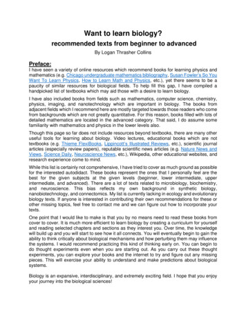
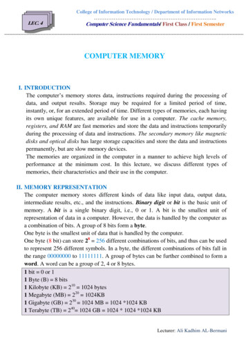
568Kandelof humans and those of a snail, a worm or a fly. Since behavior and learning is anexpression of nerve cell activity, it would be surprising if the learning capability ofpeople did not have some elementary features in common with the learning of snails,worms, or flies. And, if elementary forms of learning are common to all animalswith an evolved nervous system, there must be conserved features in the mechanismsof learning at the cell and molecular level, that can be studied effectively even insimple invertebrate animals.A SIMPLE INVERTEBRATE SYSTEM THAT LENDS ITSELF TO AREDUCTIONIST APPROACHAfter an extensive search for a suitable experimental animal, I settled on thegiant marine snail Aplysia (Fig. 1), because it offers three important experimentaladvantages: its nervous system is made up of a small number of nerve cells; manyof these are very large; and (as became evident to me later) many are uniquelyidentifiable [3,13]. Whereas the mammalian brain has a trillion central nerve cells,Aplysia has only 20,000. These cells are clustered in ten anatomical units calledFig. 1. The giant marine snail Aplysia californicabelongs to the opisthobranch subclass of the gastropod molluscs. Animals of this species may growto be 30 cm in length and weigh 1 kg.
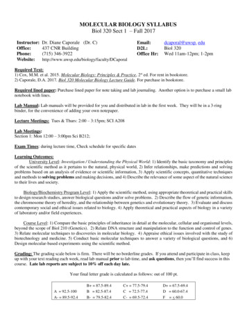
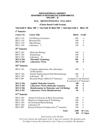
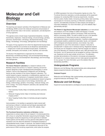
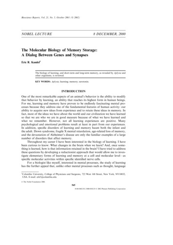
Molecular Biology of Memory Storage569Fig. 2. The human brain has a million-million neurons while the brainof Aplysia has 20,000 nerve cells. Aplysia nerve cells are clusteredtogether in five major bilateral ganglia, each containing about 2000nerve cells.ganglia, each of which contains about 2000 cells (Fig. 2). An individual ganglion,such as the abdominal ganglion, mediates not one but a family of behaviors. Thus,the simplest behaviors that can be modified by learning may involve less than 100cells. This numerical simplification made it possible to identify the specific contribution of individual neurons to the behavior in which they participate [13]. Inaddition to being few in number, these cells are the largest nerve cells in the animalkingdom, reaching up to 1000 µm in diameter, large enough to be seen with thenaked eye (Fig. 3). Because of their extraordinary size and their distinctive pigmentation, it is possible to recognize many of the cells as unique individuals. One canFig. 3. A photomicrograph of an abdominal ganglion of Aplysia shows the distinctive pigmentation and positions of its cells. The largest cells are a millimeterin diameter and can be seen with the naked eye. Scale barG1 mm.
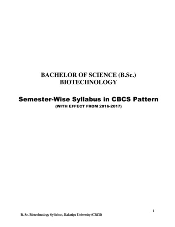
570Kandelrecord from these large cells for many hours without any difficulty, and the samecell can be returned to and recorded from over a period of days. The cells can easilybe dissected out for biochemical studies, so that from a single cell one can obtainsufficient mRNA to make a cDNA library. Finally, these identified cells can readilybe injected with labeled compounds, antibodies, or genetic construct procedureswhich opened up the molecular study of signal transduction within individual nervecells.REQUIREMENT FOR A CELL-BIOLOGICAL STUDY OF MEMORYSTORAGEGiven a technically advantageous experimental system, how does a cell biologistbegin to address the problem of learning? The strategy that my colleagues and Ideveloped involved four sequential steps: (1) We first wanted to define a simplebehavior that can be modified by learning and that gives rise to memory storage.(2) We next wanted to identify the cells that make up the neural circuit of thatbehavior. (3) Within that neural circuit we then wanted to locate the critical neuronsand interconnections that had been modified by learning and that store memory.(4) Finally, we wanted to analyze the changes that occur at those sites in responseto learning and memory storage, first on the cellular and then on the molecular level[13]. I follow this outline in the discussion below.DELINEATING A BEHAVIOR IN APLYSIA THAT IS CAPABLE OFBEING MODIFIED BY LEARNINGIrving Kupfermann and I first wanted to study the simplest possible behaviorof Aplysia [4]. We examined the animal’s behavioral capabilities and delineated avery simple defensive reflex: the withdrawal of the gill upon stimulation of thesiphon, an action that is like the quick withdrawal of a hand from a hot object. InAplysia, the gill is a respiratory organ that lies exposed in the mantle cavity. Whenthe animal is in a normal, relaxed state, the gill is partially covered by a sheet ofskin (the mantle shelf), which ends in a fleshy spout, the siphon (Fig. 1). When aweak tactile stimulus is applied to the siphon, both the siphon and gill are withdrawninto the mantle cavity and for protection under the mantle shelf [(Fig. 4); ref. 5].Kupfermann, Harold Pinsker, and later Tom Carew, Robert Hawkins and Ifound that this simple reflex could be modified by three different forms of learning:habituation, sensitization, and classical conditioning [5, 8, 9]. As we examined thesethree forms of learning, we were struck by the resemblance each had to corresponding forms of memory storage in higher vertebrates and humans. As with vertebratelearning, memory storage for each type of learning in Aplysia has two phases: atransient memory that lasts minutes and an enduring memory that lasts days. Conversion of short-term to long-term memory storage requires spaced repetition—practice makes perfect even in snails [8, 9, 10].We focused initially on one type of learning, sensitization, a form of learnedfear in which a person or an experimental animal learns to respond strongly to anotherwise neutral stimulus [5, 8, 10]. For example, if a person is suddenly exposed to
Molecular Biology of Memory Storage571Fig. 4. A dorsal view of Aplysia showing the gill, the animal’s respiratory organ.A light touch to the siphon cavity with a fine probe causes the siphon to contractand the gill to withdraw under the protection of the mantle shelf. Here, themantle shelf is shown to be retracted for a better view of the gill. Sensitizationof the gill-withdrawal reflex, produced by applying a noxious stimulus to anotherpart of the body, such as the tail, leads to an enhancement of the withdrawalreflex of both the siphon and the gill.an aversive stimulus, such as a gunshot going off nearby, that person will be sensitized by the unexpected noise. As a result, that person will be frightened and willnow startle to an otherwise innocuous stimulus like a tap on the shoulder. Similarly,on receiving an aversive shock to another part of the body such as the tail (or head),an Aplysia recognizes the stimulus as aversive and learns to enhance its defensivereflex responses to a variety of subsequent stimuli applied to the siphon, even innocuous stimuli [Fig. 4; 12]. The animal now remembers the shock, and the duration ofthis memory is a function of the number of repetitions of the noxious experience. Asingle shock gives rise to a memory lasting only minutes; this short-term memorydoes not require the synthesis of new protein. In contrast, four or five spaced shocksto the tail give rise to a memory lasting several days; this long-term memory doesrequire the synthesis of new protein. Further training gives rise to an even moreenduring memory lasting weeks, which also requires new protein synthesis [Fig.5; 10, 12].Thus, just as in the complex learning in mammals [107, 108], long-term sensitization differs from the short-term process in requiring the synthesis of new proteins.This was our first clear evidence for the conservation of biochemical mechanismsbetween Aplysia and vertebrates, and it reinforced the hope that a detailed analysisof short-term memory and its transition to long-term memory in Aplysia wouldreveal molecular mechanisms of general importance.DEFINING THE NEURAL CIRCUIT IN CELLULAR DETAILTo analyze the cellular mechanisms of sensitization, we needed to identify theneural circuit of the gill withdrawal reflex. Kupfermann and I quickly localized the
572KandelFig. 5. Spaced repetition converts short-term memory intolong-term memory in Aplysia. In the resting state, beforesensitization training, the animal withdraws its siphon andgill only briefly in response to mild touch of the siphon.After the animal receives a single noxious shock to the tail,it withdraws its siphon and gill longer in response to thesame mild touch of the siphon. Sensitization following asingle noxious stimulus lasts about one hour. After four orfive single tail shocks the animal withdraws its gill andsiphon more powerfully and the sensitization lasts morethan a day. If the animal receives four brief trains of singleshocks a day over the course of four days, it withdraws itssiphon and gill for almost eight times as long and retainsthe memory for several weeks. [Modified from 98.]central neuronal machinery for the reflex behavior in the animal’s abdominal ganglion [4, 6]. Because we soon realized that many cells could be identified in everyanimal of the species [3, 4, 6, 7], we were able to give them specific names and, mostimportant, return to the same cell time and again—in both untrained and trainedanimals. In this way Kupfermann, Castellucci, Carew, Hawkins, John Byrne, and Iwere able to work out significant components of the neural circuit gill-withdrawalreflex in terms of individual cells and cell clusters. The circuit has 24 mechanoreceptor sensory neurons that innervate the siphon skin and make direct monosynapticconnections with six gill motor cells [7, 11, 36]. The sensory neurons also madeindirect connections with the motor cells through small groups of excitatory andinhibitory interneurons [18, 19]. In addition to being identifiable, individual cells also
Molecular Biology of Memory Storage573proved to have surprisingly large effects on behavior [Fig. 6C; reviewed in 13, 22, 36].As we examined the neural circuit of this reflex in detail, we were struck by itsinvariance—the cells that make up the circuit and their interconnections are alwaysthe same. In every animal we examined, each cell connected only to certain targetcells and not to others (Fig. 7). Carew, John Koester, Wayne Hening, and I alsofound this invariance in the neural circuitry of other behaviors in Aplysia—inking,control of the circulation, locomotion [reviewed in 13, 15]—raising a key questionin the cell-biological study of learning: How can learning occur in a neural circuitthat is precisely wired?HOW DOES LEARNING AFFECT THE INVARIANT ELEMENTS OF THENEURAL CIRCUIT?In his Croonian Lecture to the Royal Society of 1894, Santiago Ramón y Cajalproposed a theory of memory storage: memory is stored in the growth of new connections [16]. This prescient idea was neglected in good part for half a century asstudents of learning fought over newer competing ideas. First, Karl Lashley, RossAdey, Wolfgang Köhler, and a number of Gestalt psychologists proposed that learning leads to changes in electric fields or chemical gradients, which they postulatedsurround neuronal populations and are produced by the aggregate activity of cellsrecruited by the learning process. Second, Alexander Forbes and Lorente de Nóproposed that memory is stored dynamically by a self-reexciting chain of neurons.This idea was later championed by Donald Hebb as a mechanism for short-termmemory. Finally, Holger Hyden proposed that learning led to changes in the basecomposition of DNA or RNA. Even though there was much discussion about themerits of each of these ideas, there was no direct evidence to support any of them[reviewed in 17].We were now in a position to address these alternative ideas by confrontingdirectly the question of how learning can occur in a circuit with fixed neuronalelements. Kupfermann, Castellucci, Carew, Hawkins, and I examined the neuralcircuit of the gill-withdrawal reflex while the animal underwent sensitization orhabituation, a form of learning in which the animal learns to ignore an innocuousstimulus to siphon when given with monotonous repetition. (We later also extendedthese studies to an examination of classical conditioning [20].) Our studies providedclear evidence for Cajal’s idea: learning results from changes in the strength ofthe synaptic connections between precisely interconnected cells [6, 7]. Thus, whilethe organism’s developmental program assures that the connections between cellsare invariant, it does not specify their precise strength. Rather, experience alters thestrength and effectiveness of these pre-existing chemical connections. Seen in theperspective of these three forms of learning, synaptic plasticity emerged as a fundamental mechanism for information storage by the nervous system, a mechanism thatis built into the very molecular architecture of chemical synapses [95].We soon appreciated that the synaptic strength of a chemical synapse couldbe modified in two ways: homosynaptically and heterosynaptically. Homosynapticchanges in synaptic strength occur in a synapse because of activity in the presynapticand postsynaptic neurons of that very synapse. During habituation, homosynaptic
574KandelFig. 6. The neural circuit of the gill-withdrawal reflex can be delineated in terms ofspecific identified cells. A. This dorsal view of the abdominal ganglion shows someidentified cells. The six identified motor cells to the gill are shaded brown; sevensensory neurons are shaded blue. In the figure a sensory neuron that synapses ongill motor neuron L7 is being stimulated electrically. A microelectrode in the motorneuron records the synaptic potential produced by the action potential in the sensoryneuron. B. The physiological demonstration of the direct connections between thesensory neuron and motor neuron. The sensory neuron receives input from thesiphon skin; the motor neuron makes direct connections onto the gill. The fact thatthe cells are large and identifiable allows for the mapping of connections betweenspecific identified cells. This part also shows the experimental arrangement for simultaneously recording in a pre- and postsynaptic cell. The sensory neuron makes adirect connection onto the motor neuron, as is evident in part C. C. Individualcells make significant contributions to the reflex. Stimulating a single motor neuronproduces a detectable change in the gill, and stimulating a single sensory neuronproduces a large synaptic potential in the motor neuron. Repeated stimulation of asingle sensory neuron increases the frequency of firing in the motor neuron, leadingto a visible reflex contraction of the gill. A single tactile stimulus to the skin normallyactivates 6–8 sensory neurons, causing each to fire 1–2 action potentials. The repetitive firing of 10 action potentials in a single sensory neuron, designed to model thefiring of the total population, in fact simulates reasonably well the reflex behavior.
Molecular Biology of Memory Storage575Fig. 7. The neural circuit of the gill-withdrawal reflex. The siphon isinnervated by 24 sensory neurons that connect directly with the sixmotor neurons. The sensory neurons also connect to populations ofexcitatory and inhibitory interneurons that in turn connect with themotor neurons. Stimulating the tail activates modulatory interneuronsthat act on the terminals of the sensory neurons as well as on thoseof the excitatory interneurons. There are three classes of modulatoryneurons activated by tail stimuli: (1) neurons that release serotonin (5HT), (2) neurons that release a peptide called the small cardioactivepeptides (SCP), and (3) the L29 cells, which release an unidentifiedmodulatory neurotransmitter. The serotonergic modulatory action isthe most important. Blocking the action of these serotonergic cellsblocks the effects of sensitizing stimuli.changes occur in the monosynaptic connections between the sensory neurons andthe motor neurons of the gill-withdrawal reflex. Heterosynaptic changes occur in asynapse where presynaptic and postsynaptic neurons are themselves not active butthere is, instead, activity in one or more modulatory interneurons that act on thepresynaptic neurons, on the postsynaptic neurons of the synapse, or on both, tomodify the strength of their synaptic connections. During sensitization, heterosynaptic changes are induced in the monosynaptic connections between the sensoryneurons and motor neurons of the gill-withdrawal reflex. Hawkins, Abrams, and Ilater found that these two types of regulation are recruited together in classicalconditioning [20, 21]. Classical conditioning therefore illustrated that the elementaryforms of homo- and heterosynaptic plasticity form an alphabet of basic mechanismsthat can produce combinations of plasticity with novel properties.
576KandelIn all three forms of learning we found particularly large changes in the synapticstrength of the direct connections between the sensory and motor neurons of thereflex. We therefore focused on this one component of the reflex and found severaladditional principles that have proven to be quite general. First, we found that thesame synaptic connection can be modulated in opposite ways by different forms oflearning. For example, habituation leads to a homosynaptic weakening of synapticconnections between the sensory neurons and their target cells, the motor neuronsand interneurons, while sensitization leads to heterosynaptic strengthening of thesesame sets of connections [7, 13, 33]. Second, learning not only leads to changes insynaptic strength, it can also affect the excitability of neurons. In the case of sensitization the excitability of the sensory (presynaptic) neurons is increased [25]. Third,the synaptic changes persist, thereby contributing to memory storage [10, 33, 94].Indeed, the same synaptic connection can store both short- and long-term memory[7, 23, 33, 43, 94, 98]. At a given synapse synaptic plasticity can either be short- orlong-lived depending on the number of spaced repetitions of the learning stimulus,and these parallel not only the behavioral changes of short-term memory [7, 43, 93].Finally, long-term memory storage involves not only a change in synaptic strength,but also anatomical changes, changes in the number of synaptic connections[57, 58, 59].The changes at the synapse between the sensory and the motor neuron are onlya part of the changes in the neural circuit of the gill-withdrawal reflex. Importantchanges occur elsewhere in the circuit, but we have studied them less. We havefocused on the monosynaptic portion of the circuit in order to probe in depth themolecular mechanisms that contribute to learning and memory.INITIAL STEPS TOWARDS A MOLECULAR CHARACTERIZATIONOF SENSITIZATIONWhat are the molecular mechanisms whereby short-term memory is established,and how it is converted to long-term memory? Initially, we focused on short-termsensitization (Fig. 8). In collaboration with James H. Schwartz, we found that thesynaptic changes, like the short-term behavior, were expressed even when proteinsynthesis was inhibited. Since the short-term changes persisted for many minutes, itseemed unlikely that they involved a simple conformational change of one or moreproteins. We therefore proposed, in 1971, that short-term memory might require aseries of sequential reactions similar to that mediated by cAMP-mediated signaling[26]. Our attention was drawn to cAMP because Sutherland, Rall and Krebs hadfound that various neurotransmitters could increase cAMP concentration in thebrain and in other tissues, and in the liver cAMP activated the cAMP-dependentprotein kinase [26]. In 1972, Schwartz, Howard Cedar, and I found that stimulationof the modulatory pathways recruited during heterosynaptic facilitation led to anincrease in cAMP in the abdominal ganglion [27]. Cedar and Schwartz next applieda number of neurotransmitter candidates and found that serotonin and dopaminecould increase levels of cAMP [29]. This made us wonder whether the population ofmodulatory interneurons, which produces the heterosynaptic facilitation that givesrise to sensitization, contains serotonergic cells.
Molecular Biology of Memory Storage577Fig. 8. The strength of the synaptic connectionsbetween the sensory and the motor neurons isincreased by sensitization, by serotonin, and bydirect activation in the presynaptic neurons of cAMPand PKA. A. The increase in synaptic strength produced by sensitizing stimuli can be achieved by electrical stimuli to the tail, to the head, or to the neuralpathways from the tail or head. B. Facilitation couldbe reproduced by stimulating individual modulatorycells such as the serotonergic cells or by the application of the serotonin to the connections betweenthe sensory neurons and motor neurons. C. Facilitation can also be produced by injecting cyclic AMPinto the sensory neurons. D. Facilitation can also beproduced by simply injecting the catalytic subu
molecular biology, and of the squid giant axon in the study of the conduction of nerve impulses. Nevertheless, when it came to the study of behavior, many investi-gators were reluctant to use a reductionist strategy. In the 1950s and 1960s many biologists and psychologists believed that behavior was the one area of biology in