Transcription
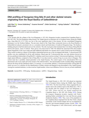
Archaeological and Anthropological Sciences (2019) 609-7ORIGINAL PAPERDNA profiling of Hungarian King Béla III and other skeletal remainsoriginating from the Royal Basilica of SzékesfehérvárJudit Olasz 1 & Verena Seidenberg 2 & Susanne Hummel 2 & Zoltán Szentirmay 1 & György Szabados 3 & Béla Melegh 4 &Miklós Kásler 1Received: 1 December 2017 / Accepted: 23 January 2018 / Published online: 10 February 2018# The Author(s) 2018. This article is an open access publicationAbstractA few decades after the collapse of the Avar Khaganate (c. 822 AD), Hungarian invaders conquered the Carpathian Basin (c.862–895 AD). The first Hungarian ruling dynasty, the Árpáds played an important role in European history during the MiddleAges. King Béla III (1172–1196) was one of the most significant rulers of the dynasty. He also consolidated Hungariandominance over the Northern Balkans. The provostry church of the Virgin Mary (commonly known as the Royal Basilica ofSzékesfehérvár) played a prominent role as a coronation church and burial place of medieval Hungarian kings. The basilica’sbuilding and graves had been destroyed over the centuries. The only royal graves that remained intact were those of King Béla IIIand his first spouse, Anna of Antioch. These graves were discovered in 1848. We defined the autosomal STR (short tandemrepeat) fingerprints of the royal couple and eight additional individuals (two females and six males) found in the Royal Basilica.These results revealed no evidence of first-degree relationship between any of the investigated individuals. Y-chromosomal STRprofiles were also established for all the male skeletons. Based upon the Y-chromosomal data, one male skeleton showed anobvious patrilineal relationship to King Béla III. A database search uncovered an existing Y-chromosomal haplotype, which had asingle-repeat difference compared to that of King Béla. It was discovered in a person living in an area close to Hungary. Thiscurrent male line is probably related paternally to the Árpád Dynasty. The control region of the mitochondrial DNA wasdetermined in the royal couple and in the remains of the inferred relative. The mitochondrial results excluded sibling relationshipbetween the King and the patrilineal relative. In summary, we successfully defined a Y-chromosomal profile of King Béla III,which can serve as a reference for the identification of further remains and disputed living descendants of the Árpád Dynasty.Among the examined skeletons, we discovered an Árpád member, whose exact affiliation, however, has not yet been established.Keywords Ancient DNA . STR typing . Kinship analysis . mtDNA . Hungarian kings . ÁrpádsIntroductionElectronic supplementary material The online version of this article(https://doi.org/10.1007/s12520-018-0609-7) contains supplementarymaterial, which is available to authorized users.* Judit Olaszolasz@oncol.hu1National Institute of Oncology, Ráth Gy. u. 7-9, Budapest 1122,Hungary2Historical Anthropology and Human Ecology,Johann-Friederich-Blumenbach Institute of Zoology andAnthropology, University of Goettingen, Buergerstr. 50,37073 Goettingen, Germany3King St. Stephen Museum, Fő u. 6, Székesfehérvár 8000, Hungary4Department of Medical Genetics, University of Pécs, Szigeti u. 12,Pécs 7624, HungaryThe Árpád Dynasty (c. 850–1301 AD) played an importantrole in European history during the Middle Ages (Hóman1940-1943). The first Great Prince Álmos organised the monarchic state in the northern region of the Black Sea c. 850. Afew decades after the collapse of the Avar Khaganate (c.822 AD), Álmos and his son Árpád conquered theCarpathian Basin (c. 862–895 AD) (Szőke 2014). Duringthe conquest, Hungarian invaders, together with Turkicspeaking Kabars assimilated the Avars and Slavonic groups(Szádeczky-Kardoss 1990). Thus, most of the population inthe Carpathian Basin originated from the Hun-Turkic culturalcommunity of the Eurasian Steppe and was accompanied bySlavonic and German-speaking groups (László 1996). Theorigin of Hungarians is still controversial, and this paper
1346cannot cover this complex subject. The Hungarian GreatPrincipality represented the Eurasian steppe empires inCentral Europe from c. 862 until 1000. Saint Stephen I, thelast Great Prince (997–1000) and first King (1000–1038) ofHungary re-organised this early Hungarian state as a Christiankingdom. Saint Stephen received the royal crown from thePope and joined the post-Roman Christian political systemand cultural commonwealth of Latin Europe (Pohl 2003;Szabados 2011). Hungary remained an independent state between the German and Byzantine empires (Makk 1989). KingBéla III (1172–1196) was one of the most significant rulers ofthe dynasty. He was the second son of King Géza II (1141–1162) and Queen Euphrosyne, the daughter of Mstislav I(1125–1132), the Great Prince of Kiev. Through the mediationof Byzantine Emperor Manuel I Komnenos, Béla marriedAnna of Châtillon from Antioch (1150–1184), the half-sisterof the Emperor’s wife in 1170. After Manuel’s death, KingBéla consolidated Hungarian dominance over the NorthernBalkans.The provostry church of the Virgin Mary (commonlyknown as the Royal Basilica of Székesfehérvár) was built bySaint Stephen I at the beginning of the eleventh century. Thebasilica played a prominent role as a church of coronation andas the main burial place of Hungarian kings in the MiddleAges. Fifteen kings, several queens, princes and princessesand clerical and secular dignitaries were buried there over fivecenturies (Engel 1987).According to sources, ten Árpáds—eight kings and twoprinces—were laid to rest in the basilica (Table 1)(Szentpétery 1937-1938). Other princes who died at an earlyage may also be entombed there.The Turks occupied the city of Székesfehérvár in1543. The Turks, and later the Christian mercenaries,plundered and destroyed the graves of the basilica. Thebuilding exploded and burned down in 1601. Followingthe liberation from Turkish rule in 1688, the remainingstones of the basilica were carried away for reconstruction of the town. Hardly any traces of the basilicaremained by the beginning of the nineteenth century.The only royal graves that remained intact were thoseof King Béla III and his first spouse, Anna of Antioch(1150–1184). These graves were lined with red marbleslabs and were discovered during sewer construction in1848. The royal couple were reliably identified based ontheir regalia and anthropological features (Érdy 1853).The following facts support the identity of the royal remains: (1) The anthropologically estimated ages of theskeletons were similar to the historically recorded ages(Érdy 1853; Éry 2008). (2) The archaeological studiesdated the grave goods (the crowns, rings, sword andcross) to the twelfth century (Kovács 1969). (3) The sarcophagi had no engraved inscriptions. These were madeonly from the thirteenth century onward. (4) The statureArchaeol Anthropol Sci (2019) 11:1345–1357of the male skeleton was remarkably tall. When theThird Crusade crossed Hungary in 1189, RichardCanonicus of London met King Béla III and noted hisexceptional tallness. (5) The Nordic features of the maleskeleton probably came from the King’s Rurik ancestors.Béla’s mother, Euphrosyne, and his paternal great-grandmother, Predslava, were both Rurikids with Viking origins. (6) The facial reconstruction of the man’s skulllooks a lot like the herm of King Ladislas I. SinceLadislas I was canonised during the reign of King BélaIII, the latter is supposed to have been the model for theherm (László 1965). (7) The female’s skeleton bears thesigns of several pregnancies. This observation is in linewith the fact that Queen Anna of Antioch gave birth toseven children. All these aspects apply only to the royalcouple of Béla and Anna (Szabados 2016).Following the discovery in 1848, further archaeologicalexcavations were conducted intermittently for more than150 years (1848, 1862, 1874, 1882, 1936–1937, 1965–2002) (Éry 2008), but no other medieval kings could be identified with certainty.The molecular genetic methods available today enablegenetic examination of ancient bones. For example, theyhave enabled identification of the remains of KingRichard III (King et al. 2014). Such studies, however,have not yet been conducted on the human remains ofthe basilica. We investigated skeletal remains excavatedby János Érdy (1848) (Érdy 1853) and Imre Henszlmann(1862 and 1874) and later reinterred in the MatthiasChurch of Buda (1862 and 1900) (Table 2, online resource 1). The remains were re-examined first by AurélTörök in 1863 and 1893 (Török 1893, 1900) and last byKinga Éry et al. in 1984 (Éry 2008). Éry et al. doubtedthe authenticity of a male skeleton believed to be foundnear Béla III (online resource 1) because of the followingobservations: First, Török and Éry estimated the person’sage to be approximately 20–22 years (Török 1900) and20–26 years,1 respectively, at the time of death. Theseestimates were in contrast to the initial estimation ofmore than 30 years of age (Érdy 1853). Second, theydiscerned that the missing bones of this skeleton weredifferent from those marked on an original drawing madein 1848 (e.g., the skull disappeared). Third, they alsonoted that the colour of this skeleton was yellowish,even though it was muddied at the time of discovery(Érdy 1853; Éry 2008). Due to these observations, theyexcluded this skeleton from further study. This skeletonis noted as II/52 in our study.1Data obtained from the documentation of human remains of theSzékesfehérvár Royal Basilica, Anthropological Repository of Saint StephenMuseum, Székesfehérvár.
Archaeol Anthropol Sci (2019) 11:1345–1357Table 11347The Árpád Dynasty members who were buried in SzékesfehérvárNameBirthDeathReignGenealogical AffiliationPrince Saint Emeric1000/071031–King Saint Stephen’s sonKing Saint Stephen IKing Coloman the LearnedPrince Álmosc. 980c. 1070c. 1071103811161127997–10381095–1116–5th descendant of Great Prince ÁlmosGreat-grandson of Prince Vazul, King Saint Stephen’s cousinKing Coloman’s younger brotherKing Béla II the BlindKing Géza IIc. 11081130114111621131–11411141–1162Son of Prince ÁlmosSon of King Béla IIKing Ladislas IIc. 113111631162–1163Younger brother of King Géza IIKing Stephen IVKing Béla III the Greatc. 1133c. 11481165119611631172–1196Younger brother of King Ladislas IISon of King Géza IIKing Ladislas IIIc. 120012051204–1205Grandson of King Béla IIIThe establishment of a dignified resting place and creationof a shrine for the House of Árpád kings were parts of theobjectives set forth by the House of Árpád Programme(1832/2013). The National Institute of Oncology undertookgenetic profiling of the human remains from the RoyalBasilica to identify skeletal remains possibly belonging tothe Royal Dynasty. The objectives of our study were to definethe Y-chromosomal STR (short tandem repeat) profile of KingBéla III and to determine the autosomal STR-based DNAfingerprints of the royal couple. Furthermore, our aim was toperform genetic profiling on some of the other skeletons fromthe Basilica to identify royal relatives. We intended to establish a molecular base for the identification of possible futurefinds of the Árpád Dynasty and for reconstruction of theirgenealogy. Therefore, our research team opened the metalcaskets of Matthias Church in March 2014.Table 2Materials and methodsSamplesThe opening of the sarcophagi, compilation of the skeletonsand sampling were carried out with the participation ofarchaeologists and anthropologists. The procedures wererecorded in a written report with photodocumentation.The sealed metal caskets and, in the case of the royal couple, additional inner glass boxes, were opened by a restorer.The innermost wooden cases, which contained the skeletalremains, were placed in new, clean plastic boxes for transportation. The person carrying out this procedure wore ascrub suit, disposable gloves and a facial mask. The skeletons were compiled in a sterile operating room of theNational Institute of Oncology. The operating table wasThe examined skeletal remainsSkeletonSexEstimated agePosition in the Royal BasilicaGraveArchaeological datingBéla III (I)Anna of Antioch (II)II/52( III?)MaleFemaleMale45-49a37-41a 30b20-22c20-26dSouth naveeSouth naveeSouth naveeStoneStoneStone12th centuryh12th centuryhNot availableGroundStoneStoneGroundGroundGroundNot available12th centuryi12th centuryiNot availableNot availableNot availableGroundNot availableFoetus 4037-41a21-27a32-38a37-42aSouth naveeNorth navefNorth navefInner church, unknowngInner church, unknowngInner church, unknowngII/109Female35-41aInner church, unknowngaaÉry 2008; b Érdy 1853; c Török 1900; d Documentation of human remains of the Székesfehérvár Royal Basilica, Anthropological Repository of SaintStephen Museum, Székesfehérvár; e From excavation by Érdy (1848); f From excavation by Henszlmann (1874); g From excavations by Henszlmann(1862 and 1873); h Kovács (1969); i Henszlmann (1876); Kralovánszky (1989)
1348prepared with a disposable surgical bed sheet for each skeleton. The sampling was carried out with sterile, surgical tools. The staff wore scrub suits, disposable glovesand facial masks. The bone samples were put in sterile,DNase- and RNase-free 50-ml centrifuge tubes. Theparticipating persons were STR genotyped. Of note isthat a number of people had come into direct contactwith the skeletons since their discovery in the nineteenth century. During the excavations, transport andinvestigations, the bones had become contaminated.The remains, especially those of the royal couple, hadbeen investigated several times (1848, 1883, 1967 and1984) without precautions against DNA contamination.The details of the examined skeletons are summarisedin Table 2. The bone samples used for DNA extractionare indicated in online resource 2. The skull and poorly preserved skeletal bones of the King and Queenwere consolidated using polyvinyl acetal (Alvar,Shawinigan Chemicals) in 1967. This consolidantproved to be a PCR (polymerase chain reaction) inhibitor in the first DNA extracts of the Göttingen laboratory (see the BDNA extraction section and online resource 3).Laboratory conditionsY-chromosomal STR and autosomal STR analyses of the samples have been performed in parallel and independently in theDepartment of Pathogenetics of the National Institute ofOncology (Budapest, Hungary) and in the Department ofHistorical Anthropology and Human Ecology of the JohannFriedrich-Blumenbach Insti tute for Zoology andAnthropology (Göttingen, Germany). Overlapping STRmarker panels were used to confirm the results and the reliability of both laboratories.Budapest Sample storage, DNA isolation and PCR setup occurred in a dedicated clean-room facility supplied with aHEPA-filter and overpressure. PCR products have never beenpresent in this area. The laboratory personnel wore disposablehooded overalls, facemasks and boot covers. Prior to experimental procedures, the working surface was decontaminatedwith 10% bleach, washed with Type 1 ultrapure water(Millipore), then UV-irradiated for 20 min. Negative controlswere included for every extraction procedure and PCR. Thegenetic profiles of the laboratory staff were also determined.Göttingen The samples were handled in a laboratory that wastwice yearly tested and certified by GEDNAP (German DNAProfiling Group). The laboratory routine (Hummel 2003) includes the following main points. The laboratories are separated strictly into a pre- and a post-PCR area, and all samplesand laboratory staff pass only in the direction from pre- toArchaeol Anthropol Sci (2019) 11:1345–1357post-PCR. The pre-PCR area is entered only by fully genetically typed personnel wearing laboratory coats, hairnets andfacemasks. The typing results from the ancient samples werecompared to the respective data from the laboratory personnel.All working surfaces and all non-disposables are cleaned withsoap (Alconox), bi-distilled water and 70% ethanol before andafter the treatment of each sample. Negative controls wereincluded in each PCR batch.DNA extractionThe bone samples used by the laboratories for DNA extractionare summarised in online resource 2.Budapest The surface of the bone samples was wiped withcotton swabs soaked in 0.5% NaOCl (Sigma-Aldrich). Thespecimens were then dipped in 0.5% NaOCl solution for15 min and washed three times with Type 1 ultrapure water(Millipore). The bone pieces were dried overnight and UVirradiated on every side for 10 min. We ground the sampleswith Freezer/Mill (Spex Sampleprep) for 30–60 s.Decalcification of 0.15–0.20 g bone powder was performedin 5 ml of 0.5 M EDTA (pH 8.0) (Sigma-Aldrich) on rollermixer at 4 C for 72 h. The EDTA solution was changed every24 h, following centrifugation at 2500g for 15 min. Thedemineralised pellet was washed in 5 ml of ultrapure water.The DNA was isolated from the pellet using DNA IQ systemkit (Promega). We followed the Bbone protocol (Promega)modified with a 3-h-long digestion step. The DNA was elutedin a first 40-μl fraction and a second 20-μl fraction. New DNAextracts for mitochondrial sequencing analysis were also prepared. An additional 10 min 0.8% NaOCl treatment of the II/52 femur powder and 7 min 0.5% NaOCl treatments of thetarsal powders of Béla III and II/52 were applied. The bonepowders were washed three times with Type 1 ultrapure water(Millipore) and processed as described above.Göttingen Initially, the DNA of all ten samples was extractedby two different extraction methods: BQiaVac MinEluteStandard and BEZ1 (online resource 3). Because some samples, in particular those of King Béla III and Queen Anna,contained too many inhibiting substances to enable successfulamplification, two new extraction methods: BQiaVacMinElute Short (online resource 3) and BQiaVac MinEluteOrganic were developed. The latter was most successful formost samples and is described below. The surface of eachbone fragment was decontaminated by incubation for15 min in commercially available bleach (6% NaOCl) followed by a 15-min rinse in bi-distilled water. The samples weredried overnight at 37 C, then crushed in a steel mortar andpowdered in a ball mill (Retsch). Approximately 0.25 g ofbone powder was incubated with rotation with 3900 μl ofEDTA (0.5 M; pH 8.0) and 100 μl of Proteinase K
Archaeol Anthropol Sci (2019) 11:1345–1357(600 mAnson-U/ml) at 37 C for 18 h. Following the 18-hincubation, an additional 50 μl of Proteinase K (600 mAnsonU/ml) was added, and the samples were rotated for 1 h at56 C. The lysate was centrifuged for 3 min at 3300. Thesupernatant was mixed with 3 ml of phenol by inverting for6 min. For phase separation, the samples were placed for10 min at 56 C. The organic phase was removed, and thesamples were mixed with 4.5 ml of chloroform by invertingfor 6 min. The phases were separated as described above. Theaqueous phase was mixed with 16 ml of PB buffer (Qiagen)and 100 μl of sodium acetate buffer (3 M; pH 5.2), centrifugedfor 3 min at 3300g and transferred to MinElute columns withlarge-volume funnels on a QIAvac 24 Plus vacuum system(both Qiagen). The lysate was pulled through by vacuum,followed by three washing steps with 700 μl of PE buffer(Qiagen). The MinElute columns were centrifuged for 1 minat 15,700g and then dried at room temperature with open lidsfor 20 min. DNA elution was performed three times with20 μl of warm RNase-free water (Qiagen).Y-chromosomal and autosomal STR analysesBudapest All the STR amplifications were performed in aGeneAmp 9700 thermal cycler (Applied Biosystems). We applied 7 μl of DNA from the first elutes in the commerciallyavailable STR kits. PCRs were composed and run accordingto the kit protocols with 34 cycles.AmpFlSTR Yfiler kit (Applied Biosystems) was used toanalyse the Y-chromosomal STRs.We used AmpFlSTR MiniFiler (Applied Biosystems),Investigator Hexaplex ESS and Investigator ESSplex Plus(both Qiagen) kits as well as self-designed tetraplex PCR toamplify autosomal STRs. Our self-designed tetraplex PCRincluded the markers D2S441, vWA, D10S1248 and TH01(online resource 4). These PCR mixes contained 5 μl ofDNA extract, 1 GoTaq Flexi Buffer (Promega), 2 mMMgCl2, 0.2 mM dNTPs, 3.2 μg of BSA, 2.5 U GoTaq HotStart Polymerase (Promega) and the PCR primers (online resource 4) in a final volume of 20 μl. The cycling conditionswere 1 cycle of 2 min at 95 C, 45 cycles of 30 s at 94 C,1 min at 58 C, 1 min at 72 C and a final extension of 50 minat 60 C. Simplex reactions targeting D2S441 and D3S1358(online resource 4) (Urquhart et al. 1995; Krenke et al. 2002)were also run under the above conditions.The PCR fragments were separated on a 3130 GeneticAnalyzer (Applied Biosystems) using POP7 in a 36-cm capillary array. We evaluated the results using the GeneMapperSoftware v.4.0 (Applied Biosystems).Göttingen The amplification of Y-chromosomal STRs wascarried out using the Powerplex Y kit (Promega) and a labinternal decaplex Y-miniSTR-kit (for primer sequences seeonline resource 4). The decaplex reaction setup contained 1 1349Qiagen Multiplex PCR Master Mix plus, 0.25 μl of ammonium sulphate (3 M) and 2.25 μl of primer set. The reactionswere performed using 3–4 μl of DNA extract in a final volumeof 25 μl. The cycling was performed in a Mastercycle(Eppendorf) and consisted of 5 min at 95 C; 10 cycles of1 min at 94 C, 1.5 min at 62 C and 1 min at 70 C; 30 cyclesof 1 min at 90 C, 1.5 min at 59 C and 1 min at 70 C. At theend of the cycling, a final elongation of 45 min at 60 C wasadded.Amplifications with the Powerplex Y kit (Promega)were performed with 1–3 μl of DNA extract. The cyclingconsisted of 11 min at 94 C, 1 min at 96 C, 10 cycles of1 min at 94 C, 1 min at 60 C, 1.5 min at 70 C and30 cycles of 1 min at 90 C, 1 min at 58 C and 1.5 min at70 C. At the end of the cycling, a final elongation of30 min at 60 C was added.Self-designed heptaplex and decaplex miniSTR assays andthe commercially available Investigator ESSplex SE plus andInvestigator ESSplex SE QS kits (both Qiagen) were used foramplification of the autosomal STRs. The self-designedheptaplex miniSTR assay was used as described inSeidenberg et al. (2012), except that amelogenin was labelledwith 6-FAM. The amplifications were performed for 40 or45 cycles in a Mastercycler (Eppendorf) using 0.1–5 μl ofancient DNA extracts. The decaplex miniSTR assay was usedas described by Fehren-Schmitz et al. (2015) with 40 cyclesusing 0.5–5 μl of ancient DNA extracts. Further amplifications were performed using the Investigator ESSplex SE plusand the Investigator ESSplex SE QS (both Qiagen). The PCRreactions used 0.1–5 μl of DNA extracts and ran for 40 cyclesin a Mastercycler (Eppendorf).The PCR products were checked for quality and quantityon a 2.5% agarose gel. Afterwards, the products were separated on a 3500 Genetic Analyzer (Applied Biosystems)using POP7 in 50-cm capillaries. The 3500 series DataCollection Software v2.0 was used for data collection, andthe GeneMapper Software v.5.0 was used for alleledetermination.Consensus profiles were generated for the samples at eachlaboratory. We considered an allele valid if it was present in atleast two independent replicates. In the case of incompleteprofiles, the results of the two laboratories were pooled beforeestablishing a consensus profile.Y haplogroups were statistically predicted by Athey’shaplogroup predictor (http://www.hprg.com/hapest5/) (Athey2005).Kinship analysisWe used the BFamilias 3 software (http://familias.no) (Klinget al. 2014) to perform kinship analysis based on the autosomal STR data of the persons and the population allele frequencies (Molnár et al. 2011; Rak et al. 2010).
1350Database searchY-profiles were searched against the Y ChromosomeHaplotype Reference Database (YHRD) (http://yhrd.org)(Willuweit and Roewer 2015) and US Y-STR Database(https://www.usystrdatabase.org) (Fatolitis and Ballantyne2008).Archaeol Anthropol Sci (2019) 11:1345–1357samples of Queen Anna’s rib revealed slightly lower qualityDNA, which was possibly because of the thin cortical layer.Unfortunately, former contamination of the bone samplescould not be controlled (see the BSamples section). We experienced better preservation of DNA in samples from theground graves, although the exact ages of these graves arenot known.Mitochondrial DNA analysisY-chromosomal and autosomal STR analysesBudapest The complete mitochondrial control region (the hypervariable region) was determined in the samples of Béla III(tarsal), Anna of Antioch (rib) and II/52 (tarsal, femur). Thecontrol region was covered by ten overlapping PCRs usingprimers described by Eichmann and Parson (2008). We usedthe second elutes of DNA extracts as PCR templates. NewDNA extracts were also prepared following an additionalNaOCl treatment to eliminate residual mitochondrial DNAcontamination (see the BDNA extraction—Budapest section). The PCR mixes contained 2–4 μl of DNA extract, 1 Phire Reaction Buffer (Thermo Scientific), 0.2 mM dNTPs,0.4 μl of Phire Hot Start II DNA Polymerase (ThermoScientific) and 0.2 μM of each primer in a final volume of20 μl. The cycling conditions are given in online resource 5.The PCR products were purified with ExoSAP-IT(Affymetrix) and sequenced using BigDye Terminator v3.1cycle sequencing kit (Applied Biosystems). The reactionproducts were purified with BigDye XTerminatorPurification Kit and resolved on ABI PRISM 3130 GeneticAnalyzer (Applied Biosystems). Data analysis was performedby the Sequencing Analysis v5.2 software. The SNPs wereidentified by comparison to the revised Cambridge ReferenceSequence (rCRS). Sequences were confirmed by at least twodifferent amplification products. We used the EMPOP database (http://empop.online) (Parson and Dür 2007) andHaploGrep2 (https://haplogrep.uibk.ac.at/) (KlossBrandstätter et al. 2011) to infer the mitochondrialhaplogroups.ResultsDNA extractionDNA was extracted successfully from all investigated persons.During DNA extraction from the samples of person II/52, theBudapest laboratory experienced a striking dark browndiscolouration of the decalcification buffer (online resource6). Parts of this skeleton (rib and femur), perhaps because offlooding of the grave (see the BIntroduction andBDiscussion sections), had less preserved DNA. The DNAMost of the Y-chromosomal and autosomal STR results revealed very good and full profiles due to the clearly outstanding DNA preservation of most skeletal remains.Y-STR analysis revealed a perfect match between Béla IIIand II/52 (Table 3, Fig. 1). All other male persons showed nopatrilineal kinship with the King or with each other. The Ychromosomal STR results for each run are enclosed in online resource 7. The most likely haplogroups according toAthey (http://www.hprg.com/hapest5/, accessed 09/03/2015)are presented in Table 3.The consensus results for autosomal STRs are shown inTable 4 and online resource 8. The detailed results for eachrun are also enclosed in online resource 8. Since the DNAsamples of Anna of Antioch were of slightly poorer quality,we determined an incomplete consensus profile from thepooled data of the rib samples of both laboratories (Table 4).A partial STR profile could also be defined for the foetus(online resource 8) because only limited bone material wasavailable. The autosomal STR results obtained from the femurof II/52 were inconsistent and not reproducible. Tarsal and ribsamples of this person, however, yielded consistent autosomaland Y-chromosomal STR data. Following an additional10 min 0.8% NaOCl treatment of the femur powder, someof the mitochondrial PCRs ran efficiently and the sequencesof the amplified fragments were identical with those of thetarsal samples (online resource 10). Presumably, the femurof II/52 was grossly contaminated. The autosomal markerswere not amplifiable after the additional NaOCl treatment.The Y-chromosomal and autosomal STR results of II/53 obtained from the sternum and rib samples in the Göttingenlaboratory showed mixed haplotypes and genotypes of at leasttwo individuals (online resources 7 and 8). Since no maleindividual was involved in the processing of the samples inthis lab, the results can be attributed to past human contamination. In the Budapest laboratory, we were able to extractDNA of appropriate quality from the rib and vertebra samplesof this skeleton to establish a full Y-chromosomal and an almost complete autosomal STR profile (Table 3 and online resource 8). The morphological and molecular sex matched inall cases.
Archaeol Anthropol Sci (2019) 11:1345–1357Table 3 Y-STR consensushaplotypes of the male skeletons1351Béla IIIII/52I/3GI/4HII/53II/54II/55STR lociDYS1916161514131614DYS385 a, bDYS389 I11, 131311, 131312, 171411, 161315, 191311, 141311, 1413DYS389 9.21617151517151518DYS635GATA H4Athey’s HaplogroupaNameProbabilityFrequencies in Y-HRDbWorldwide (in 136,433)Eastern Europe (in x10-30Hungary (in 937)Frequency in US-YSTRcCaucasian (in 7449)2.1x10-3000008.1x10-40Consensus result in brackets means that the result occurred only once.ahttp://www.hprg.com/hapest5/ (accessed 09/03/2015); b http://yhrd.org (Release 54; 06/06/2017); c https://www.usystrdatabase.org (Release 4.2; 02/18/2017)The autosomal STR results of II/52 showed the closestrelationship to Béla III (Fig. 2). He could not, however, bethe son or father of Béla III because of five excluding markers(Table 4). The simplex autosomal STR results of D2S441 andDS1358 markers confirmed the exclusion of paternity (online resource 9). The STR of Queen Anna did not supporthim as a son. The auto
Prince Saint Emeric 1000/07 1031 - King Saint Stephen'sson King Saint Stephen I c. 980 1038 997-1038 5th descendant of Great Prince Álmos King Coloman the Learned c. 1070 1116 1095-1116 Great-grandson of Prince Vazul, King Saint Stephen'scousin Prince Álmos c. 1071 1127 - King Coloman's younger brother