Transcription
DIGEST GuidelinesTo the Extracorporeal Shock Wave TherapyUpdated 05/2019Table of Contents1Physics and Technology of ESWTDr. Vinzenz Auespergp. 032Practical ApplicationTendopathies Upper ExtremitiesTendinosis calcareaRadial epicondylopathyUlnare epicondylopathyMorbus DupuytrenProf. Dr. Ludger GerdesmeyerSergej ThieleSergej ThieleProf. Dr. Karsten Knoblochp. 20p. 26p. 30p. 33Tendopathies Lower ExtremitiesTrochanteric pain syndromePlantar fasciitisAchilles tendon tendinopathyPatellar tip syndromeTibial stress syndromeHamstring tendinopathyMorbus LedderhosePD Dr. Jörg HausdorfPD Dr. Jörg HausdorfDr. Martin RingeisenDr. Martin RingeisenDr. Martin RingeisenDr. Martin RingeisenProf. Dr. Karsten Knoblochp. 37p. 40p. 44p. 49p. 56p. 59p. 62Cartilage and BoneArthrosisOsteochondrosis dissecansBone marrow edema syndromePseudarthrosisStress fracturesSergej ThieleSergej ThieleSergej Thiele / Dr. W. SchadenDr. Wolfgang SchadenDr. Wolfgang Schadenp. 65p. 69p. 73p. 77p. 82SkinESWT on the skinESWT on celluliteProf. Dr. Karsten KnoblochProf. Dr. Karsten Knoblochp. 90p. 97Myofascial pain therapyMyofascial pain therapyDr. Hannes Müller-Ehrenbergp. 102UrologyESWT urological diseasesDr. Hannes Müller-Ehrenbergp. 1082
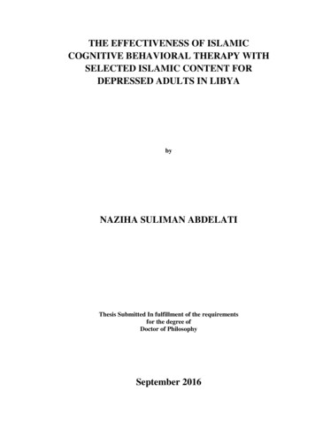
1Physics and Technology of ESWTPhysical BasicsThe Encyclopedia Britannica online (www.britannica.com) definition of shock wave is "astrong pressure wave in any elastic medium such as air, water, or a solid substance,produced by supersonic aircraft, explosions, lightning, or other phenomena that createviolent changes in pressure. Shock waves differ from sound waves in that the wave front, inwhich compression takes place, is a region of sudden and violent change in stress, density,and temperature. Because of this, shock waves propagate in a manner different from that ofordinary acoustic waves. In particular, shock waves travel faster than sound and their speedincreases as the amplitude is raised. Furthermore, the intensity of a shock wave alsodecreases faster than does that of a sound wave because some of the energy of the shockwave is expended to heat the medium in which it travels. The amplitude of a strong shockwave, as created in air by an explosion, decreases almost as the inverse square of thedistance until the wave has become so weak that it obeys the laws of acoustic waves. Shockwaves alter the mechanical, electrical, and thermal properties of solids and thus, can beused to study the equation of state (a relation between pressure, temperature, and volume)of any material".Although shock waves have some characteristics similar to those of other waves, they differconsiderably. Here these differences and similarities will be discussed.Characteristic Properties and Parameters of Shock WavesThe energy of a shock wave is released as pressure on the environment. This pressure is veryhigh, builds up extremely fast and with a classical shock wave the pressure amplitude ismainly positive pressure. The negative part is much weaker and longer and corresponds tothe tension wave.The graphic should show the classical form of a shock wave, the following properties arecharacteristic: Extremely fast rise of the curveVery high pressureLow negative wave compared to very high peak pressure3
Shock waves are mechanical pressure impulses which propagate in the medium in a wave-like manner.To better illustrate the differences to other wave types, here is a graph showing shockwaves, pressure waves and ultrasound in the same coordinates.4
Which parameters of the shock waves are the most suitable to describe the sound fields ofthe devices used, is not yet completely clear today. Again and again one comes up againstlimits, because one cannot characterize the applied impulses with one or the otherparameter. In accordance with the extracorporeal shock wave lithotripsy in urology (ESWL),the technicians of the manufacturers have especially chosen the EFD (energy flux density),the peak pressure (P) and the total energy (E) in the -6 dB focus. It is now agreed thatfurther parameters should be documented, but the process of determination is not yetcomplete.The following list lists the most important parameters currently in use:Positive pressureMPa-6 dB Focusmm5 MPa FocusmmPositive energy flux density (EFD )mJ/mm²Total energy flux densitymJ/mm²Positive energy in -6dB focusmJTotal energy in -6dB focusmJPositive energy in 5 MPa focusmJTotal energy in 5 MPa focusmJPositive energy in 5 mm focusmJTotal energy in 5 mm focusmJIn the graph it can beseen that the size of thefocus is obviously verydifferent. Some of thesedevices are no longeravailable on the market,so it is pointless to discusstheir superiority, which isonly noteworthy herethat all devices definethemselves as highenergy devices.Finally, it should be noted here that the documentation must not only include the deviceused, but also the energy levels applied and the number of impulses in the individual energylevels. This is the only way to retrospectively calculate a value for the total applied energy.On the homepage of the ISMST (International Society for Medical Shockwave Treatment) a5
corresponding recommendation is placed, which requires the documentation of the devicedata and the treatment parameters. This recommendation was developed with thecooperation of DIGEST and applies to all devices equally! recommendations/Generation of Shock WavesThe illustration shows that there are apparently very different devices. These differences aredue to the different ways in which shock waves are generated.The shock waves are generated for medical applications in a therapy head and coupled bymeans of a usually flexible coupling piece in the form of a membrane or a three-dimensionalbody. In order to achieve a suitable coupling, it is necessary to introduce a coupling mediumsuch as ultrasonic gel between the interfaces. Otherwise even the smallest air bubbles willinterfere with the propagation of the sound. Since any layer of air effectively prevents thepropagation of the shock waves.Electrohydraulic Generation PrincipleThe oldest principle used in medicine is that in which the shock waves are generated by aspark plug, the electrohydraulic principle. The shock waves propagate in a medium (water)and are also focused in one place by a parabolic mirror.6
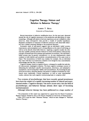
Piezoelectric Generation PrincipleThe piezoelectric principle is based on a focusing of pressure waves, which is produced bythe electrical activation of piezoelectric crystals. The piezoelectric crystals are mounted in ashell in such a way that the pressure waves of each crystal meet in a focus.Scheme of piezoelectric shock wave generationElectromagnetic Generation PrincipleIn electromagnetic shock wave generation the shock wave is emitted by a flat coil or acylindrical coil by electrical pulses. In the flat coil a membrane is deflected which compressesa medium (water). The pulse is focused by means of an acoustic lens. In the cylindrical coilthe shock wave is focused by a reflector.The electromagnetic and piezoelectric systems produce pressure waves that only becomeshock waves in the focus by summing the energy. The electrohydraulic systems produceshock waves already in focus 1, but in focus 2 the energies are significantly higher.7
A) The light blue membrane is deflected by the coil, which increases the pressure in the vessel betweenmembrane and lens. The pressure is focused by the lens. B) One sees that only in the center (focus) the wavesare divided and summed up in such a way that they get the classical shock wave form, outside the center thereare more or less energetic pressure waves.Radial or Ballistic Generation PrincipleThe techniques described in the previous chapters are opposed to the r-ESWT technique. Itwould be more correct not to speak of shock waves in this technique. However, in medicalusage the term radial shock wave therapy has established itself and it is generally acceptedthat this technique is called so even if other terms are also used; ("radial shock wavetherapy", RSWT; "Extracorporeal Pulse Activation Therapy", EPAT; "radial pressure wavetherapy").8
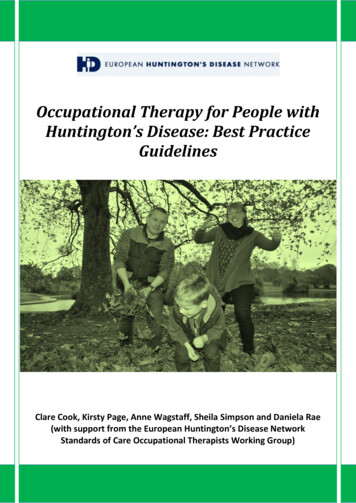
The generation of radial pressure waves is generated by the collision of a projectileaccelerated by compressed air or electromagnetic induction on an impact body (applicator).The projectile has a velocity of approx. 5 to 25 m / s. The impact body is deflected byapproximately 0.6 mm by the impact of the projectile. Most of the energy is transferred as apressure wave into the adjacent medium. The wave propagates radially from the applicator,with the square of the distance the energy decreases.The different devices generate sound fields which can differ fundamentally from each other.When the pressure pulses are generated radially, the rapid division of the wave front andthe characteristic significantly lower negative and positive deflection of the waves are neverdiscernible, as can be seen in high-energy devices with maximum energy levels.9
Fig.: Diagram of a pressure wave profile of a radial "shock waveThe different types of generation of shock waves (including pressure waves) differ in manyrespects, as is impressively shown in the graph "Comparison of three different acousticwaves" (see chapter Characteristic Properties and Parameters of Shock Waves).To date, the significance of the various parameters in orthopaedic shock wave therapy isunclear, but it is useful to document those that are known in order to compare the clinicalresults and perhaps arrive at the ideal physical settings.It is important to note at this point that the vast majority of treatments in (orthopaedic)indications are performed with more or less energetic pressure waves and not with"classical" shock waves, as shown graphically above. It would be very helpful if the differentsound fields could be better characterized with parameters, but we are not there yet.10
Physical Mechanisms of ActionHere different observations are arranged in a way which seems logical for the author. Itshould be noted in advance that many questions are open.Speed and acoustic impedance of shock waves in different mediaWhen understanding the effect of shock waves one recognizes the similarity to other waves.Shock waves pass through a medium in a characteristic speed. The speed in the differentmedia is a typical feature for shock waves, exemplarily some are listed here.Density (kg / m3) Speed of Sound (m / s) Acoustic Impedance (kg / m2s)MaterialAir1.2330161.44 x 106Water100014371.44 x 106Fat97014801.66 x 106Muscle10601570Bones:6.12 x 10⁶Cortical170036001.4 x 106Cancellous 10001450Table 1: The table shows how differently the properties of the media affect the shock waves. Impedance varieswith water content of the tissue.As with other waves, the energy is released at interfaces, at those places where the wavesfrom one medium meet another medium. The following phenomena occur at the interfaces,which we also know from classical wave theory:Transmission (transverse wave, longitudinal wave)(Partial) reflectionDiffraction and / or deflection, scatteringWave velocity x density of the medium acoustic impedancePressure / tension wave, shear forces11
The higher the impedance jump, i. e. the greater the differences in sound conduction in theadjacent media, the more energy is released at this point. In order to minimise the loss ofenergy, ultrasound gel or similar is used as the coupling medium; The first treatments werecarried out in a bathtub.CavitationIf one looks at Wikipedia, one reads: "Cavitation (lat. cavitare "hollow out") is the formationand dissolution of vapour-filled cavities (vapour bubbles) in liquids. There are two borderlinecases between which there are many transitional forms. In steam cavitation or hard(transient) cavitation, the cavities mainly contain steam from the surrounding liquid. Suchcavities collapse under the influence of external pressure by bubble implosion and cangenerate an intense shock wave (microscopic steam impact). In soft or stable gas cavitation,gases dissolved in the liquid enter the cavities and attenuate or prevent their collapse".Due to the extreme pressure differences, bubbles form in the medium, the bubbles collapseand the released energy generates further shock waves (jet streams) and photons or freeradicals. This phenomenon can be proven in experiments, but today it is not possible to sayhow necessary cavitation is in order to produce a biological effect.As described above, there are also bubbles in liquids which are pre-existent but, similar tothe hard (transient) cavitation bubbles, change under the influence of a shock wave,causing similar phenomena but only at a much lower level. They are also known as pseudocavitation bubbles.12
In the following, different effects are described which can be observed in the medium or inthe tissue: Direct, mechanical effect or tearing (stone disintegration, bone fissures,tissue tearing)The direct, mechanical effect of high-energy shock waves we know from nature; It is theforce that leads to the destruction of houses in an earthquake or, as is well known, destroyseverything on the coast in a tsunami; Part of the energy of a lightning bolt is also emitted tothe environment as a shock wave, which can be heard as thunder further away and can havea destructive effect. The bang of an airplane when breaking through the speed of sound isalso a shock wave.In medicine, we use the destructive effect of shock waves to disintegrate concrements(kidney stones, bladders, ureter stones, gallstones or saliva stones).At very high shock wave energies, the connective tissue parts of the body such as skin,muscles, lungs, parenchymatous organs or blood vessels can also rupture. This effect is notsought in orthopaedics, which is why such high energies are not used in orthopaedics.The effect occurs at sites of high impedance jumps of different tissues that are penetratedby the shock wave, i. e. at the interfaces of different tissues and structures embedded in thetissue.13
The shattering, destructive effect is only aimed at in urology today (or surgery). In thestimulation of non-healing or poorly healing bone fractures, so-called pseudoarthrosis ordelayed bone fracture healing, it was also discussed whether the shattering or fracture ofthe bony structures leads to healing, but as we know from recent studies, one can assumethat disintegration is not needed in orthopedics.If the shock wave energy would be selected accordingly high, one could observe thedestructive effects. In animal experiments, these effects of high shock wave energies ontendons have been impressively demonstrated. Within the framework of an animalexperiment, for bone examinations, bone fragments ("bone embolism") could even befound by chance in the lungs of the experimental animal. We are therefore really carefulnot to produce any damaging effect of ESWT.With the same idea of disintegration (comminution), the treatment of calcium deposits onthe shoulder was started in Hamburg at the beginning of the 1990s. However, it is alsoknown in the case of calcium treatment that it is not the disintegrative effects that lead tohealing, but rather molecular biological changes in the treated tissue (see below). Thermal and chemical effectsThe significance of this ESWT effect is unclear. Thermal and chemical effects are detectablebut their influence on tissue changes is unclear. Probably these effects have a much greatersignificance in very high energy shock waves. The thermal effects are similar to those ofultrasound but shock waves are emitted at such a low frequency in orthopedic applicationsthat the thermal effects appear negligible. Biological effects of shock wavesMuch of what is described here is still little researched today. One should go so far and evenwrite that the biological mechanisms of action described here are largely hypotheses, butthere are some publications that give rise to the following analyses.A cascade of reactions to the mechanical stimulus of the shock wave is most likely to betriggered:14
MechanotransductionThe "vibration" of the tissue by the shock waves is a mechanotransduction, a transformationof a mechanical stimulus on the tissue. Mechanical energy is transferred to the cell byaltering the cytoskeleton and extracellular molecules connected to the cytoskeleton. It isprobable that stimulus transmission occurs particularly in the cell nucleus, but also in othercellular structures (endoplasmic reticulum, mitochondria, etc.).Illustration of mechanotransduction by Wang, Tytell, Ingber (Nature Rev 2009) Gene expression:The stimulation of the cell nucleus via the excited cytoskeleton might result in geneexpression. In any case, Wang et al. 2004 have shown that ESWT stimulates the RAS systemin rats and that, for example, m-RNA is released for VEGF, a performance that is onlyachieved in the cell nucleus.15
Wang et al. (2004 J.Biolchemistry): Ras Induction of Superoxide Activates ERK-dependentAngiogenicTranscription Factor HIF-1 and VEGF-A Expression in ShockWave-stimulated Osteoblasts Enzymatic tissue responseAnalogous to gene therapy, a tissue response is found that can also be measured bydifferent enzyme changes. As with gene therapy, enzymatic changes can be observed longafter gene therapy or gene expression.In animal experiments (rats, rabbits, dogs) changes of the following substances could befound by shock wave effect:- Nitric oxide (NO)- Prostaglandin E2- COX-2- Substance P16
- PGP (phosphoglycolate phosphatase)- CGRP (calcitonin gene related peptide)- eNOS (endothelial nitric oxide synthetase)- VEGF (vessel endothelial growth factor)- PCNA (proliferating cell antinuclear antigen)- Signal-regulated kinase (ERK)- p38 kinaseThe formation of heat shock proteins was also found in connection with NO.Animal experiments are not transferable 1 : 1 to humans. The list of the many substancesthat were successfully tested in animal experiments should show that obviously extremelycomplex changes in tissue are induced by shock waves. Macroscopically recognizable healingThe healing of the tissue is a process that takes months and can therefore be detected latemacroscopically and microscopically.Today, we assume that neovascularisation plays a central role in the healing of ESWT-treatedareas (formation of new blood vessels).In addition, changes in the nerve cells can also be detected, which would explain a change inpain after ESWT (reduction in the number of non-myelinated nerve fibres by ESWT). In thelast 5 years, there has been a flood of basic ESWT knowledge which, piece by piece,represents an interesting counterpart to the clinical research results. Hyperstimulation analgesia (gate-control mechanism)The importance of this ESWT effect has never been confirmed by animal testing and is nowconsidered to be an unlikely hypothesis:Repeated nerve irritation can cause a change in pain processing in the spinal cord or centralnervous system (gate-control). In the past, it was thought that such a "gate-controlmechanism" would lead to pain relief (according to Melzak). Today, the molecular biologicalchanges described above are much more likely.17
Contraindications and adverse effects of ESWTThe following list shows the contraindications for which ESWT is not performed:Epiphyseal plate in focusCoagulopathy (high-energy ESWT is associated with a risk of bleeding, also withmarcumarization)Acute infection (the chronic infection was dropped as a contraindication)Lung in focusBrain or nerve in focusPregnancyMalignant tumor in focusThere is no definite evidence of persistent complications from ESWT, but there have beentendon ruptures after ESWT, for example, and ESWT has been accused of this. Rompe andMaier had carried out impressive experiments with tendons, which showed that at energies 0.6 mJ / mm2 such tendons suffer damages. The tendon ruptures described have alwaysoccurred after repeated cortisone infiltrations, so ESWT should therefore rather not be ableto prevent the rupture any more than it was the cause of it.Among other things, pain (including headaches - migraines), redness of the skin (blistering)and bruising (haematomas) can usually occur during and after treatment. Tendon loosening(edemas, occasionally also tendon tears) were observed after the treatment. Complicationsnot previously known may also occur.Procedure of shock wave treatment:Other treatment options (immobilization, splints, bandages, tape bandages, cold,heat, electricity, ultrasound, stretching and physiotherapy exercises, medication asointment, for ingestion or infiltration, acupuncture, alternative methods, surgery, .)are explained before ESWT.Patients give their written consent to the ESWT.The treatment is carried out by the doctor!The treatment is painful! Nevertheless, pain anesthesia (anesthesia or localanesthesia) is usually not necessary for the treatment.The shock waves are introduced with an applicator (with contact gel) through theskin. The adjustment is made before and during the treatment by means of a clinicalexamination and pain description of the patient (feedback), ultrasound, X-ray or laser.A fixed position is important, an interruption of the treatment e. g. with pain isat any time possible. Usually 1000 - 3000 single shocks (1 - 5 impulses per second) areapplied during one treatment, with small wounds at least 500 impulses pertreatment. The duration of treatment is about 10 - 60 minutes.18
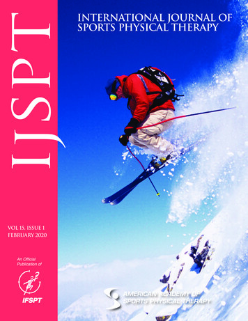
Depending on the disease, one or more treatments are necessary (see also thechapter where the indications are described). The treatment data are recordedprecisely.As post-treatment, medication (painkillers) may be recommended, furtherimmobilization (plaster, fixator, .) with relief and corresponding additionalthrombosis prevention in the case of bone fracture treatment, controls should beagreed after ESWT.The chances of success are indicated differently in the literature, a listing and exactdiscussion of the same goes beyond the scope here. In this respect every user should dealwith the recent literature or keep up to date.1: ESWT at the elbow in epicondylitishumeri lateralis2: ESWT in plantar fasciitis with / withoutheel spur3: ESWT on the shoulder atTend.calc.4: ESWT in pseudoarthrosis19
2Practical ApplicationTendinosis calcareaProf. Dr. Ludger GerdesmeyerICD-10: M-75.3SynonymsKalkschulter, Tendinitis calcarea, calcifying tendinitis, calcific lesionKey wordsPainful shoulder, shoulder joint, rotator cuff, calcium deposit, calcium shoulder, shock wavetherapy, ESWTDefinitionRotator cuff calcification as a consequence of dystrophic tendon disease with optionalchondroid metaplasia.AetiologyThe tendinosis calcarea of the rotator cuff is a common cause of shoulder pain. Data on theincidence of the disease show a considerable range of variation. It is given with 2.5 % to 20%. It is usually a self-limiting disease with high spontaneous healing rates. In a clinicallysymptomatic calcific shoulder with calcific deposits with radiologically sharp edges and ahomogeneous or inhomogeneous structure (type Gärtner 3 and 2), spontaneous resorptionoccurs within 3 years in 33 % of cases. The spontaneous resolution rate is considerablyhigher for calcium deposits with soft edges and inhomogeneous X-ray structure (typeGärtner 3) with up to 85 %. Within one year, 6.4 % of asymptomatic calcific shoulders arespontaneously resorbed. There is no data for a chronic and conservatively unsuccessfullytreated tendinosis calcarea. There is no reliable correlation between tendinosis calcarea androtator cuff lesions. A bilateral occurrence is observed between 9 % - 40 % of cases. Thesupraspinatus tendon is affected in 82 % - 94.5 % of cases. The classical calcium deposit islocated in the hypovascular zone, approx. 1.5 cm proximal to the attachment zone.PathogenesisA distinction is made between an acute phase and a chronic phase. The acute phasesuddenly begins with severe pain over a period of 2 - 3 weeks with swelling, overheating andclear pain at night and at rest. Then the pain gradually subsides until it is completely free ofsymptoms. Residual pain can persist for months (post calcification tendinitis).Macroscopically, a pasty milky emulsion is found which mineralogically consists of poorlycrystallised carbonate apatite. The crystals are formed in the tendon or resorbed afterbreakthrough into the bursa subacromialis / subdeltoidea (resorption stage). The chronicphase of tendinosis calcarea is characterized by slowly increasing pain. The self-limiting cycliccourse of the disease, which leads from a pre-calcification phase into the calcification phase20
and finally into a post-calcification phase, is interrupted. Chronic patients are in thecalcification phase for years. Mechanical, vascular and biochemical factors are discussed aspossible causes of calcification.Local pressure increases lead to reduced blood flow and hypoxia of the tendon tissue withdegeneration of the tendon cells and dystrophic calcification.ClassificationThe classification is based on size on the one hand and radiological criteria on the other. Theclassification according to gardener has established itself.Classification according to GärtnerGrade ISharp edges, homogeneous structure,radiopaqueGrade IISharp edges, inhomogeneous structure,less radiopaqueGrade IIIFuzzy edges, inhomogeneous structure,less radiopaqueMedical key systemsICD10M75.3Tendinitis calcarea in the shoulder areaS46.0Injury to a tendon of the rotator cuffS46.7Injury to several muscles and tendons at shoulder and upper arm levelAnamnesisSpecial anamnesisDisease durationAccident anamnesisDirect, indirect impact of violence, pseudoparalysisPainLocalisation, radiation, painful movement restriction, night rest painFunctional restriction mobilityFunctional handlesGeneral diseases and risk factorsSkeletal or connective tissue diseasesMetabolic diseasesPre-treatments21
DiagnosicsClinical diagnosticInspection: Muscle relief, symmetry, skin rednessPalpation: Palpable resistance in the area of the attachment zone of the rotator cuffFindings:Extent of movement (active and passive)Pain caused by movementSpecific positive tests for differential diagnosis, rotator cuff rupture and subacromialimpingement (drop arm sign), jobe test, patte test, palm-up test, O'Brian test, lift off test,Neer's Matsen's Hawkins's impingement signsAssessment of blood circulation, motor skills and sensitivityApparative diagnosticsSonography of the shoulderX-ray of the shoulder in 3 planesy-view image according to NeerOptional investigationMRX-ray of adjacent joints (e. g. cervical spine)Clinical chemistry laboratoryDifferential diagnosesImpingement syndrome (mechanical outlet, secondary or functional impingement)Frozen shoulderVertebragene, vascular, neurovascular shoulder painNeuralgic shoulder amyotrophyRotator cuff lesionsGout arthropathyClinical scoresConstant scoreTherapyMyofascial shoulder painObjectivesPain relief and restoration of shoulder functionInduction of calcium depot resorptionTherapeutic principleTreatment of tendinosis calcarea of the shoulder can be conservative or surgical.Conservative treatments must be carried out before surgical procedures are performed.Shock wave therapy is a method of first choice.22
Conservative therapySystemic NSARSystemic analgesicsLocal infiltrationInfiltration with PRP (Platelet Riched Plasma)Passive movement exercisesActive muscle strengthening for depression and centering of the humeral headErgotherapyMedical training therapySurgical therapySonographically or radiologically controlled needling of the calcium depositArthroscopic resectionOpen resectionShock wave therapyIndication: Indication by the expert physician.Contraindication: Malignant tumor in focus, osteomyelitis.Room requirements: Certification criteria of a medical practice e. g. hygiene plan,emergency management available according to DIN standard.Patient preparation: Differentiated and documented education and information (onset ofeffect after weeks). Explicit information about the risk of tendon rupture in the case ofprevious damage and premature sports exertion after treatment.Doctor and assistant staff: ESWT is a service to be provided personally by a doctor qualifiedby means of specialist knowledge.Implementation of the therapy: For the treatment of tendinosis calcarea, high-energytreatment with focused shock wave is used.Documentation:Designation of the shock wave source and the parameters used.Localisation:The treatment is controlled by imaging procedures (X-ray or ultrasound). The treatmenttarget area is the calcium depot.23
- Focused ESWT:The mean energy flux density of the shock waves should be between 0.18 and 0.32 mJ /mm2. Up to 5 treatment sessions are performed with an interval between 1 - 2 weeks.Between 1500 and 2000 shock waves with a frequency of up to 5 Hz are applied pertreatment session.Complications:Possible complications are: Haematoma discoloration, petchial skin bleeding, temporarypain enhancement, vascular injury, nerve injury.Literature:BosworthB.Calciumdeposits in the shoulder and subacromialbursitis:a survey 008HarmonPH.Methods and results in the treatment of 2580 painful shoulders with special reference tocalcific tendinitis and the frozen shoulder.AmJSurg 1958;95:527-544GärtnerJ.Tendinosis calcarea-Behandlungsergebnisse mit dem D, BuchM, GerdesmeyerL, HaakeM, LoewM, MaierM, HeineJ. MuskuloskeletaleStosswellenapplikation- Aktueller Stand der klinischen Forschung zu 0:267-274GreenS, BuchbinderR, GlazierR, ForbesA. Systematic review of randomised controlled trials ofinterventions for painful shoulder: selection criteria, outcome assessment, and efficacy.B
Fat 970 1480 1.44 x 10⁶ Muscle 1060 1570 1.66 x 10⁶ Bones: Cortical 1700 3600 6.12 x 10⁶ Cancellous 1000 1450 1.4 x 10⁶ Table 1: The table shows how differently the properties of the media affect the shock waves. Impedance varies with water content of the tissue.