Transcription
Animal Cell Culture and TechnologySecond Edition
The BasicsSeries Advisors:Rob Beynon UMIST, Manchester, UKChris Howe Department of Biochemistry, University of Cambridge, Cambridge, UKMonoclonal AntibodiesPCRAnalyzing ChromosomesSeparating CellsBiological CentrifugationPlant Cell CultureAnimal Cell Culture and TechnologyForthcoming titlesAnalyzing Gene ExpressionGene MappingReconstructing Evolutionary Trees
Animal Cell Culture andTechnologySecond EditionMichael ButlerLONDON AND NEW YORK
Garland Science/BIOS Scientific Publishers, 2004First published 2004This edition published in the Taylor & Francis e-Library, 2005.“To purchase your own copy of this or any of Taylor & Francisor Routledge's collection of thousands of eBooksplease go towww.eBookstore.tandf.co.uk.”All rights reserved. No part of this book may be reprinted or reproduced or utilised in any form orby anyelectronic, mechanical, or other means, now known or hereafter invented, including photocopyingandrecording, or in any information storage or retrieval system, without permission in writing from thepublishers.A CIP catalogue record for this book is available from the British Library.ISBN 0-203-42783-1 Master e-book ISBNISBN 0-203-44656-9 (Adobe e-reader Format)ISBN 1 859960499 (Print Edition)Garland Science/BIOS Scientific Publishers4 Park Square, Milton Park, Abingdon, Oxon, OX14 4RN, UK and29 West 35th Street, New York, NY 10001–2299, USAWorld Wide Web home page: www.bios.co.ukGarland Science/BIOS Scientific Publishers is a member of the Taylor & Francis Group.Distributed in the USA byFulfilment CenterTaylor & Francis10650 Toebben DriveIndependence, KY 41051, USAToll Free Tel.: 1 800 634 7064; E-mail: taylorandfrancis@thomsonlearning.comDistributed in Canada byTaylor & Francis74 Rolark DriveScarborough, Ontario M1R 4G2, CanadaToll Free Tel.: 1 877 226 2237; E-mail: tal fran@istar.caDistributed in the rest of the world byThomson Publishing ServicesCheriton HouseNorth WayAndover, Hampshire SP1O 5BE, UKTel.: 44 (0)1264 332424; E-mail: oduction Editor: Georgia Bushell
ContentsAbbreviationsPrefaceChapter 1 Introduction: The use of animal cell cultureChapter 2 Characteristics of cells in cultureChapter 3 Basic equipment and laboratory design: what you need to getstarted in cell cultureChapter 4 Growth and maintenance of cells in cultureChapter 5 Cell line and culture monitoringChapter 6 Genetic engineering of animal cells in cultureChapter 7 The glycosylation of proteins in cell cultureChapter 8 Hybridomas—sources of antibodiesChapter 9 Scaling up animal cell cultureChapter 10 Modes of culture for high cell densitiesChapter 11 Production from cell cultureChapter 12 Mammalian cell products: established and 226237271277
AbbreviationsADAadenosine deaminaseADCCantibody-dependent cellular cytotoxicityATCCAmerican Type Culture CollectionBHKbaby hamster kidneyBMEEagle’s basal mediumBSAbovine serum albuminCAMRCentre for Applied Microbiology ResearchCDRcomplementarity determining regionCHOChinese hamster minoethylDHFRdihydrofolate reductaseDMEMDulbecco’s modification of Eagle’s mediumDMSOdimethyl sulfoxideDOdissolved oxygenECACCEuropean Collection of Animal Cell CultureEDTAethylene-diaminetetraacetic acidELISAenzyme-linked immunosorbent assayEMEMEagle’s minimum essential mediumEPOerythropoietinERendoplasmic reticulumFACEfluorescence-activated carbohydrate electrophoresisFCSfetal calf serumG-6Pglucose 6-phosphateG6PDglucose 6-phosphate dehydrogenaseGalgalactoseGalNACN-acetyl galactosamineGlcNACN-acetyl glucosamineGMEMGlasgow’s Modification of Eagle’s MediumGODglucose oxidaseGPIglycosyl phosphatidylinositolHAMAhuman anti-murine antibodyHAThypoxanthine, aminopterin, thymidine
HEPAhigh-efficiency particulate nic acidHGPRThypoxanthine-guanine phosphoribosyl transferaseHIVhuman immunodeficiency virusHPLChigh-performance liquid chromatographyhTRThuman telomerase reverse transcriptaseLDHlactate dehydrogenaseMabmonoclonal antibodymoimultiplicity of infectionMOPSmorpholinopropane sulfonic acidMSmultiple nyltetrazoliumbromideNANAN-acetyl-neuraminic acidNESPnovel erythropoiesis-stimulating proteinNGNAN-glycolyl-neuraminic acidNKnatural killerOTRoxygen transfer rateOURoxygen utilization ratePBSphosphate buffered salinePEGpolyethylene glycolpfuplaque-forming unitsPHLSPublic Health Laboratory idasePVApolyvinyl alcoholPVPpolyvinyl pyrrolidoneRIAradioimmunoassayrphrevolutions per hourRPMIRoswell Park Memorial Institute mediumRSVrespiratory syncytial virusSAsialic acidSBMserum-based mediumSCIDsevere combined immunodeficiencySDSsodium dodecyl sulfateSFMserum-free mediumSSEAstage-specific embryonic antigen
STRstirred tank reactorTCAtricarboxylic acidTCAAtrichloroacetic acidTKthymidine kinaset-PAtissue plasminogen activatorTRHthyrotropin-releasing hormoneXGPRTxanthine-guanine phosphoribosyl transferaseYACyeast artificial chromosome
PrefaceAnimal cell culture has become an essential laboratory technique for the study ofbiochemical and physiological processes. Homogeneous cell populations can be producedby controlled growth over several generations and this offers an important alternative forsuch studies to the use of homogenized animal tissue. Whereas such cultures were onceconsidered to be difficult to set up and impossible to maintain without contamination,now most biological laboratories can perform the technique routinely with minimalequipment.In this book, the essential background information is provided to enable animal cellcultures to be established. The early chapters emphasize the historical development ofculture processes. It is important to understand that many of the ideas and terms now usedhave slowly evolved with a greater understanding arising through continued experimentalfindings. The original idea held by Carrel that all cells could be grown indefinitely waschallenged by the experimental work of Hayflick and Moorhead in the 1960s whodefined the finite life span of mammalian cells. This phenomenon can now be explainedby molecular biology in terms of the gradual reduction of the telomeres at the end of thechromosomes. Terms like ‘immortalization’ and ‘anchorage dependence’ are explained inthe book so that these cell properties can be understood and allow appropriate choicesbefore using a particular cell line. The basic equipment required to establish a cell culturelaboratory is described so as to meet the needs of most research students and scientists.Animal cell technology has become an important area because mammalian cellcultures have become the systems of choice for large-scale production operations fordesired proteins, mainly in health-care. At one time it was thought that most of theseproteins could be produced by appropriate genetic manipulation of bacteria such asE.coli. However, although such cells are simpler to culture because of their limitedsubstrate requirements and fast growth rates, it became clear that they do not have themetabolic network to support the synthesis of large glycoproteins. The glycosylationprocess that is exclusive to eukaryotic cells is required to ensure the complete productionof most biologically active proteins. Genetic engineering and cell hybridization havebecome important techniques in manipulating and designing mammalian cells,particularly for the biosynthesis of biologicals such as recombinant proteins ormonoclonal antibodies. The principles behind such cell manipulations are explained aswell as the techniques to ensure that highly productive cell lines are isolated. Examples ofa wide range of bioreactor designs are described. Some of these are currently being usedin commercial bioprocesses, but in other cases they are included to show whatalternatives could be considered.Viral vaccines represented the major group of commercial products from mammaliancell cultures for years. These are generally produced from well-defined anchoragedependent cells grown in microcarriers. However, there is now a growing list of otherimportant commercial products such as monoclonal antibodies and recombinant proteins
that have been licensed for human therapeutic use. Monoclonal antibodies can now be‘humanized’ by appropriate molecular recombination of mouse antibodies and humanimmunoglobulin regions. This has created a huge potential for the use of antibodies in thetreatment of human disease. These developments account for the growing expansion ofthe biotechnology industry and the need for increased capacity for large-scale cell culturebioprocesses. The demand for biomanufacturing is predicted to increase even further asgeneric copies of the originally patented biological molecules are allowed.The commercial developments of animal cell technology can be followed through thearchives of two major conference groups—the European Society of Animal CellTechnology (ESACT) and the Cell Culture Engineering (CCE) meetings. Theseorganizations host meetings on alternate years and each has become a showcase for thebiotechnology industry and the cell culture technology that underpins it. The increasingdemand for attendance at these meetings is evidence of the growing expansion of thetechnology The subject area has also become popular at universities as the demand forexpertise in the technology is high and can be translated to good work prospects forstudents. The course in Industrial Microbiology at the University of Manitoba is one suchcourse that includes a high content of the material outlined in this bookIt is hoped that this book may act as an easily readable introduction to animal cellculture and technology to undergraduates, graduates, technicians or experiencedindustrial researchers who are about to start projects or courses involving the use of cellculture. The explanations are kept basic, in keeping with the rest of the series, so that theminimal amount of background knowledge is assumed. In this way it is hoped that eventhose students with little background in the biological sciences may find this informationuseful.Michael ButlerUniversity of ManitobaSeptember 2003
Chapter 1Introduction: The use of animal cell culture1.Tissue culture, organ culture and cell cultureCells, removed from animal tissue or whole animals, will continue to grow if suppliedwith nutrients and growth factors. This process is called cell culture. It occurs in vitro (‘inglass’) as opposed to in vivo (‘in life'). The culture process allows single cells to act asindependent units much like any microorganism such as bacteria or fungi. The cells arecapable of division by mitosis and the cell population can continue growth until limitedby some parameter such as nutrient depletion.Animal cells selected for culture are maintained as independent units. Culturesnormally contain cells of one type (e.g. fibroblasts). The cells in the culture may begenetically identical (homogeneous population) or may show some genetic variation(heterogeneous population). A homogeneous population of cells derived from a singleparental cell is called a clone. Therefore all cells within a clonal population aregenetically identical. Cell culture is a widely used technique and is the main subject ofthis book.Cell culture is quite different from organ culture. Organ culture means the maintenanceof whole organs or fragments of tissue with the retention of a balanced relationshipbetween the associated cell types as exists in vivo. This idea of the maintenance of tissuewas important in the early development of culture techniques. However, it was soonrealized that this was extremely difficult over long periods because of the differinggrowth potential of individual cell types within a tissue.Nowadays the culture of individual cells is the preferred technique because conditionscan be controlled to allow some degree of consistency and reproducibility in cell growth.Although ‘cell culture’ is the most appro priate and logical term for this, it is still widelyreferred to as ‘tissue culture’ - a term that can be misleading, as it is also used to refer toorgan culture.2.Why grow animal cells in culture?There are a number of applications for animal cell cultures: to investigate the normal physiology or biochemistry of cells. For example, metabolicpathways can be investigated by applying radioactively labeled substrates andsubsequently looking at products; to test the effects of compounds on specific cell types. Such compounds may be
Animal cell culture and technology2metabolites, hormones or growth factors. Similarly, potentially toxic or mutageniccompounds may be evaluated in cell culture; to produce artificial tissue by combining specific cell types in sequence. This hasconsiderable potential for production of artificial skin for use in treatment of burns. to synthesize valuable products (biologicals) from large-scale cell cultures. Thebiologicals encompass a broad range of cell products and include specific proteins orviruses that require animal cells for propagation. The number of such commerciallyvaluable biologicals has increased rapidly over the last decade and has led to thepresent widespread interest in animal cell technology. Proteins that are present inminute quantities in vivo can be synthesized in gram (or kilogram) quantities bygrowing genetically engineered cells in vitro.3.The advantages and disadvantages of using cell cultureIn all the situations listed above we could use animal tissue (e.g. liver) or whole animals(e.g. mice). Biochemists have traditionally used homogenized liver as a source of cells forenzyme or metabolic studies. So, why use animal cell culture which may require far moretime in preparation and may require specialized equipment?The major advantage to using cell culture is the consistency and reproducibility ofresults that can be obtained by using a batch of cells of a single type and preferably ahomogeneous population (clonal). For example, we may be doing a biochemical analysiswhere it is important to relate a particular metabolic pathway to a certain cell type. Thiswould be possible in a culture containing a homogeneous cell population which can bemonitored for biochemical and genetic characteristics, but very difficult in a tissuehomogenate which would contain a heterogeneous mix of cells at different stages ofgrowth and viability.Toxicological testing procedures have been well established in laboratory animals.However, the use of cell culture techniques may allow a greater understanding of theeffects of a particular compound on a specific cell type such as liver cells (hepatocytes).Furthermore, routine toxicity tests are far less expensive in cell culture than in wholeanimals.In the production of biological products on a large scale, the avoidance of contaminantssuch as unwanted viruses or proteins is important. This can be more easily performed in awell-characterized cell culture than when dealing with pooled animal tissue.The major disadvantage to the use of cell culture is that after a period of continuousgrowth, cell characteristics can change and may be quite different from those originallyfound in the donor animal. Cells can adapt to different nutrients. This adaptation involveschanges in intracellular enzyme activities. Furthermore, culturing favors the survival offast-growing cells which are selectively retained in a mixed cell population.The changes in the growth and biochemical characteristics of a cell population may bea particular problem when using cultures to develop an understanding of the behavior ofcells in vivo. After a period of culture, the cells may be significantly different from thosethat are highly differentiated in vivo where growth has ceased. Intracellular enzyme
Introduction: The use of animal cell culture3activities change dramatically in response to nutrient depletion and by-productaccumulation in a culture.Some of the characteristics of cells in culture are discussed further in Chapter 2.4.The early history of cell cultureThe techniques necessary to allow animal cells to grow in culture were graduallydeveloped over the course of the last century (see Table 1.1). The original impetus for thedevelopment of cell culture was to study, under a microscope, normal physiologicalevents such as nerve development.One of the earliest experiments showing cell culture in vitro was conducted by RossHarrison in 1907 who developed the ‘hanging drop’ technique which is illustrated inFigure 1.1. In the depression of a microscope slide he entrapped small pieces of frogembryo tissue in clotted lymph fluid (Harrison, 1907). By this method he was able toobserve the elongation of nerve fibers over several weeks.From his experiments he demonstrated some of the characteristics and requirements forthe maintenance of cells.Table 1.1. Milestones in the development of animal cell technology1880Roux maintained embryonic chick cells in saline solution18901900Harrison grew frog nerve cells by the ‘hanging drop’ technique1910Carrel used aseptic techniques for long-term cell culturesRous and Jones used trypsin for subculture of adherent cells1920The ‘Carrel’ flask was designed for cell culture19301940
Animal cell culture and technology4Antibiotics were added to culture mediumEarle isolated mouse L fibroblastsEnders grew polio virus on cultured human cells1950Gey cultured HeLa cellsEagle developed a chemically defined culture medium1960Hayflick and Moorhead showed that human cells have a finite lifespanHam grew cells in a serum-free mediumHarris and Watkins fused human and mice cells1970Kohler and Milstein produced an antibody-secreting hybridomaSato developed serum-free media from hormones and growth factors1980Human insulin was produced from bacteriaMonoclonal antibody (OKT3) used for human therapyRecombinant tPA licensed for human therapy1990Humanized chimeric antibodies used for human therapyStem cells isolated
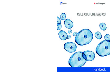
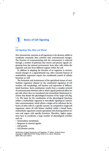
Introduction: The use of animal cell culture5Figure 1.1 Hanging drop technique of cell culture. The cells require an anchor for support which in this case was provided by thecoverslip and the fibrous matrix of the lymph clot. Cells that are bound to a tissuematrix in vivo are normally anchorage-dependent when grown in culture. Severalproteins are responsible for glueing the cells to a solid surface. The best characterizedis fibronectin. The cells require nutrients which are provided by the biological fluid contained in theclot. The growth rate is relatively slow (compared to bacteria) and this makes the culturevulnerable to contamination. A small number of bacteria would soon outgrow a largerpopulation of animal cells. The doubling time for animal cells is normally between 18and 24 hours but bacterial cells can double in 30 minutes.Some of these early culture experiments were continued by Alexis Carrel who wastrained as a surgeon (Carrel, 1912). He showed that the application of strict asepticcontrol enabled the prolonged subculture of the cells. He used chick embryo extractsmixed with blood plasma to support cell growth. In 1923 Carrel designed a flask suitablefor routine cell culture work (Figure 1.2). This facilitated subculture under asepticconditions and was the forerunner of the modern cell culture flask. Sterile manipulation isfacilitated by the narrow-angled glass neck of the Carrel flask which can be flamed by aBunsen burner prior to inoculation or sampling.However, Carrel’s application of surgical procedures for aseptic manipulation of cellcultures was elaborate and difficult to repeat by others. Consequently cell culture was notadopted as a routine laboratory technique until much later (reviewed by Witkowski,1979). Of course, today cell culture is widely used and can be performed in mostmoderately equipped laboratories.
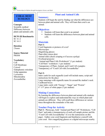
Animal cell culture and technology6Figure 1.2 The Carrel flask.Carrel is particularly well known for his 34-year culture experiment. In 1912 he isolated apopulation of chick embryo fibroblasts and these cells were grown in culture until 1946.From this, Carrel concluded that the cells were immortal. However, this claim was laterrefuted by the work of Hayflick and Moorhead who showed the finite lifespan of isolatedanimal cells (see below). It is now thought that Carrel’s technique of using plasma andhomogenized tissue as growth medium most likely allowed continuous reinoculation ofcells into his culture.5.How long will primary cultures survive?In 1961, Hayflick and Moorhead studied the growth potential of human embryonic cells.They showed that these cells could be grown continuously through repeated subculturefor about 50 generations (Hayflick and Moorhead, 1961). After this time, the cells enter asenescent phase and are incapable of further growth. Figure 1.3 illustrates the pattern ofgrowth during the lifespan of these cells.
Introduction: The use of animal cell culture7Figure 1.3 Normal and transformed growth of human embryonic cells over anextended number of passages.A finite growth capacity is a characteristic of all cells derived from normal animal tissue.The cells appear to pass through a series of age-related changes until the final stage whenthe cells are incapable of dividing further. The finite number of generations of growth is acharacteristic of the cell type, age and species of origin and is referred to as the ‘Hayflicklimit’.The capacity for growth is related to the origin of the cells—those derived from
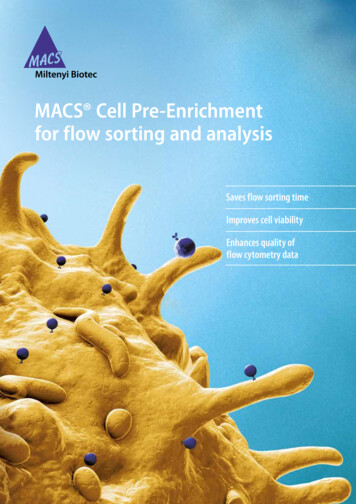
Animal cell culture and technology8embryonic tissue have a greater growth capacity than those derived from adult tissue.Each cell appears to have an inherent ‘biological clock′ defined by the number ofdivisions from the original stem cell. Even if cells are stored by cryopreservation the totalcapacity for cell division is not altered.6.The biological clockThe molecular mechanism that limits the growth capacity of normal cells was a mysteryfor a number of years until some key observations were made regarding chromosomallength. In 1986 Howard Cooke made the observation that the caps at the end ofchromosomes of human germline cells were longer than somatic cells (Cooke and Smith,1986). These caps known as telomeres are repeats of the nucleotide sequenceTTAGGG/CCCTAA and are shortened at each generation of growth of somatic cells.This is not surprising given the semiconservative mechanism of DNA replication whichoperates in one direction (5′ to 3' end). This generates a fork of replication that cannotwork at the end of double-stranded DNA. Therefore, at each mitotic division there is asmall segment of DNA that is not replicated, thus shortening the telomeric cap.However, in germ cells the telomere is maintained at 15 kilobases apparently by aribonucleoprotein enzyme, telomerase. The telomerase is expressed in germ cells and hasmoderate activity in stem cells but is absent in somatic cells. The finite lifespan of normalsomatic cells is regulated by 10 or more ‘senescence’ genes that suppress the expressionof the telomerase gene. This causes the human telomeres to gradually shorten at a rate ofaround 100 basepairs per cell division. From the telomere hypothesis, cells becomesenescent and stop dividing when progressive telomeric shortening produces a thresholdtelomere length. If oncogenes are activated then cells escape the negative control of thecell cycle and re-express telomerase.The critical experiment to validate this hypothesis came with a publication in 1998.Bodnar et al (1998) described the introduction of human telomerase reverse transcriptase(hTRT) into normal human cells (retinal pigment epithelial cells and foreskin fibroblasts).This converted the normal hTRT state to hTRT with the capability for expression ofthe telomerase enzyme. This process succeeded in increasing the lifespan of the cellssignificantly. The mean population doubling capacity of both cell types increased by atleast 20 generations. Figure 1.4 shows that for clones of the epithelial cells the meanpopulation doubling of 54 generations increased to a value of 73 generations by theintroduction of the telomerase enzyme. Similarly for the fibroblasts, there was an increaseof the mean population doubling of 64 to 100. This experiment established a causalrelationship between telomere shortening and in vitro cell senescence which is dependentupon a mitotic clock.
Introduction: The use of animal cell culture9Figure 1.4 The effect of telomerase activity on the growth span of cells. Theinitial mean doubling time (a) of the cell clones was extended (b) bythe introduction of the telomerase gene. Modified from Bodnar et al.,1998.7.The first products of animal cell technologyThe impetus for the application of cell culture to large-scale production came with theneed for viral vaccines. Major epidemics of polio in the 1940s and 1950s prompted a lotof effort to develop an effective vaccine. In 1949, Enders showed that the poliomyelitisvirus could be grown in cultures of human embryonic cells (Enders et al., 1949). Thepolio vaccine, which could be produced from the de-activated virus, became one of thefirst commercial products of cultured animal cells. Initially the virus was propagated onprimary cells, which are defined as those taken directly from an animal into culture. Laterprimary monkey kidney cells were replaced by human diploid lung fibroblast cells withdefined characteristics (e.g. WI-38, MRC-5). These were widely accepted because oftheir apparent ‘normality’ Since the 1950s a range of human and veterinary vaccines havebeen produced routinely from large-scale mammalian cell cultures.During the 1950s, several cell types with good growth characteristics were isolated andcharacterized. These included the chemically transformed mouse L cells and the humancarcinoma cell line, HeLa, which derives from the name of the donor—Henrietta Lach,although some texts designate the name as Helen Lane. Earle and Eagle focused theirattention on the development of chemically defined nutrient formulations for the growthof these cells and to replace the undefined biological extracts previously used (Eagle,1955). The chemically defined media, Eagle’s Minimum Essential Medium (EMEM)developed in the 1950s was the first widely used culture medium. EMEM had several
Animal cell culture and technology10advantages over previously used media: consistency between culture batches; ease of sterilization; reduced chance of contamination.Basic research in the 1960s using cell culture techniques included the classical work ofHayflick and Moorhead which is described in Section 5 of this chapter. This establishedthat there is a finite lifespan intrinsic to the genetic make-up of all normal animal cells.Recombinant DNA technology was developed in the 1970s. This allowed DNA to beisolated and inserted in any cell. Initially this was used for the expression of mammaliangenes in bacteria (particularly E. coli). It was considered at the time that such geneticallyengineered bacteria could be developed for the production of any desired mammalianprotein. However, it soon became apparent that large complex proteins (particularly thosewith carbohydrate groups attached) could only be synthesized with all theirposttranslational modifications in eukaryotic cells. Bacteria do not have the appropriatemetabolism to complete these modifications which are necessary for full activity of manymammalian proteins. Therefore, genetically engineered animal cells were developed forthe large-scale commercial synthesis of such important complex macromolecules asplasminogen activator, factor VIII and erythropoietin. These compounds have beenlicensed for human therapeutic use.Fusion techniques were pioneered in the 1960s by Harris and Watkins who allowedcells of different origin to be fused to produce hybrids. The initial value of this work wasto be able to assign specific genes to specific chromosomes. However, a novelapplication of this fusion technique was described by Kohler and Milstein in 1975 whoproduced hybridoma cells capable of the continuous production of a single type ofantibody (Kohler and Milstein, 1975). These monoclonal antibodies have beenparticularly valued as diagnostic and therapeutic agents because of their ability toselectively bind specific compounds. They are now produced commercially in kilogramquantities from the large-scale culture of hybridomas.The three major categories of valuable products from animal cells that we can identifyover the last 40 years are: viral vaccines; monoclonal antibodies; recombinant glycoproteins.The range of these biologicals has increased rapidly over the last few years. This isparticularly so for the glycoproteins that are of use as pharmaceutical, medical orveterinary products. They are generally synthesized as secreted products from selected orgenetically engineered mammalian cell lines. They are needed in large quantities and thisrequires a careful study of the control of the production processes. Such a study shouldstart with an examination of the principles of biochemistry, cell biology and biochemicalengineering that underlie such production processes.
Introduction: The use of animal cell culture11ReferencesBodnar, A.G., Ouellette, M., Frolkis, M., Holt, S.E., Chiu, C.-P.,Morin, G.B., Harley, C.B., Shay, J.W., Lichtsteiner, S. and Wright,W.E. (1998) Extension of life-span by introduction of telomerase intonormal human cells. Science 279:349–352.Carrel, A. (1912) On the permanent life of tissues outside the organism. J.Exp. Med. 15:516–528.Cooke, H.J. and Smith, B.A. (1986) Variability at the telomeres of thehuman X/Y pseudoautosomal region. Cold Spring Harbor Symp. Quant.Biol. 51Pt1: 213–219.Eagle, H. (1955) Nutrition needs of mammalian cells in tissue culture.Science 122: 501–50
the book so that these cell properties can be understood and allow appropriate choices before using a particular cell line. The basic equipment required to establish a cell culture laboratory is described so as to meet the needs of most research students and scientists. Animal cell technology has become