Transcription
3D U-Net: Learning Dense VolumetricSegmentation from Sparse AnnotationÖzgün Çiçek1,2 , Ahmed Abdulkadir1,4 , Soeren S. Lienkamp2,3 , ThomasBrox1,2 , and Olaf Ronneberger1,2,51Computer Science Department, University of Freiburg, GermanyBIOSS Centre for Biological Signalling Studies, Freiburg, Germany3University Hospital Freiburg, Renal Division, Faculty of Medicine, University ofFreiburg, GermanyDepartment of Psychiatry and Psychotherapy, University Medical Center Freiburg,Germany5Google DeepMind, London, UKcicek@cs.uni-freiburg.de24Abstract. This paper introduces a network for volumetric segmentation that learns from sparsely annotated volumetric images. We outline two attractive use cases of this method: (1) In a semi-automatedsetup, the user annotates some slices in the volume to be segmented.The network learns from these sparse annotations and provides a dense3D segmentation. (2) In a fully-automated setup, we assume that a representative, sparsely annotated training set exists. Trained on this dataset, the network densely segments new volumetric images. The proposednetwork extends the previous u-net architecture from Ronneberger etal. by replacing all 2D operations with their 3D counterparts. The implementation performs on-the-fly elastic deformations for efficient dataaugmentation during training. It is trained end-to-end from scratch, i.e.,no pre-trained network is required. We test the performance of the proposed method on a complex, highly variable 3D structure, the Xenopuskidney, and achieve good results for both use cases.Keywords: Convolutional Neural Networks, 3D, Biomedical Volumetric Image Segmentation, Xenopus Kidney, Semi-automated, Fully-automated,Sparse Annotation1IntroductionVolumetric data is abundant in biomedical data analysis. Annotation of suchdata with segmentation labels causes difficulties, since only 2D slices can beshown on a computer screen. Thus, annotation of large volumes in a slice-by-slicemanner is very tedious. It is inefficient, too, since neighboring slices show almostthe same information. Especially for learning based approaches that require asignificant amount of annotated data, full annotation of 3D volumes is not aneffective way to create large and rich training data sets that would generalizewell.
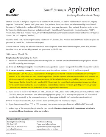
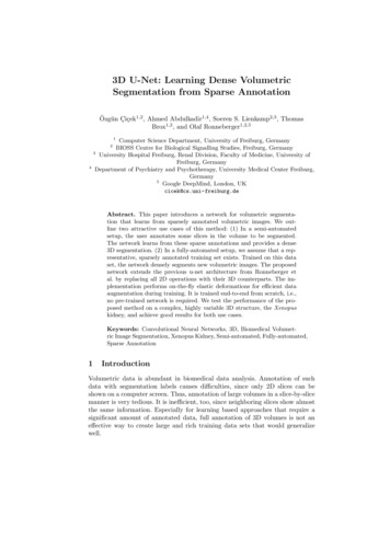
2Volumetric Segmentation with the 3D U-Netatrain andapply3D u-netraw imagemanual sparse annotationdense segmentationbapply trained 3D u-netraw imagedense segmentationFig. 1: Application scenarios for volumetric segmentation with the 3D u-net.(a) Semi-automated segmentation: the user annotates some slices of each volume to be segmented. The network predicts the dense segmentation. (b) Fullyautomated segmentation: the network is trained with annotated slices from arepresentative training set and can be run on non-annotated volumes.In this paper, we suggest a deep network that learns to generate dense volumetric segmentations, but only requires some annotated 2D slices for training.This network can be used in two different ways as depicted in Fig. 1: the firstapplication case just aims on densification of a sparsely annotated data set; thesecond learns from multiple sparsely annotated data sets to generalize to newdata. Both cases are highly relevant.The network is based on the previous u-net architecture, which consists of acontracting encoder part to analyze the whole image and a successive expandingdecoder part to produce a full-resolution segmentation [11]. While the u-net is anentirely 2D architecture, the network proposed in this paper takes 3D volumesas input and processes them with corresponding 3D operations, in particular,3D convolutions, 3D max pooling, and 3D up-convolutional layers. Moreover, weavoid bottlenecks in the network architecture [13] and use batch normalization[4] for faster convergence.In many biomedical applications, only very few images are required to traina network that generalizes reasonably well. This is because each image alreadycomprises repetitive structures with corresponding variation. In volumetric images, this effect is further pronounced, such that we can train a network on justtwo volumetric images in order to generalize to a third one. A weighted loss function and special data augmentation enable us to train the network with only fewmanually annotated slices, i.e., from sparsely annotated training data.
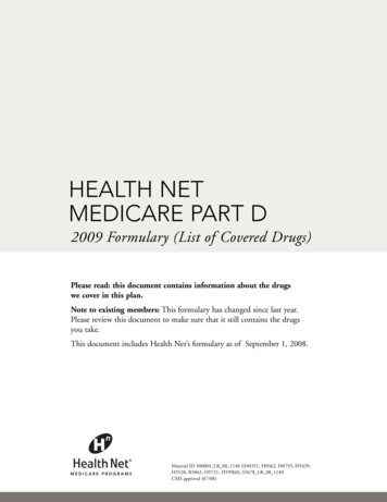
Volumetric Segmentation with the 3D U-Net3We show the successful application of the proposed method on difficult confocal microscopic data set of the Xenopus kidney. During its development, theXenopus kidney forms a complex structure [7] which limits the applicability ofpre-defined parametric models. First we provide qualitative results to demonstrate the quality of the densification from few annotated slices. These resultsare supported by quantitative evaluations. We also provide experiments whichshows the effect of the number of annotated slices on the performance of our network. The Caffe[5] based network implementation is provided as OpenSource1 .1.1Related WorkChallenging biomedical 2D images can be segmented with an accuracy closeto human performance by CNNs today [11,12,3]. Due to this success, severalattempts have been made to apply 3D CNNs on biomedical volumetric data.Milletari et al. [9] present a CNN combined with a Hough voting approach for3D segmentation. However, their method is not end-to-end and only works forcompact blob-like structures. The approach of Kleesiek et al. [6] is one of fewend-to-end 3D CNN approaches for 3D segmentation. However, their networkis not deep and has only one max-pooling after the first convolutions; therefore, it is unable to analyze structures at multiple scales. Our work is based onthe 2D u-net [11] which won several international segmentation and trackingcompetitions in 2015. The architecture and the data augmentation of the u-netallows learning models with very good generalization performance from onlyfew annotated samples. It exploits the fact that properly applied rigid transformations and slight elastic deformations still yield biologically plausible images.Up-convolutional architectures like the fully convolutional networks for semanticsegmentation [8] and the u-net are still not wide spread and we know of onlyone attempt to generalize such an architecture to 3D [14]. In this work by Tranet al., the architecture is applied to videos and full annotation is available fortraining. The highlight of the present paper is that it can be trained from scratchon sparsely annotated volumes and can work on arbitrarily large volumes dueto its seamless tiling strategy.2Network ArchitectureFigure 2 illustrates the network architecture. Like the standard u-net, it has ananalysis and a synthesis path each with four resolution steps. In the analysispath, each layer contains two 3 3 3 convolutions each followed by a rectifiedlinear unit (ReLu), and then a 2 2 2 max pooling with strides of two ineach dimension. In the synthesis path, each layer consists of an upconvolutionof 2 2 2 by strides of two in each dimension, followed by two 3 3 3convolutions each followed by a ReLu. Shortcut connections from layers of equalresolution in the analysis path provide the essential high-resolution features /opensource/unet.en.html
4Volumetric Segmentation with the 3D U-NetFig. 2: The 3D u-net architecture. Blue boxes represent feature maps. The number of channels is denoted above each feature map.the synthesis path. In the last layer a 1 1 1 convolution reduces the number ofoutput channels to the number of labels which is 3 in our case. The architecturehas 19069955 parameters in total. Like suggested in [13] we avoid bottlenecksby doubling the number of channels already before max pooling. We also adoptthis scheme in the synthesis path.The input to the network is a 132 132 116 voxel tile of the image with 3channels. Our output in the final layer is 44 44 28 voxels in x, y, and z directionsrespectively. With a voxel size of 1.76 1.76 2.04µm3 , the approximate receptivefield becomes 155 155 180µm3 for each voxel in the predicted segmentation.Thus, each output voxel has access to enough context to learn efficiently.We also introduce batch normalization (“BN”) before each ReLU. In [4],each batch is normalized during training with its mean and standard deviationand global statistics are updated using these values. This is followed by a layerto learn scale and bias explicitly. At test time, normalization is done via thesecomputed global statistics and the learned scale and bias. However, we havea batch size of one and few samples. In such applications, using the currentstatistics also at test time works the best.The important part of the architecture, which allows us to train on sparseannotations, is the weighted softmax loss function. Setting the weights of unlabeled pixels to zero makes it possible to learn from only the labelled ones and,hence, to generalize to the whole volume.33.1Implementation DetailsDataWe have three samples of Xenopus kidney embryos at Nieuwkoop-Faber stage36-37 [10]. One of them is shown in Fig. 1 (left). 3D Data have been recorded in
Volumetric Segmentation with the 3D U-Net5four tiles with three channels at a voxel size of 0.88 0.88 1.02µm3 using a ZeissLSM 510 DUO inverted confocal microscope equipped with a Plan-Apochromat40x/1.3 oil immersion objective lens. We stitched the tiles to large volumes usingXuvTools [1]. The first channel shows Tomato-Lectin coupled to Fluorescein at488nm excitation wavelength. The second channel shows DAPI stained cell nucleiat 405 nm excitation. The third channel shows Beta-Catenin using a secondaryantibody labelled with Cy3 at 564nm excitation marking the cell membranes. Wemanually annotated some orthogonal xy, xz, and yz slices in each volume usingSlicer3D2 [2]. The annotation positions were selected according to good datarepresentation i.e. annotation slices were sampled as uniformly as possible in all3 dimensions. Different structures were given the labels 0: “inside the tubule”; 1:“tubule”; 2: “background”, and 3: “unlabeled”. All voxels in the unlabelled slicesalso get the label 3 (“unlabeled”). We ran all our experiments on down-sampledversions of the original resolution by factor of two in each dimension. Therefore,the data sizes used in the experiments are 248 244 64, 245 244 56 and246 244 59 in x y z dimensions for our sample 1, 2, and 3, respectively.The number of manually annotated slices in orthogonal (yz, xz, xy) slices are(7, 5, 21), (6, 7, 12), and (4, 5, 10) for sample 1, 2, and 3, respectively.3.2TrainingBesides rotation, scaling and gray value augmentation, we apply a smooth densedeformation field on both data and ground truth labels. For this, we samplerandom vectors from a normal distribution with standard deviation of 4 in agrid with a spacing of 32 voxels in each direction and then apply a B-splineinterpolation. The network output and the ground truth labels are comparedusing softmax with weighted cross-entropy loss, where we reduce weights for thefrequently seen background and increase weights for the inner tubule to reacha balanced influence of tubule and background voxels on the loss. Voxels withlabel 3 (“unlabled”) do not contribute to the loss computation, i.e. have a weightof 0. We use the stochastic gradient descent solver of the Caffe [5] framework fornetwork training. To enable training of big 3D networks we used the memoryefficient cuDNN3 convolution layer implementation. Data augmentation is doneon-the-fly, which results in as many different images as training iterations. We ran70000 training iterations on an NVIDIA TitanX GPU, which took approximately3 days.4Experiments4.1Semi-Automated SegmentationFor semi-automated segmentation, we assume that the user needs a full segmentation of a small number of volumetric images, and does not have dia.com/cudnn
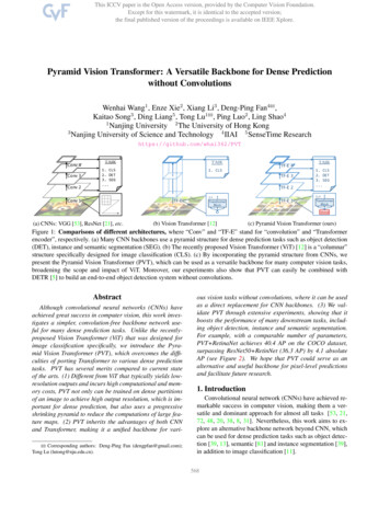
6Volumetric Segmentation with the 3D U-Netsegmentations. The proposed network allows the user to annotate a few slicesfrom each volume and let the network create the dense volumetric segmentation.For a qualitative assessment, we trained the network on all three sparselyannotated samples. Figure 3 shows the segmentation results for our 3rd sample.The network can find the whole 3D volume segmentation from a few annotatedslices and saves experts from full volume annotation.(a)(b)Fig. 3: (a) The confocal recording of our 3rd Xenopus kidney. (b) Resultingdense segmentation from the proposed 3D u-net with batch normalization.To assess the quantitative performance in the semi-automated setup, we uniformly partitioned the set of all 77 manually annotated slices from all 3 samplesinto three subsets and did a 3-fold cross validation both with and without batchnormalization. To this end, we removed the test slices and let them stay unlabeled. This simulates an application where the user provides an even sparserannotation. To measure the gain from using the full 3D context, we compare theresults to a pure 2D implementation, that treats all labelled slices as independentimages. We present the results of our experiment in Table 1. Intersection overUnion (IoU) is used as accuracy measure to compare dropped out ground truthslices to the predicted 3D volume. The IoU is defined as true positives/(true positives false negatives false positives). The results show that our approach isable to already generalize from only very few annotated slices leading to a veryaccurate 3D segmentation with little annotation effort.We also analyzed the effect of the number of annotated slices on the networkperformance. To this end, we simulated a one sample semi-automated segmentation. We started using 1 annotated slice in each orthogonal direction andincreased the number of annotated slices gradually. We report the high performance gain of our network for each sample (S1, S2, and S3) with every fewadditional ground truth (“GT”) slices in Table 2. The results are taken fromnetworks which were trained for 10 hours with batch normalization. For testingwe used the slices not used in any setup of this experiment.
Volumetric Segmentation with the 3D U-NetTable 1: Cross validation results forsemi-automated segmentation (IoU)7Table 2: Effect of # of slices forsemi-automated segmentation (IoU)test3D3D2Dslices w/o BN with BN with BNsubset 1 0.8220.8550.785subset 2 0.8570.8710.820subset 3 0.8460.8630.782average 830.5790.8080.849IoUS30.4750.7380.8350.872Table 3: Cross validation results for fully-automated segmentation (IoU)testvolume123average4.23Dw/o BN0.6550.7340.7790.7233Dwith BN0.7610.7980.5540.7042Dwith BN0.6190.6980.3250.547Fully-automated SegmentationThe fully-automated segmentation setup assumes that the user wants to segmenta large number of images recorded in a comparable setting. We further assumethat a representative training data set can be assembled.To estimate the performance in this setup we trained on two (partially annotated) kidney volumes and used the trained network to segment the thirdvolume. We report the result on all 3 possible combinations of training and testvolumes. Table 3 summarizes the IoU as in the previous section over all annotated 2D slices of the left out volume. In this experiment BN also improves theresult, except for the third setting, where it was counterproductive. We thinkthat the large differences in the data sets are responsible for this effect. Thetypical use case for the fully-automated segmentation will work on much largersample sizes, where the same number of sparse labels could be easily distributedover much more data sets to obtain a more representative training data set.5ConclusionWe have introduced an end-to-end learning method that semi-automatically andfully-automatically segments a 3D volume from a sparse annotation. It offers anaccurate segmentation for the highly variable structures of the Xenopus kidney.We achieve an average IoU of 0.863 in 3-fold cross validation experiments forthe semi-automated setup. In a fully-automated setup we demonstrate the performance gain of the 3D architecture to an equivalent 2D implementation. Thenetwork is trained from scratch, and it is not optimized in any way for this application. We expect that it will be applicable to many other biomedical volumetricsegmentation tasks. Its implementation is provided as OpenSource.
8Volumetric Segmentation with the 3D U-NetAcknowledgments. We thank the DFG (EXC 294 and CRC-1140 KIDGEMProject Z02 and B07) for supporting this work. Ahmed Abdulkadir acknowledges funding by the grant KF3223201LW3 of the ZIM (Zentrales Innovationsprogramm Mittelstand). Soeren S. Lienkamp acknowledges funding from DFG(Emmy Noether-Programm). We also thank Elitsa Goykovka for the useful annotations and Alena Sammarco for the excellent technical assistance in imaging.References1. Emmenlauer, M., Ronneberger, O., Ponti, A., Schwarb, P., Griffa, A., Filippi, A.,Nitschke, R., Driever, W., Burkhardt, H.: Xuvtools: free, fast and reliable stitchingof large 3d datasets. J Microscopy 233(1), 42–60 (2009)2. Fedorov, A., Beichel, R., Kalpathy-Cramer, J., Finet, J., Fillion-Robin, J.C., Pujol,S., Bauer, C., Jennings, D., Fennessy, F., Sonka, M., et al.: 3D slicer as an imagecomputing platform for the quantitative imaging network. J. Magn Reson Imaging30(9), 1323–1341 (2012)3. Hariharan, B., Arbeláez, P., Girshick, R., Malik, J.: Hypercolumns for object segmentation and fine-grained localization. In: Proc. CVPR. pp. 447–456 (2015)4. Ioffe, S., Szegedy, C.: Batch normalization: Accelerating deep network training byreducing internal covariate shift. CoRR abs/1502.03167 (2015)5. Jia, Y., Shelhamer, E., Donahue, J., Karayev, S., Long, J., Girshick, R., Guadarrama, S., Darrell, T.: Caffe: Convolutional architecture for fast feature embedding.In: Proc. ACMMM. pp. 675–678 (2014)6. Kleesiek, J., Urban, G., Hubert, A., Schwarz, D., Maier-Hein, K., Bendszus, M.,Biller, A.: Deep mri brain extraction: A 3d convolutional neural network for skullstripping. NeuroImage (2016)7. Lienkamp, S., Ganner, A., Boehlke, C., Schmidt, T., Arnold, S.J., Schäfer, T.,Romaker, D., Schuler, J., Hoff, S., Powelske, C., Eifler, A., Krönig, C., Bullerkotte,A., Nitschke, R., Kuehn, E.W., Kim, E., Burkhardt, H., Brox, T., Ronneberger, O.,Gloy, J., Walz, G.: Inversin relays frizzled-8 signals to promote proximal pronephrosdevelopment. PNAS 107(47), 20388–20393 (2010)8. Long, J., Shelhamer, E., Darrell, T.: Fully convolutional networks for semanticsegmentation. In: Proc. CVPR. pp. 3431–3440 (2015)9. Milletari, F., Ahmadi, S., Kroll, C., Plate, A., Rozanski, V.E., Maiostre, J.,Levin, J., Dietrich, O., Ertl-Wagner, B., Bötzel, K., Navab, N.: Hough-cnn: Deeplearning for segmentation of deep brain regions in MRI and ultrasound. CoRRabs/1601.07014 (2016)10. Nieuwkoop, P., Faber, J.: Normal Table of Xenopus laevis (Daudin)(Garland, NewYork) (1994)11. Ronneberger, O., Fischer, P., Brox, T.: U-net: Convolutional networks for biomedical image segmentation. In: MICCAI. LNCS, vol. 9351, pp. 234–241. Springer(2015)12. Seyedhosseini, M., Sajjadi, M., Tasdizen, T.: Image segmentation with cascadedhierarchical models and logistic disjunctive normal networks. In: Proc. ICCV. pp.2168–2175 (2013)13. Szegedy, C., Vanhoucke, V., Ioffe, S., Shlens, J., Wojna, Z.: Rethinking the inception architecture for computer vision. CoRR abs/1512.00567 (2015)14. Tran, D., Bourdev, L.D., Fergus, R., Torresani, L., Paluri, M.: Deep end2endvoxel2voxel prediction. CoRR abs/1511.06681 (2015)
3D segmentation. However, their method is not end-to-end and only works for compact blob-like structures. The approach of Kleesiek et al. [6] is one of few end-to-end 3D CNN approaches for 3D segmentation. However, their network is not deep and has only one