Transcription
Hindawi Publishing CorporationCritical Care Research and PracticeVolume 2015, Article ID 675041, 8 pageshttp://dx.doi.org/10.1155/2015/675041Research ArticleImpact of a 2-Day Critical Care Ultrasound Course duringFellowship Training: A Pilot StudyVi Am Dinh,1,2 Paresh C. Giri,1 Inimai Rathinavel,1 Emilie Nguyen,3 David Hecht,2,4Ihab Dorotta,4 H. Bryant Nguyen,1,2 and Ara A. Chrissian11Division of Pulmonary and Critical Care, Department of Medicine, Loma Linda University, Loma Linda, CA, USADepartment of Emergency Medicine, Loma Linda University, Loma Linda, CA, USA3School of Medicine, Loma Linda University, Loma Linda, CA, USA4Division of Critical Care, Department of Anesthesia, Loma Linda University, Loma Linda, CA, USA2Correspondence should be addressed to Vi Am Dinh; vadinh@llu.eduReceived 27 May 2015; Accepted 27 July 2015Academic Editor: Robert BootsCopyright 2015 Vi Am Dinh et al. This is an open access article distributed under the Creative Commons Attribution License,which permits unrestricted use, distribution, and reproduction in any medium, provided the original work is properly cited.Objectives. Despite the increasing utilization of point-of-care critical care ultrasonography (CCUS), standards establishingcompetency for its use are lacking. The purpose of this study was to evaluate the effectiveness of a 2-day CCUS courseimplementation on ultrasound-naı̈ve critical care medicine (CCM) fellows. Methods. Prospective evaluation of the impact of a twoday CCUS course on eight CCM fellows’ attitudes, proficiency, and use of CCUS. Ultrasound competency on multiple organ systemswas assessed including abdominal, pulmonary, vascular, and cardiac systems. Subjects served as self-controls and were assessed justprior to, within 1 week after, and 3 months after the course. Results. There was a significant improvement in CCM fellows’ writtentest scores, image acquisition ability, and pathologic image interpretation 1 week after the course and it was retained 3 monthsafter the course. Fellows also had self-reported increased confidence and usage of CCUS applications after the course. Conclusions.Implementation of a 2-day critical care ultrasound course covering general CCUS and basic critical care echocardiography usinga combination of didactics, live models, and ultrasound simulators is effective in improving critical care fellows’ proficiency andconfidence with ultrasound use in both the short- and long-term settings.1. IntroductionPoint-of-care ultrasonography is a rapidly developing fieldembraced by multiple medical specialties including primarycare, emergency medicine, and critical care medicine departments [1]. Common point-of-care critical care ultrasound(CCUS) applications include assessing the vascular, cardiopulmonary, and abdominal systems, as well as guidinginvasive procedures and hemodynamic management [1–3].With the increased availability and practicality of bedsideultrasound, intensivists worldwide are incorporating thistechnology into regular practice [4–6].Despite the rapid increase of point-of-care ultrasounduse in the ICU and the recognition by critical care fellowship programs for the need of formal CCUS trainingprograms, standardized education does not yet exist [7, 8].Even though CCUS competency requirements for imageacquisition and interpretation have been outlined [9–11],there remains no consensus on how the education, training,and evaluation of these competencies should be achieved[12]. Consequently, CCUS education during fellowship canbe inconsistent between training programs and may leadto varied ultrasound proficiency among graduating fellowsand practicing intensivists [1, 11, 13]. This creates the risk ofinappropriate ultrasound utilization in critical situations.The implementation of an ICU ultrasound curriculumbased on published guidelines and positive evidence-basededucational outcomes is important for the standardization oftraining among critical care fellowships [9, 10]. Herein, wepresent such a course and hypothesize that it will serve as avalid educational tool for critical care ultrasonography. Thepurpose of this study is to evaluate the short- and long-term
2impact of a formal 2-day critical care ultrasound courseduring fellowship training on critical care fellows’ ultrasoundknowledge, skills, and attitudes.2. Materials and Methods2.1. Study Design and Setting. This was a prospective observational cohort study examining the effect of a 2-day CCUScourse on ICU fellows from August 1, 2013, to November30, 2013, at a university-based tertiary care medical center.Inclusion criteria were that the subject needed to be currentlyenrolled in a medical ICU (MICU) or surgical ICU (SICU)fellowship at the study institution. Subjects were excluded ifthey had any ultrasound certification or attended a formalCCUS course within the previous 12 months. A total of eightICU fellows (6 MICU and 2 SICU) were enrolled in the study.Subjects were used as self-controls and examined at threepredetermined time points defined as PRE-CCUS (within 1week prior to CCUS course), POST-CCUS (within 1 weekafter CCUS course), and 3MO-CCUS (3 months after CCUScourse). The study was approved by the Institutional ReviewBoard and was considered to present minimal risk to thesubjects. Subjects were not notified of any of the test answersor scores until after study completion.We aimed to assess the effect of our course contentand delivery on learners using the validated Kirkpatrick 4level model of evaluating training outcomes [14, 15]. Thefirst level, effects on “reaction,” was examined in the formof surveys assessing comfort level of scanning. Level 2,effects on “learning,” was assessed by scores on written,image acquisition, and pathologic image interpretation tests.Level 3, effect on “behavior,” was assessed by comparing thenumber of learner scans performed 3 months after the courseto the number of reported scans immediately before thecourse. Finally, Level 4, “results,” was assessed by evaluatingretention scores on written, image acquisition, and pathologicimage interpretation tests 3 months after the course.2.2. Course Curriculum. The 2-day course (Table 1) includedboth general CCUS and basic critical care echocardiography(CCE) with content consistent with national guidelines [9,10]. The format of the course was multimodal and consistedof 1.5–2 hours of lectures, a live demonstration on the lecturetopic by an expert lecturer, and a focused session on thelecture topic with ultrasound practicum on live healthymodel volunteers, followed by learning pathologic imageinterpretation with cases using an ultrasound simulator(SonoSim Ultrasound Trainings Solution, Santa Monica,CA). The live demonstration by the lecturer consisted of using2 projectors, with one projector connected to the ultrasoundmachine showing a real-time ultrasound image and with theother projector simultaneously connected to a camcorderprojecting the instructor’s hand position, movements, andtransducer manipulation on a live model.Critical Care Research and Practicea 50-question test during the PRE-CCUS, POST-CCUS,and 3MO-CCUS time points. The question distribution wasfive physics, fifteen cardiology, twelve pulmonary, fourteenabdominal, and four vascular domains. To minimize recallbias, the test questions were randomly reordered at each of thestudy time points. CCUS instructors, with formal ultrasoundcertification as registered diagnostic medical sonographer(RDMS), registered diagnostic cardiac sonographer (RDCS),and/or Advanced Perioperative Transesophageal Echocardiography (PTEeXAM), developed the physics, cardiology,abdominal, and vascular questions. The director of interventional pulmonology with significant lung ultrasound experience developed the pulmonary questions. All questions werepeer-reviewed amongst the CCUS instructors prior to finaldistribution.2.3.2. Normal Image Acquisition on Healthy Volunteers. Toassess ultrasound image acquisition ability, all ICU fellowswere evaluated by a CCUS faculty instructor during livescanning of a healthy model patient during the PRE-CCUS,POST-CCUS, and 3MO-CCUS time points. Fellows wererandomly assigned to the faculty instructor; however, thesame faculty tested the fellows at all time points. The testconsisted of an 84-point checklist, and organ systems testedwere abdominal, pulmonary, vascular, and cardiac. The testevaluated the subject on proper patient positioning, correctuse of the machine and transducers, ability to acquire standard ultrasound images, and quality of image acquisition.2.3.3. Pathologic Image Interpretation with Ultrasound Simulator. To assess pathologic image interpretation, all ICUfellows were tested on the ultrasound simulator using 4 casesduring the PRE-CCUS, POST-CCUS, and 3MO-CCUS timepoints. Each case had 20 questions testing the following organsystems: abdominal, pulmonary, vascular, and cardiac. Therewas a possible total of 80 points for all four cases. Case 1 wasa patient with decompensated congestive heart failure withseverely depressed ejection fraction and pulmonary edema.Case 2 was a patient with a large pericardial effusion and tamponade. Case 3 was a trauma patient with hemoperitoneum,right hemothorax, and left pneumothorax. Case 4 was apatient with massive pulmonary embolism with findings ofright ventricular dilation/hypokinesis, noncollapsible IVC,and right lower extremity deep vein thrombosis.2.3. Course Assessment2.3.4. Survey on Ultrasound Comfort Level and Use. Toassess the changes in reaction and behavior, all ICU fellowscompleted a 16-question survey during the PRE-CCUS,POST-CCUS, and 3MO-CCUS time points. The questionsassessed how many self-reported scans the fellows performedeach week in the ICU as well as comfort level of specificCCUS applications including general CCUS, abdominalultrasound, pulmonary ultrasound, lower extremity vascular ultrasound, echocardiography, ultrasound-guided centralline, ultrasound-guided peripheral line, and ultrasoundguided thoracentesis.2.3.1. Ultrasound Knowledge: Written Test. To assess CCUSknowledge and retention, each ICU fellow completed2.4. Data Collection. All fellows were given a subject codenumber for deidentification for data analysis. Data collected
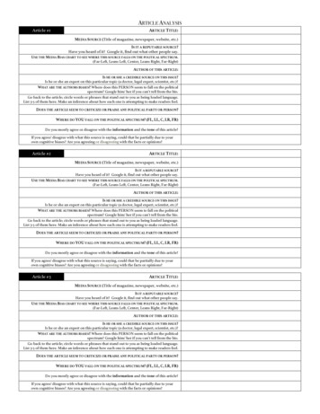
Critical Care Research and Practice3Table 1: Two-day critical care ultrasound course curriculum.Day 1General critical care ultrasoundDidactic lectures (2 hours)(i) General ultrasound principles (30 minutes)(ii) Abdominal vasculature (30 minutes)(iii) Hepatobiliary/renal (30 minutes)(iv) FAST scan (30 minutes)Live demonstration (15 minutes)Abdominal vasculature, hepatobiliary, renal, FAST scanHands-on rotation on live model (60 minutes)Abdominal vasculature, hepatobiliary, renal, FAST scanPathologic ultrasound simulator cases (60 minutes)Table 1: Continued.Lunch break (30 minutes)Didactic lectures (1.5 hours)(i) Valvular applications (30 minutes)(ii) Hemodynamics (30 minutes)(iii) Advanced applications and limitations (30 minutes)Live demonstration (30 minutes)Hemodynamics and valvular applicationsHands-on rotation on live model (60 minutes)Hemodynamics: cardiac output and ejection fractionassessmentHands-on rotation on live model (60 minutes)Valvular applications with use of Doppler (color, pulsed,continuous)(i) Case 1: ruptured abdominal aortic aneurysm(ii) Case 2: fluid overload: noncollapsible inferior vena cava(iii) Case 3: abnormal FAST scan: positive intra-abdominalfree fluid(iv) Case 4: cholecystitis: thickened gallbladder wall(v) Case 5: ascites and renal calculiLunch break (30 minutes)Didactic lectures (2 hours)(i) Pulmonary ultrasound (60 minutes)(ii) Deep vein thrombosis (30 minutes)from surveys and pre/posttests were transferred to electronicformat using an online secure resource (Qualtrics.com). Thesite is password protected and data is only available toauthorized study personnel.2.5. Statistical Analysis. Student’s 𝑡-test, chi-square, orANOVA with repeated measures was performed to determinethe changes in performance skills, test scores, and surveyresults between precourse, postcourse, and 3-month followup. Data analysis was performed using STATA 13.1.(iii) Vascular access (30 minutes)Live demonstration (15 minutes)Pulmonary, deep vein thrombosis, vascular accessHands-on rotation on live model (60 minutes)Pulmonary, deep vein thrombosis, vascular accessPathologic ultrasound simulator cases (60 minutes)(i) Case 1: right pneumothorax: absence of lung sliding(ii) Case 2: right pleural effusion(iii) Case 3: pulmonary edema: diffuse B-lines(iv) Case 4: right lower extremity DVT: noncompressibleveinDay 2Basic critical care echocardiographyDidactic lectures (1.5 hours)(i) Echocardiography: technique and standard views (45minutes)(ii) Common echocardiography applications (45 minutes)Live demonstration (30 minutes)Demonstration of standard echocardiography viewsHands-on rotation on live model (60 minutes)Practice obtaining standard echocardiography viewsPathologic ultrasound simulator cases (60 minutes)(i) Case 1: pericardial effusion with no tamponade(ii) Case 2: depressed left ventricular ejection fraction(iii) Case 3: right ventricular strain(iv) Case 4: pericardial effusion with tamponade3. ResultsSix MICU and two SICU fellows were enrolled in the study.All eight fellows completed 100% of the surveys and testsat all three assessment time points. Results are expressed aspercentage standard deviation. 𝑝 values convey significancewhen compared to the PRE-CCUS group. There were nostatistically significant differences among baseline resultsbetween the 1st-, 2nd-, and 3rd-year fellows (Table 2).3.1. Ultrasound Knowledge: Written Test. The total writtentest score in the PRE-CCUS group was 37.3% 6.3%. Thetotal written test scores in the POST-CCUS and 3MO-CCUSgroups were 64.8% 13.6% (𝑝 0.05) and 64.8% 12.9%(𝑝 0.05), respectively. Analysis of the five components ofthe test showed a statistically significant increase in scores ofthe POST-CCUS and 3MO-CCUS groups with respect to thefollowing three organ systems: abdominal, lower extremityvascular, and echocardiography (Figure 1). Nonsignificantincrease in score was noted in the physics and pulmonarycomponents.3.2. Normal Image Acquisition on Healthy Volunteers. Thetotal image acquisition score in the PRE-CCUS group was28.6% 15.5%. The total image acquisition scores in thePOST-CCUS and 3MO-CCUS groups were 84.1% 8.5%(𝑝 0.05) and 81.9% 15.0% (𝑝 0.05), respectively. Inaddition, when compared to the PRE-CCUS group, scores inall individual organ systems tested (abdominal, pulmonary,
4Critical Care Research and PracticeTable 2: Baseline test results for the 1st-, 2nd-, and 3rd-year criticalcare fellows. Results reported as percent SD.2nd year𝑛 213rd year𝑛 2Overall written34% 6.6%37% 1.0%test scoreOverall normalimage20.7% 16.1% 40.0% 3.7%acquisitionOverallpathologic67.6% 9.5% 70.2% 0.0%imageinterpretation40% 8.0%38.4 8.0%74.4 3.0%Cardiac1PRE-CCUSPOST-CCUS3MO-CCUS3.3. Pathologic Image Interpretation. The total pathologicimage interpretation score in the PRE-CCUS group was69.9% 7.9%. The total pathologic image interpretationscores in the POST-CCUS and 3MO-CCUS groups were82.7% 6.6% (𝑝 0.05) and 80.1% 8.2% (𝑝 0.05),respectively. Compared to the PRE-CCUS group, there wasalso a statistically significant increase in scores of the POSTCCUS and 3MO-CCUS groups in the lower extremity vascular scores (Figure 3).3.4. Attitudes and Behavior: Questionnaire. Comfort leveland self-reported scans regarding CCUS were surveyedusing a 16-point questionnaire at the three time points.Comfort level increased significantly in the POST-CCUSCardiacLE vascular 0.80.70.6Cardiac0.5Total scorelower extremity vascular, and echocardiography) were significantly higher in the POST-CCUS and 3MO-CCUS groups(Figure 2).0.9Mean scoreFigure 1: Written test score results with mean scores and standarddeviation. “PRE-CCUS” group was tested within 1 week beforeCCUS course, “POST-CCUS” group was tested within 1 week afterCCUS course, and “3MO-CCUS” group was tested 3 months afterCCUS course. LE: lower extremity. Statistical significance whencompared to the PRE-CCUS group.Pathologic image interpretationusing ultrasound simulator LE vascularLE vascular0.2Pulmonary0.4Figure 2: Normal image acquisition test score results with meanscores and standard deviation. “PRE-CCUS” group was testedwithin 1 week before CCUS course, “POST-CCUS” group was testedwithin 1 week after CCUS course, and “3MO-CCUS” group wastested 3 months after CCUS course. LE: lower extremity. Statisticalsignificance when compared to the PRE-CCUS group.Abdominal minalAbdominalTotal score Physics 0 Total score 0.4 Mean score 0.21 0.6Written test scores0.8 0.8Mean score1st year𝑛 4Normal image acquisitionusing healthy volunteersPRE-CCUSPOST-CCUS3MO-CCUSFigure 3: Pathologic image interpretation test score results withmean scores and standard deviation. “PRE-CCUS” group was testedwithin 1 week before CCUS course, “POST-CCUS” group was testedwithin 1 week after CCUS course, and “3MO-CCUS” group wastested 3 months after CCUS course. LE: lower extremity. Statisticalsignificance when compared to the PRE-CCUS group.and 3MO-CCUS groups in general bedside ultrasound,abdominal ultrasound, pulmonary ultrasound, lower extremity vascular ultrasound, and echocardiography (Table 3).There was a significant increase in the number of selfreported scans at 3 months in general bedside ultrasound,
Critical Care Research and Practice5Table 3: Comfort level of performing specific ultrasound applications using 5-point Likert scale (1 strongly disagree, 2 disagree,3 neither disagree nor agree, 4 agree, and 5 strongly agree).Results reported as median (25th to 75th percentiles). “PRE-CCUS”group was tested within 1 week before CCUS course, “POST-CCUS”group was tested within 1 week after CCUS course, and “3MOCCUS” group was tested 3 months after CCUS course. 𝑝 0.05when compared to PRE-CCUS data.Median (25%–75%)General soundLower extremityvascularEchocardiographyCentral lineplacementPeripheral lineplacementThoracentesisPRE-CCUSPOST-CCUS 3MO-CCUS3 (3-4) 2 (1-2)4 (3-4)1 (1-2)3.5 (3-4) 2.5 (2–3.5) 2 (2–2.5)4 (4-5) 4 (3.5–4.5) 1 (1-2)4 (4-4) 3 (2.5–4) 2 (2-2)4 (3-4) 3.5 (3-4) 4 (4-5)5 (5-5) 5 (5-5) 3.5 (2–4.5)4.5 (4-5)4 (3.5–5)4 (1.5–4)4.5 (3.5–5)4.5 (3.5–5)pulmonary ultrasound, lower extremity vascular ultrasound,and echocardiography (Table 4).4. DiscussionOur study was a self-evaluation of a critical care ultrasoundcourse taught by a multidisciplinary team of expert instructors. We implemented a formal 2-day CCUS course forcritical care fellows that met published societal requirementsfor basic CCUS competency [9, 10]. Our results showed thatsuch a course could improve skills in ultrasound knowledge,normal image acquisition, pathologic image interpretation,and comfort with ultrasound technique. These improvementswere apparent immediately after the course, persisted 3months after, and led to increased ultrasound usage in clinicalpractice. Consequently, we believe our course representsone possible model of implementing CCUS education andprovide evidence that it might be a useful launch pad formedical staff wishing to start using ultrasound in their dailypractice.Several obstacles may be contributing to lagging CCUSeducation in the United States [6–8]. These include thelack of regulation in training requirements, inconsistentformatting of course delivery, variable focus and breadth ofeducational material, and a paucity of proficient faculty toprovide instruction. Furthermore, the potential educationalbenefits of many proposed training programs have not beenvalidated [1, 13, 16–18]. To help establish more uniform CCUStraining and enhance the ultrasound proficiency of practicingintensivists, implementation of a course rooted in evidencebased, learner-oriented outcomes may be helpful.We used a comprehensive method of self-assessment,Kirkpatrick’s four-level training evaluation model, toTable 4: Number of self-reported ultrasound examinations performed per ICU month at precourse and 3-month follow-up. Resultsreported as mean SD. “PRE-CCUS” group was surveyed within 1week before CCUS course and “3MO-CCUS” group was surveyed3 months after CCUS course. 𝑝 0.05 when compared to PRECCUS data.General bedside ultrasoundAbdominal ultrasoundPulmonary ultrasoundLower extremity vascularEchocardiographyCentral line placementPeripheral line placementThoracentesisPRE-CCUS24.5 13.22.0 3.96.8 7.80.13 0.357.1 4.463.6 29.127.5 45.06.6 5.33MO-CCUS58.6 32.8 7.3 6.837.8 32.4 6.2 7.1 29.8 23.6 67.5 30.127 40.710.5 7.6highlight the positive educational impact of our course[14, 15]. To judge the first level, the “reaction” of our studentsto the course, we demonstrated their enhanced comfortwith ultrasound technique after taking our course. Thesecond Kirkpatrick level, “learning,” was judged by showingimmediate postcourse improvements on several levels,including theoretical ultrasound knowledge, normal imageacquisition, and pathologic image interpretation (Figures1–3). By reporting increased ultrasound usage during theirICU rotations, our students confirmed the positive impactof our course on the third Kirkpatrick level, “behavior”(Table 4). To evaluate the fourth and perhaps most importantKirkpatrick level, “results,” we assessed the persistence oflevel 2 learned material. Indeed, our trainees showed bothacquired theoretical ultrasound knowledge and practicalskill maintained three months after course (Figures 1–3).This was a novel finding, since most other studies have onlyreported on immediate benefits of their training programs[19]. In short, our critical care ultrasound course seemedto have consistent and lasting beneficial effects across allareas deemed important for an effective training program,according to the Kirkpatrick system.Although this was a pilot study involving a relativelysmall group of subjects, it was comprehensive in scope. Thecontent and time spent on each topic adhered to publishedguidelines [9, 10]. More importantly, in contrast to previousinvestigations that highlight educational effects of programsfocusing on specific organ systems such as critical careechocardiography on ultrasound-naı̈ve trainees [19–22], weutilized a more inclusive model. The rapidly expandingcritical care ultrasound literature overwhelmingly supportsa multisystem approach to the evaluation and managementof the critically ill patient. Established protocols suggestthe integration of bedside pulmonary, cardiac, abdominal,and vascular ultrasound to quickly assess patients in acuterespiratory and/or circulatory failure and to combine theresults in formulating management plans [23–27]. We organized our course around these major organ systems, whilealso teaching, in parallel, the ultrasound applications forcorresponding common procedures in the ICU (Table 1).Limited studies exist on looking at comprehensive CCUS
6training, with one study examining the benefits of a webbased curriculum covering general CCUS without CCE usinga web-based and simulation format, while another studylooked at the educational value of a 6-week general CCUSand basic CCE course [28, 29]. Our study differs because webelieve that a CCUS course should include echocardiographygiven its benefit in the evaluation of unstable ICU patients. Inaddition, it may not be feasible to have a 6-week ultrasoundcurriculum at many institutions given the varied schedules ofcritical care fellows. The use of a multidisciplinary instructionfaculty, with a varied ultrasound background and expertise,also helped create a well-rounded experience for the courseattendees. Furthermore, this “wide-net” approach may aidother institutions in establishing similar courses. Since recentdata suggest only 7–33% of faculty teaching in academic CCMfellowships are trained in CCUS [7], recruiting potentialultrasound instructors may need to target a range of disciplines, including radiology, emergency medicine, cardiology,pulmonology, surgery, and cardiothoracic anesthesia.Another novel aspect of our course was the use of anultrasound simulator to teach pathologic image interpretation and acquisition. It is difficult to incorporate and standardize live pathology into a course given the impracticalityof finding and recruiting patients with specific disease states.Some ultrasound courses use case-based group presentationsin which faculty present clinical scenarios and correspondingultrasound findings to groups of learners [30]. While thisapproach may be resource-efficient, memory retention hasbeen shown to be low and students are deprived of the tactileimage association we feel is essential to learning applicableultrasound technique [30]. With a simulator, the studentengages in both acquiring the image and interpreting theabnormal finding, while assimilating muscle memory withcognitive learning [31]. This is more reflective of a real clinicalsetting and enhances the retention of newly learned skills andinformation [31, 32].Perhaps the aspect of our course most responsible forits effectiveness was the utilization of an “active learning”format, since this approach has been shown to improvelearning outcomes [33]. While several course designs exist,ranging from short introductory sessions to yearlong longitudinal classes, we felt our interactive and multimodal twoday program was time-, resource-, and yield-efficient. To bestaccomplish our desired educational goals, our course adheredto the following format and order for each organ system:(1) a short 1-2-hour didactic session, (2) demonstrationof ultrasound technique and relevant image acquisition bythe expert instructor, (3) ultrasound scanning and normalimage acquisition by the learner on a healthy volunteer,with direct observation and feedback by the instructor, and(4) pathologic image interpretation utilizing the simulator(Table 1). We believe this format not only is comprehensive,but achieves better skill imprinting by allowing the studentto practice it immediately after observing expert didacticsessions and practical simulation.There are several limitations to our study. The samplewas not randomized into two groups (one receiving a courseand one not), and hence there was no true control group.However, the number of fellows made this design impractical,Critical Care Research and Practiceand we did not want to deprive our trainees of what wefeel is an essential part of critical care education. Second,some of the positive effects of the study at 3 months may besubject to recall bias and varied levels of clinical experienceamong the fellows. The latter is less likely, however, sinceour results showed that 2nd- and 3rd-year fellows with moreclinical experience did not perform better at baseline than1st-year fellows. In addition, it is possible that a periodlonger than 3 months is needed to more appropriately assessthe persistence of the positive effects of a course, thoughwe felt that this was a reasonable amount of time for thetrainees to have assimilated (or forgotten) their courserelated knowledge. The 50 multiple choice questions were thesame questions asked throughout the three time points andmotivated individuals may score higher after the initial sittingdue to further reading material as the study was intended toprovide a focal point for learning. Finally, we did not examinethe effects of improved ultrasound proficiency on changes inclinical decisions or patient outcomes or the impact of thecourse on the diverse, ultrasound-naı̈ve critical care facultythat attended as learners. These end points would serve asintriguing targets for future investigations of the benefits ofa CCUS course.5. ConclusionOur results suggest that the introduction of a critical careultrasound course has both a positive short- and longterm impact on fellows’ confidence and proficiency withultrasound use. Utilizing tools such as written tests to assessbasic knowledge, live models to teach practical skills, andultrasound simulators to teach pathological image identification can help standardize critical care ultrasound training.The proposed course and self-assessment methods presentedherein can serve as a model for other institutions lookingto implement a formal CCUS curriculum as part of theirfellowship training program.DisclosureThis study was presented at the American Thoracic SocietyInternational Conference, May 2014, San Diego, CA.Conflict of InterestsAll the authors report no conflict of interests in performingthis study.AcknowledgmentsThe authors are grateful to Ramesh Bansal, MD, for serving asan instructor and lending his expertise on echocardiographyto this study. They thank Heather Tassone, DO, for servingas an instructor. They acknowledge Dottie Acosta’s effortsin helping administer testing. Finally, they thank the LomaLinda University Critical Care Center for helping support thiscurriculum.
Critical Care Research and PracticeReferences[1] L. Neri, E. Storti, and D. Lichtenstein, “Toward an ultrasoundcurriculum for critical care medicine,” Critical Care Medicine,vol. 35, no. 5, pp. S290–S304, 2007.[2] S.-Y. Wu, Q. Ling, L.-H. Cao, J. Wang, M.-X. Xu, and W.A. Zeng, “Real-time two-dimensional ultrasound guidance forcentral venous cannulation: a meta-analysis,” Anesthesiology,vol. 118, no. 2, pp. 361–375, 2013.[3] P. Ferrada, S. Murthi, R. J. Anand, G. V. Bochicchio, and T.Scalea, “Transthoracic focused rapid echocardiographic examination: real-time evaluation of fluid status in critically ill traumapatients,” Journal of Trauma—Injury, Infection and Critical Care,vol. 70, no. 1, pp. 56–64, 2011.[4] G. A. Schmidt, “ICU ultrasound. The coming boom,” Chest, vol.135, no. 6, pp. 1407–1408, 2009.[5] M. Antonelli, M. Bonten, J. Chastre et al., “Year in reviewin Intensive Care Medicine 2011. II.
Research Article Impact of a 2-Day Critical Care Ultrasound Course during Fellowship Training: A Pilot Study ViAmDinh, 1,2 PareshC.Giri, 1 InimaiRathinavel, 1 EmilieNguyen, 3 DavidHecht, 2,4 IhabDorotta, 4 H.BryantNguyen, 1,2 andAraA.Chrissian 1 Division of Pulmonary and Critical Care, Departmen