Transcription
ALBERTRER TS WALTEESSENTIAL CELL BIOLOGYS BRAYESSENTIALCELL BIOLOGYGARLAND SCIENCEESSENTIALCELL BIOLOGY ROBFOURTHEDITIONFOUrTh EDiTiONFOURTH EDITION HOINJORAFFPKALBERTS BRAY HOPKIN JOHNSONLEWIS RAFF ROBERTS WALTER HNSON IWELS ISBN 978-0-8153-4455-19 780815 344551ecb4 cover soft.indd 1ECB4 interactive DVD-ROM inside11/09/2013 13:25
FOURTH EDITIONESSENTIALCELL BIOLOGYALBERTS BRAY HOPKIN JOHNSON LEWIS RAFF ROBERTS WALTER
Garland ScienceVice President: Denise SchanckSenior Editor: Michael MoralesProduction Editor and Layout: Emma Jeffcock of EJ PublishingServicesIllustrator: Nigel OrmeDevelopmental Editor: Monica ToledoEditorial Assistants: Lamia Harik and Alina YurovaCopy Editor: Jo ClaytonBook Design: Matthew McClements, Blink Studio, Ltd.Cover Illustration: Jose OrtegaAuthors Album Cover: Photography, Christophe Carlinet;Design, Nigel OrmeIndexer: Bill Johncocks 2014 by Bruce Alberts, Dennis Bray, Karen Hopkin,Alexander Johnson, Julian Lewis, Martin Raff, Keith Roberts,and Peter Walter 2010 by Bruce Alberts, Dennis Bray, Karen Hopkin,Alexander Johnson, Julian Lewis, Martin Raff, Keith Roberts,and Peter Walter 2004 by Bruce Alberts, Dennis Bray, Karen Hopkin,Alexander Johnson, Julian Lewis, Martin Raff, Keith Roberts,and Peter Walter 1998 by Bruce Alberts, Dennis Bray, Alexander Johnson,Julian Lewis, Martin Raff, Keith Roberts, and Peter WalterThis book contains information obtained from authentic andhighly regarded sources. Every effort has been made to tracecopyright holders and to obtain their permission for the use ofcopyright material. Reprinted material is quoted with permission, and sources are indicated. A wide variety of references arelisted. Reasonable efforts have been made to publish reliabledata and information, but the author and the publisher cannotassume responsibility for the validity of all materials or for theconsequences of their use.Essential Cell Biology WebsiteArtistic and Scientific Direction: Peter WalterNarrated by: Julie TheriotProducer: Michael MoralesAbout the AuthorsBruce Alberts received his PhD from Harvard Universityand is the Chancellor’s Leadership Chair in Biochemistryand Biophysics for Science and Education, University ofCalifornia, San Francisco. He was the editor-in-chief ofScience magazine from 2008–2013, and for twelve yearshe served as President of the U.S. National Academy ofSciences (1993–2005).Dennis Bray received his PhD from Massachusetts Instituteof Technology and is currently an active emeritus professorat the University of Cambridge.Karen Hopkin received her PhD in biochemistry fromthe Albert Einstein College of Medicine and is a sciencewriter in Somerville, Massachusetts. She is a contributor toScientific American’s daily podcast, 60-Second Science, and toE. O. Wilson’s digital biology textbook, Life on Earth.Alexander Johnson received his PhD from HarvardUniversity and is Professor of Microbiology and Immunologyat the University of California, San Francisco.Julian Lewis received his DPhil from the University ofOxford and is an Emeritus Scientist at the London ResearchInstitute of Cancer Research UK.Martin Raff received his MD from McGill University and isat the Medical Research Council Laboratory for MolecularCell Biology and Cell Biology Unit at University CollegeLondon.Keith Roberts received his PhD from the University ofCambridge and was Deputy Director of the John InnesCentre, Norwich. He is currently Emeritus Professor at theUniversity of East Anglia.Peter Walter received his PhD from The RockefellerUniversity in New York and is Professor of the Departmentof Biochemistry and Biophysics at the University ofCalifornia, San Francisco, and an Investigator of the HowardHughes Medical Institute.All rights reserved. No part of this book covered by the copyright hereon may be reproduced or used in any format in anyform or by any means—graphic, electronic, or mechanical, including photocopying, recording, taping, or information storageand retrieval systems—without permission of the publisher.ISBNs: 978-0-8153-4454-4 (hardcover); 978-0-8153-4455-1(softcover).Published by Garland Science, Taylor & Francis Group, LLC,an informa business, 711 Third Avenue, New York, NY 10017,USA, and 3 Park Square, Milton Park, Abingdon, OX14 4RN, UK.Printed in the United States of America15 14 13 12 11 10 9 8 7 6 5 4 3 2 1Visit our website at http://www.garlandscience.comLibrary of Congress Cataloging-in-Publication DataAlberts, Bruce.Essential cell biology / Bruce Alberts [and seven others].-- Fourth edition.pages cm.ISBN 978-0-8153-4454-4 (hardback)1. Cytology. 2. Molecular biology. 3. Biochemistry. I. Title.QH581.2.E78 2013571.6--dc232013025976
CHAPTER SEVEN7From DNA to Protein:How Cells Read the GenomeOnce the double-helical structure of DNA (deoxyribonucleic acid) hadbeen determined in the early 1950s, it became clear that the hereditaryinformation in cells is encoded in the linear order—or sequence—of thefour different nucleotide subunits that make up the DNA. We saw inChapter 6 how this information can be passed on unchanged from a cellto its descendants through the process of DNA replication. But how doesthe cell decode and use the information? How do genetic instructionswritten in an alphabet of just four “letters” direct the formation of a bacterium, a fruit fly, or a human? We still have a lot to learn about how theinformation stored in an organism’s genes produces even the simplestunicellular bacterium, let alone how it directs the development of complex multicellular organisms like ourselves. But the DNA code itself hasbeen deciphered, and we have come a long way in understanding howcells read it.Even before the DNA code was broken, it was known that the informationcontained in genes somehow directed the synthesis of proteins. Proteinsare the principal constituents of cells and determine not only cell structure but also cell function. In previous chapters, we encountered someof the thousands of different kinds of proteins that cells can make. Wesaw in Chapter 4 that the properties and function of a protein moleculeare determined by the sequence of the 20 different amino acid subunitsin its polypeptide chain: each type of protein has its own unique aminoacid sequence, which dictates how the chain will fold to form a moleculewith a distinctive shape and chemistry. The genetic instructions carriedby DNA must therefore specify the amino acid sequences of proteins. Wewill see in this chapter exactly how this is done.FROM DNA TO RNAFROM RNA TO PROTEINRNA AND THE ORIGINS OF LIFE
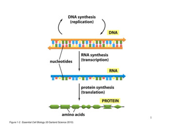
224CHAPTER 7From DNA to Protein: How Cells Read the GenomegeneDNA5′3′3′5′nucleotidesRNA SYNTHESISTRANSCRIPTIONRNA3′5′PROTEIN SYNTHESISTRANSLATIONPROTEINCOOHH2Namino acidsFigure 7–1 Genetic information directsthe synthesis of proteins. The flow ofgenetic information from DNA to RNA(transcription) and from RNA to protein(translation) occurs in all living cells. Itwas Francis Crick who dubbed this flowof information “the central dogma.” Thesegments of DNA that are transcribed intoRNA are called genes.ECB4 E7.01/7.01DNA does not synthesize proteins itself, but it acts like a manager, delegating the various tasks to a team of workers. When a particular proteinis needed by the cell, the nucleotide sequence of the appropriate segmentof a DNA molecule is first copied into another type of nucleic acid—RNA(ribonucleic acid ). That segment of DNA is called a gene, and the resulting RNA copies are then used to direct the synthesis of the protein. Manythousands of these conversions from DNA to protein occur every second in each cell in our body. The flow of genetic information in cells istherefore from DNA to RNA to protein (Figure 7–1). All cells, from bacteria to humans, express their genetic information in this way—a principleso fundamental that it has been termed the central dogma of molecularbiology.In this chapter, we explain the mechanisms by which cells copy DNAinto RNA (a process called transcription) and then use the informationin RNA to make protein (a process called translation). We also discussa few of the key variations on this basic scheme. Principal among theseis RNA splicing, a process in eukaryotic cells in which segments of anRNA transcript are removed—and the remaining segments stitched backtogether—before the RNA is translated into protein. In the final section,we consider how the present scheme of information storage, transcription, and translation might have arisen from much simpler systems in theearliest stages of cell evolution.FROM DNA TO RNATranscription and translation are the means by which cells read out, orexpress, the instructions in their genes. Many identical RNA copies can bemade from the same gene, and each RNA molecule can direct the synthesis of many identical protein molecules. This successive amplificationenables cells to rapidly synthesize large amounts of protein whenevernecessary. At the same time, each gene can be transcribed, and its RNAtranslated, at different rates, providing the cell with a way to make vastquantities of some proteins and tiny quantities of others (Figure 7–2).Moreover, as we discuss in Chapter 8, a cell can change (or regulate) theexpression of each of its genes according to the needs of the moment.In this section, we discuss the production of RNA, the first step in geneexpression.QUESTION 7–1gene Agene BDNAConsider the expression “centraldogma,” which refers to the flowof genetic information from DNAto RNA to protein. Is the word“dogma” appropriate in ATIONTRANSLATIONFigure 7–2 A cell can express differentgenes at different rates. In this and laterfigures, the untranscribed portions of theDNA are shown in gray.AAAAAAAAAAAAAAAAAAAAAAAAAproteinBBBprotein
225From DNA to RNA(A)HOCH2 OOHHHOHHOHH(B)5′ endSUGAR DIFFERENCESHOCH2 OH–OHHPHOHribosedeoxyriboseused in CHOHONHuracilused in RNACOH 2CBASE DIFFERENCESCOOHused in RNAsugar–phosphatebackboneOOHOO–Oused in DNAFigure 7–3 The chemical structure of RNA differs slightly fromthat of DNA. (A) RNA contains the sugar ribose, which differs fromdeoxyribose, the sugar used in DNA, by the presence of an additional–OH group. (B) RNA contains the base uracil, which differs fromthymine, the equivalent base in DNA, by the absence of a –CH3 group.(C) A short length of RNA. The chemical linkage between nucleotidesin RNA—a phosphodiester bond—is the same as that in DNA.PPUOH2CriboseO–OOHPPOGOOH 2COOH3′ end(C)3′5′HHCAlthough their chemical differences are small, DNA and RNA differ quitee7.03/7.03dramatically in overall structure. Whereas DNA always occursECB4in cellsas a double-stranded helix, RNA is single-stranded. This difference hasimportant functional consequences. Because an RNA chain is singlestranded, it can fold up into a variety of shapes, just as a polypeptidechain folds up to form the final shape of a protein (Figure 7–5); doublestranded DNA cannot fold in this fashion. As we discuss later, the abilityto fold into a complex three-dimensional shape allows RNA to carry outvarious functions in cells, in addition to conveying information betweenDNA and protein. Whereas DNA functions solely as an information store,some RNAs have structural, regulatory, or catalytic roles.Figure 7–4 Uracil forms a base pair with adenine. The hydrogenbonds that hold the base pair together are shown in red. Uracil has thesame base-pairing properties as thymine. Thus U-A base pairs in RNAclosely resemble T-A base pairs in DNA (see Figure 5–6A).OOPortions of DNA Sequence Are Transcribed into RNAThe first step a cell takes in expressing one of its many thousands ofgenes is to copy the nucleotide sequence of that gene into RNA. The process is called transcription because the information, though copied intoanother chemical form, is still written in essentially the same language—the language of nucleotides. Like DNA, RNA is a linear polymer madeof four different nucleotide subunits, linked together by phosphodiesterbonds. It differs from DNA chemically in two respects: (1) the nucleotides in RNA are ribonucleotides—that is, they contain the sugar ribose(hence the name ribonucleic acid) rather than deoxyribose; (2) although,like DNA, RNA contains the bases adenine (A), guanine (G), and cytosine (C), it contains uracil (U) instead of the thymine (T) found in DNA(Figure 7–3). Because U, like T, can base-pair by hydrogen-bonding withA (Figure 7–4), the complementary base-pairing properties described forDNA in Chapter 5 apply also to �sugar–phosphate backbone
226CHAPTER 7From DNA to Protein: How Cells Read the GenomeGUAUGCCAGUUAGCCGCAUACGUCCC UG GAC(C)(B)Figure 7–5 RNA molecules can form intramolecular base pairs and fold into specific structures. RNA is singlestranded, but it often contains short stretches of nucleotides that can base-pair with complementary sequencesfound elsewhere on the same molecule. These interactions—along with some “nonconventional base-pairinteractions (e.g., A-G)—allow an RNA molecule to fold into a three-dimensional structure that is determined by itssequence of nucleotides. (A) A diagram of a hypothetical, folded RNA structure showing only conventional (G-C andA-U) base-pair interactions. (B) Incorporating nonconventional base-pair interactions (green) changes the structure ofthe hypothetical RNA shown in (A). (C) Structure of an actual RNA molecule that is involved in RNA splicing. This RNAcontains a considerable amount of double-helical structure. The sugar–phosphate backbone is blue and the bases arered; the conventional base-pair interactions are indicated by red “rungs” that are continuous, and nonconventionalbase pairs are indicated by broken red rungs. For an additional view of RNA structure, see Movie 7.1.ECB4 e7.05/7.05Transcription Produces RNA That Is Complementary toOne Strand of DNAcoding strandDNA5′3′3′5′template strandTRANSCRIPTION5′3′RNAFigure 7–6 Transcription of a geneproduces an RNA complementary to onestrand of DNA. The transcribed strandof the gene, the bottom strand in thisexample, is called the template strand.The nontemplate strand of the gene(here, shown at the top) is sometimes calledthe coding strandbecauseits sequenceECB4e7.06/7.06is equivalent to the RNA product, asshown. Which DNA strand serves as thetemplate varies, depending on the gene,as we discuss later. By convention, an RNAmolecule is always depicted with its5 end—the first part to be synthesized—to the left.All the RNA in a cell is made by transcription, a process that has certainsimilarities to DNA replication (discussed in Chapter 6). Transcriptionbegins with the opening and unwinding of a small portion of the DNAdouble helix to expose the bases on each DNA strand. One of the twostrands of the DNA double helix then acts as a template for the synthesis of RNA. Ribonucleotides are added, one by one, to the growing RNAchain; as in DNA replication, the nucleotide sequence of the RNA chainis determined by complementary base-pairing with the DNA template.When a good match is made, the incoming ribonucleotide is covalentlylinked to the growing RNA chain by the enzyme RNA polymerase. TheRNA chain produced by transcription—the RNA transcript—is thereforeelongated one nucleotide at a time and has a nucleotide sequence exactlycomplementary to the strand of DNA used as the template (Figure 7–6).Transcription differs from DNA replication in several crucial respects.Unlike a newly formed DNA strand, the RNA strand does not remainhydrogen-bonded to the DNA template strand. Instead, just behind theregion where the ribonucleotides are being added, the RNA chain is displaced and the DNA helix re-forms. For this reason—and because onlyone strand of the DNA molecule is transcribed—RNA molecules aresingle-stranded. Further, because RNAs are copied from only a limitedregion of DNA, RNA molecules are much shorter than DNA molecules;DNA molecules in a human chromosome can be up to 250 million nucleotide pairs long, whereas most mature RNAs are no more than a fewthousand nucleotides long, and many are much shorter than that.
From DNA to RNARNA polymerase5′3′3′5′DNA templatestrand5′newly synthesizedRNA e site ofpolymeraseribonucleosidetriphosphatetunnelLike the DNA polymerase that carries out DNA replication (discussedin Chapter 6), RNA polymerases catalyze the formation of the phosphodiester bonds that link the nucleotides together and form thesugar–phosphate backbone of the RNA chain (see Figure 7–3). The RNAECB4alonge7.07/7.07polymerase moves stepwisethe DNA, unwinding the DNA helixjust ahead to expose a new region of the template strand for complementary base-pairing. In this way, the growing RNA chain is extended byone nucleotide at a time in the 5 -to-3 direction (Figure 7–7). The incoming ribonucleoside triphosphates (ATP, CTP, UTP, and GTP) provide theenergy needed to drive the reaction forward (see Figure 6–11).The almost immediate release of the RNA strand from the DNA as it is synthesized means that many RNA copies can be made from the same genein a relatively short time; the synthesis of the next RNA is usually startedbefore the first RNA has been completed (Figure 7–8). A medium-sizedgene—say, 1500 nucleotide pairs—requires approximately 50 seconds fora molecule of RNA polymerase to transcribe it (Movie 7.2). At any giventime, there could be dozens of polymerases speeding along this singlestretch of DNA, hard on one another’s heels, allowing more than 1000transcripts to be synthesized in an hour. For most genes, however, theamount of transcription is much less than this.Although RNA polymerase catalyzes essentially the same chemical reaction as DNA polymerase, there are some important differences betweenthe two enzymes. First, and most obviously, RNA polymerase uses ribonucleoside for phosphates as substrates, so it catalyzes the linkage ofribonucleotides, not deoxyribonucleotides. Second, unlike the DNApolymerase involved in DNA replication, RNA polymerases can start anRNA chain without a primer. This difference likely evolved because transcription need not be as accurate as DNA replication; unlike DNA, RNA isnot used as the permanent storage form of genetic information in cells,so mistakes in RNA transcripts have relatively minor consequences for acell. RNA polymerases make about one mistake for every 104 nucleotidescopied into RNA, whereas DNA polymerase makes only one mistake forevery 107 nucleotides copied.Cells Produce Various Types of RNAThe vast majority of genes carried in a cell’s DNA specify the amino acidsequences of proteins. The RNA molecules encoded by these genes—which1 μm227Figure 7–7 DNA is transcribed into RNAby the enzyme RNA polymerase. RNApolymerase (pale blue) moves stepwisealong the DNA, unwinding the DNA helix infront of it. As it progresses, the polymeraseadds ribonucleotides one by one to theRNA chain, using an exposed DNA strand asa template. The resulting RNA transcript isthus single-stranded and complementary tothis template strand (see Figure 7–6). As thepolymerase moves along the DNA template(in the 3 -to-5 direction), it displaces thenewly formed RNA, allowing the two strandsof DNA behind the polymerase to rewind.A short region of hybrid DNA/RNA helix(approximately nine nucleotides in length)therefore forms only transiently, causing a“window” of DNA/RNA helix to move alongthe DNA with the polymerase (Movie 7.2).QUESTION 7–2In the electron micrograph in Figure7–8, are the RNA polymerasemolecules moving from right to leftor from left to right? Why are theRNA transcripts so much shorterthan the DNA segments (genes) thatencode them?Figure 7–8 Transcription can be visualizedin the electron microscope. Themicrograph shows many molecules of RNApolymerase simultaneously transcribing twoadjacent ribosomal genes on a single DNAmolecule. Molecules of RNA polymeraseare barely visible as a series of tiny dotsalong the spine of the DNA molecule;each polymerase has an RNA transcript (ashort, fine thread) radiating from it. TheRNA molecules being transcribed from thetwo ribosomal genes—ribosomal RNAs(rRNAs)—are not translated into protein, butare instead used directly as components ofribosomes, macromolecular machines madeof RNA and protein. The large particles thatcan be seen at the free, 5 end of each rRNAtranscript are believed to be ribosomalproteins that have assembled on the endsof the growing transcripts. (Courtesy ofUlrich Scheer.)
228CHAPTER 7From DNA to Protein: How Cells Read the Genomeultimately direct the synthesis of proteins—are called messenger RNAs(mRNAs). In eukaryotes, each mRNA typically carries information transcribed from just one gene, which codes for a single protein; in bacteria, aset of adjacent genes is often transcribed as a single mRNA, which therefore carries the information for several different proteins.The final product of other genes, however, is the RNA itself. As we seelater, these nonmessenger RNAs, like proteins, have various roles, serving as regulatory, structural, and catalytic components of cells. They playkey parts, for example, in translating the genetic message into protein:ribosomal RNAs (rRNAs) form the structural and catalytic core of the ribosomes, which translate mRNAs into protein, and transfer RNAs (tRNAs) actas adaptors that select specific amino acids and hold them in place on aribosome for their incorporation into protein. Other small RNAs, calledmicroRNAs (miRNAs), serve as key regulators of eukaryotic gene expression, as we discuss in Chapter 8. The most common types of RNA aresummarized in Table 7–1.In the broadest sense, the term gene expression refers to the processby which the information encoded in a DNA sequence is translated intoa product that has some effect on a cell or organism. In cases wherethe final product of the gene is a protein, gene expression includes bothtranscription and translation. When an RNA molecule is the gene’s finalproduct, however, gene expression does not require translation.Signals in DNA Tell RNA Polymerase Where to Start andFinish TranscriptionThe initiation of transcription is an especially critical process because itis the main point at which the cell selects which proteins or RNAs are tobe produced. To begin transcription, RNA polymerase must be able torecognize the start of a gene and bind firmly to the DNA at this site. Theway in which RNA polymerases recognize the transcription start site of agene differs somewhat between bacteria and eukaryotes. Because thesituation in bacteria is simpler, we describe it first.When an RNA polymerase collides randomly with a DNA molecule, theenzyme sticks weakly to the double helix and then slides rapidly along itslength. RNA polymerase latches on tightly only after it has encountereda gene region called a promoter, which contains a specific sequenceof nucleotides that lies immediately upstream of the starting point forRNA synthesis. Once bound tightly to this sequence, the RNA polymeraseopens up the double helix immediately in front of the promoter to exposethe nucleotides on each strand of a short stretch of DNA. One of the twoexposed DNA strands then acts as a template for complementary basepairing with incoming ribonucleoside triphosphates, two of which areTABLE 7–1 TYPES OF RNA PRODUCED IN CELLSType of RNAFunctionmessenger RNAs (mRNAs)code for proteinsribosomal RNAs (rRNAs)form the core of the ribosome’s structure andcatalyze protein synthesismicroRNAs (miRNAs)regulate gene expressiontransfer RNAs (tRNAs)serve as adaptors between mRNA and amino acidsduring protein synthesisother noncoding RNAsused in RNA splicing, gene regulation, telomeremaintenance, and many other processes
From DNA to A polymerase5′3′DNAterminatortemplate strandRNA SYNTHESISBEGINS3′5′5′SIGMA FACTOR RELEASEDPOLYMERASE CLAMPS DOWN ON DNA;RNA SYNTHESIS CONTINUES5′3′3′5′5′growing RNA transcriptTERMINATION AND RELEASE OFBOTH POLYMERASE ANDCOMPLETED RNA EBINDSjoined together by the polymerase to begin synthesis of the RNA chain.Chain elongation then continues until the enzyme encounters a secondsignal in the DNA, the terminator (or stop site), where the polymerasehalts and releases both the DNA template and the newly made RNA transcript (Figure 7–9). This terminator sequence is contained within thegene and is transcribed into the 3 end of the newly made RNA.ECB4 e7.09/7.09Because the polymerase must bind tightly before transcription can begin,a segment of DNA will be transcribed only if it is preceded by a promoter.This ensures that those portions of a DNA molecule that contain a genewill be transcribed into RNA. The nucleotide sequences of a typical promoter—and a typical terminator—are presented in Figure 7–10.In bacteria, it is a subunit of RNA polymerase, the sigma ( ) factor (seeFigure 7–9), that is primarily responsible for recognizing the promotersequence on the DNA. But how can this factor “see” the promoter, giventhat the base-pairs in question are situated in the interior of the DNAdouble helix? It turns out that each base presents unique features to theoutside of the double helix, allowing the sigma factor to find the promotersequence without having to separate the entwined DNA strands.The next problem an RNA polymerase faces is determining which ofthe two DNA strands to use as a template for transcription: each strandhas a different nucleotide sequence and would produce a different RNAtranscript. The secret lies in the structure of the promoter itself. Everypromoter has a certain polarity: it contains two different nucleotidesequences upstream of the transcriptional start site that position the RNApolymerase, ensuring that it binds to the promoter in only one orientation229Figure 7–9 Signals in the nucleotidesequence of a gene tell bacterial RNApolymerase where to start and stoptranscription. Bacterial RNA polymerase(light blue) contains a subunit called sigmafactor (yellow) that recognizes the promoterof a gene (green). Once transcription hasbegun, sigma factor is released, and thepolymerase moves forward and continuessynthesizing the RNA. Chain elongationcontinues until the polymerase encounters asequence in the gene called the terminator(red ). There the enzyme halts and releasesboth the DNA template and the newlymade RNA transcript. The polymerase thenreassociates with a free sigma factor andsearches for another promoter to begin theprocess again.
230CHAPTER 7From DNA to Protein: How Cells Read the GenomeFigure 7–10 Bacterial promotersand terminators have specificnucleotide sequences that arerecognized by RNA polymerase.(A) The green-shaded regionsrepresent the nucleotide sequencesthat specify a promoter. Thenumbers above the DNA indicatethe positions of nucleotidescounting from the first nucleotidetranscribed, which is designated 1.The polarity of the promoter orientsthe polymerase and determineswhich DNA strand is transcribed.All bacterial promoters containDNA sequences at –10 and –35that closely resemble those shownhere. (B) The red-shaded regionsrepresent sequences in the genethat signal the RNA polymerase toterminate transcription. Note thatthe regions transcribed into RNAcontain the terminator but not thepromoter nucleotide sequences. Byconvention, the sequence of a geneis that of the non-template strand, asthis strand has the same sequenceas the transcribed RNA (with Tsubstituting for U).(A)35PROMOTER5′3′10 1T A startsite5′(B)3′DNA5′template ′C CCACAGCCGCCAGTTCCGCTGGCGGCATTTTAACTTTCTTTAATGAG SCRIPTIONtemplate strand5′3′5′DNAstopsiteC C CACAGCCGCCAGUUCCGCUGGCGGCAUUUU3′RNA(see Figure 7–10A). Because the polymerase can only synthesize RNAin the 5 -to-3 direction once the enzyme is bound it must use the DNAstrand oriented in the 3 -to-5 direction as its template.This selection of a template strand does not mean that on a given chromosome, transcription always proceeds in the same direction. WithECB4 e7.10/7.10respect to the chromosome as a whole, the direction of transcription varies from gene to gene. But because each gene typically has only onepromoter, the orientation of its promoter determines in which directionthat gene is transcribed and therefore which strand is the template strand(Figure 7–11).Initiation of Eukaryotic Gene Transcription Is a ComplexProcessMany of the principles we just outlined for bacterial transcription alsoapply to eukaryotes. However, transcription initiation in eukaryotes differs in several important ways from that in bacteria:bacteria contain a single type of RNA polymerase, eukaryotic cellshave three—RNA polymerase I, RNA polymerase II, and RNA polymerase III. These polymerases are responsible for transcribing differenttypes of genes. RNA polymerases I and III transcribe the genes encoding transfer RNA, ribosomal RNA, and various other RNAs that playstructural and catalytic roles in the cell (Table 7–2). RNA polymeraseII transcribes the vast majority of eukaryotic genes, including all thosethat encode proteins and miRNAs (Movie 7.3). Our subsequent discussion will therefore focus on RNA polymerase II.Figure 7–11 On an individualchromosome, some genes are transcribedusing one DNA strand as a template,and others are transcribed from theother DNA strand. RNA polymerasealways moves in the 3 -to-5 directionand the selection of the template strandis determined by the orientation of thepromoter (green arrowheads) at thebeginning of each gene. Thus the genestranscribed from left to right use the bottomDNA strand as the template (see Figure7–10); those transcribed from right to leftuse the top strand as the template.
fourth edition garland science essential cell biology fourth edition essential cell biology ecb4 interactive dvd-rom inside a l b e r t s b r a y h o p k i n j o h n s on l e w i s r a f f r o b e r t s w a l t e r essential cell biology