Transcription
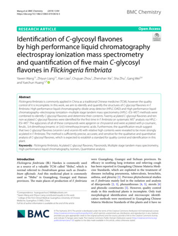
(2019) 13:94Wang et al. BMC RESEARCH ARTICLEBMC ChemistryOpen AccessIdentification of C‑glycosyl flavonesby high performance liquid chromatographyelectrospray ionization mass spectrometryand quantification of five main C‑glycosylflavones in Flickingeria fimbriataYawen Wang1†, Zhiyun Liang1†, Xian Liao2, Chujuan Zhou1, Zhenshan Xie1, Sha Zhu1, Gang Wei3,4and Yuechun Huang1,2*AbstractFlickingeria fimbriata is commonly applied in China as a traditional Chinese medicine (TCM), however the qualitycontrol of it is incomplete. In this work, we aim to identify and quantify the structures of C-glycosyl flavones in F.fimbriata. High performance liquid chromatography-diode array detector (HPLC-DAD) and High performance liquidchromatography–electrospray ionization–multiple stage tandem mass spectrometry (HPLC–ESI–MSn) methods werecombined to identify C-glycosyl flavones and determine their contents. Twenty acylated C-glycosyl flavones and tennon-acylated C-glycosyl flavones were identified for the first time in F. fimbriata on systematic M Sn analysis via HPLC–nESI–MS . The aglycones of all of these compounds were apigenin or chrysoeriol and were acylated with p-coumaric,ferulic, 3,4-dimethoxycinnamic or 3,4,5-trimethoxycinnamic acids. Furthermore, the quantification result suggestthat two C-glycosyl flavones (vicenin-I and vicenin-III) with relative high contents were revealed to be more stronglyacylated in F. fimbriata. The method is sufficiently precise, accurate, and sensitive for the qualitative and quantitativeanalysis of C-glycosyl flavones, which is expected to establish a standard for quality control and identification in thisplant.Keywords: Flickingeria fimbriata, Acylated C-glycosyl flavones, Flavonoids, Multiple stage tandem mass spectrometry,High-performance liquid chromatography, Isomers, Quantitative analysisIntroductionFlickingeria fimbriata (Bl.) Hawkes is commonly usedas a source of a valuable TCM called “Shihu”, which isnormal referred to Dendrobium genus such as Dendrobium officinale. And this medicinal plant is commonlyused as “Shihu” in Guangdong, Guangxi and Hainanprovinces. The main places of production of F. fimbriata*Correspondence: huangyuechun218686@outlook.com†Yawen Wang and Zhiyun Liang contributed equally to this work1College of the First Clinical Medical, Guangzhou University of ChineseMedicine, Guangzhou 510405, ChinaFull list of author information is available at the end of the articlewere Guangdong, Guangxi and Sichuan provinces. Itsefficacy in soothing lung irritation and relieving coughhas been reported in Guangdong Chinese Materia Medicine Standards, which are exploited in the treatment ofdiseases including pneumonia, tuberculosis, bronchitis,asthma, and pleurisy [1]. Previous phytochemical studieson F. fimbriata mainly lied in the isolation and analysisof diterpenoids [2, 3], phenanthrenes [4, 5], sterols [6]and phenolic constituents [5]. However, quality controlstudy in this medicinal plants is incomplete. Only traitmorphological identification and microscopic identification methods were mentioned in Guangdong ChineseMateria Medicine Standards of this plants and it have no The Author(s) 2019. This article is distributed under the terms of the Creative Commons Attribution 4.0 International License(http://creat iveco mmons .org/licen ses/by/4.0/), which permits unrestricted use, distribution, and reproduction in any medium,provided you give appropriate credit to the original author(s) and the source, provide a link to the Creative Commons license,and indicate if changes were made. The Creative Commons Public Domain Dedication waiver (http://creat iveco mmons .org/publi cdoma in/zero/1.0/) applies to the data made available in this article, unless otherwise stated.
Wang et al. BMC Chemistry(2019) 13:94specific method to control quality. The previous studyof F. fimbriata in our laboratory showed that 7–8 stablecommon peaks of flavonoids in the characteristic spectrawere found using HPLC [7], five of which were characterized as non-acylated C-glycosyl flavones (vicenin-II,vicenin-I, schaftoside, isoschaftoside and vicenin-III) byion-trap mass spectrometer. The other uncharacterizedpeaks still required further investigation to complete aquality control study. It was proved that flavone is a suitable compound for quality control study of F. fimbriata.Flavonoids, as common and widespread secondaryplant metabolites, distributed in all parts of plants. Theypresent as glycosides in the vacuoles, leaves, stem, androots of flowers [8]. Sugar substitution on the flavonoidskeleton may occur through hydroxyl groups, in the caseof O-glycosides (O-glycosyl flavones), or directly to carbon atoms in the A ring in C-glycosides (C-glycosyl flavones) [9]. Contents and types of C-glycosyl flavoneswere ideal index for identifying plants from the same species for its high specificity [10]. Generally, the flavonoidsclassification depends on the nature of aglycones, sugars,and acylate groups. Some secondary plant metabolitesoccur in the form of acylated glycosyl flavones with benzoic acid and/or cinnamic acid moiety. The cinnamoylgroups including p-coumaroyl, feruloyl, 3,4-dimethoxycinnamoyl and 3,4,5-trimethoxycinnamoyl [11–13], andtheir differences lie in the number and/or position ofhydroxy and methoxy substituents. Many compoundslike diterpenoids and phenanthrenes in F. fimbriata areacylated with aromatic acids, such as trans-cinnamoylacid [14] and methoxybenzoyl acid derivatives [3, 5, 14,15], suggesting that the aromatic acids could be synthesized in this plants. Moreover, O-methyltransferase(OMT) genes revealed the internal relations of cinnamic acids with different substituents, it was possiblethat OMTs might be associated with the formation of 3,4-dimethoxycinnamate and 3, 4, 5-trimethoxycinnamatein biosynthesis of plants [16]. Additionally, acylated flavonoids have several health beneficial effects including anti-inflammatory [17, 18] and antioxidant activity[19], and the acylation position on glucose is regard as apotential approach for the antioxidant and cytoprotectiveeffects of flavonoid glycosides [20].MS is important due to its applicability for analyzing herbal medicines. The application of electrosprayionization (ESI) enabled the analysis of flavonoid glycosides without derivatization [21]. Although distinctionbetween glycosidic and aromatic acidic substituents offlavonoids is problematic, such as deoxyhexoside andcoumaric acid, both of which lose a fragment of 146 Da[9], high-performance liquid chromatography (HPLC)combined with a diode array detector (DAD) couldprovide online UV spectrum for each individual peakPage 2 of 20in a chromatogram which displays different spectrumsbetween the glycosidic and aromatic acid substituentsof flavonoids. Additionally, HPLC–ESI–MSn, equippedwith an ion trap (IT) mass analyzer can obtain a largenumber of fragmentation patterns and typical losses upto MS4 [22] which could be used to identify many complex isomers of C-glycoside flavones [23]. By this way, thenature of aglycones and sugars as well as the position ofsugar and acyl groups could be deduced in C-glycosideflavones. The determination by MS fragmentation ofacylated O-glycosyl flavones is possible [24], and theO-glycosylation at 2′′ and at 6′′ positions could bededuced from it [25]. However, few systematic analysisof acylated C-glycosyl flavones in plants via HPLC–ESI/MSn combining with HPLC-DAD method was affordedbefore this study.To date, 20 acylated C-glycosyl flavones and 10 nonacylated C-glycosyl flavones were identified by HPLC–ESI–MSn and HPLC-DAD. These 30 compounds havenot been reported yet in F. fimbriata. In addition, 12batches of F. fimbriata were successfully quantitativelyanalyzed, which is expected to establish a standard forquality control and identification.Materials and ucoside(vicenin-1) were isolated from the leaves of D. officinale, andthese compounds were identified by comparing their UV,IR, HPLC–MS and NMR data with those in publishedreports [10], and purity was determined to be higherthan 98% by the normalization of the peak area side(vicenin-3) was purchased from Shanghai StandardTechnology Co., Ltd. (Shanghai, China), and its puritywas over 98%; schaftoside 92.5% was purchased fromthe National Institutes for Food and Drug Control (Beijing, China). Isoschaftoside was purchased from Extrasynthese (Genay, France), and its purity was over 95%.HPLC-grade acetonitrile was purchased from Merck(Darmstadt, Germany). Analytical grade methanol, formic acid, and phosphoric acid were obtained from FuyuFine Chemical Co., Ltd. (Tianjin, China). Distilled waterwas purchased from A.S, Watson Group Co., Ltd. (Hongkong, China). YUHUA SHZ-D (III) was purchased fromYUHUA Instrument Co., Ltd. (Gongyi, China).Twelve samples of natural medicinal parts of F. fimbriata were collected from different regions of China(Guangdong, Guangxi, and Sichuan provinces). Of these,6 batches were from Guangdong province (No. FF1–FF6), 4 batches were from Guangxi province (No. FF7–FF10), and 2 batches were from Sichuan province (No.
Wang et al. BMC Chemistry(2019) 13:94FF11–FF12) (Table 1). The tested samples of F. fimbriata(12 batches) were authenticated by professor YuechunHuang from The First Affiliated Hospital of GuangzhouUniversity of Chinese Medicine, Guangzhou, China. Thevoucher specimens (No. FF20190701) were deposited inthe School of Pharmaceutical Science, Guangzhou University of Chinese Medicine, Guangzhou.Preparation of standard solutionsThe standard samples of vicenin-II, vicenin-I, schaftoside,isoschaftoside and vicenin-III were accurately weightedand then dissolved with methanol to produce concentrations of 121.2, 120.0, 121.6, 118.4, and 122.0 μg/mL,respectively. A mixed standard solution was prepared bymixing the standard solution with methanol to obtain acertain injection amount in the range of 0.030–1.782 μg,0.029–3.234 μg, 0.028–1.687 μg, 0.028–1.687 μg, and0.030–3.288 μg, respectively.Preparation of sample extractionThe air-dried and smashed F. fimbriata (0.5 g) samplesfrom each batch material were accurately weighted andextracted with 50 mL of methanol, after being weightedwith a vessel, in the case of the volatilization of methanolthen refluxed for 4 h at 90 C using a Jie Rui Er HH-4 constant temperature water bath (Jiang Su Jie Rui Er electricCo., Ltd., Jiang Su, China). The extractions were removedand cooled down. The extraction was weighted again, andmethanol was added into the vessel to compensate for thelost weight. Of the filtrated extraction, 25 mL was accurately transferred into an evaporation pan. The resultantconcentrated extractions were transferred to a 2 mL volumetric flask and diluted to the indicated volume (2 mL).The obtained extract was filtrated through a 0.22 μmPage 3 of 20pore-size nylon filter for MS analysis and 0.45 μm poresize nylon filters for quantitative analysis.HPLC–ESI–MSn and HPLC‑DAD analysis conditionAnalysis was performed on an HPLC system equippedwith a vacuum degasser, quaternary pump, auto-sampler,and ultraviolet detector (Thermo Separation ProductsInc., Riviera Beach, FL, USA) and coupled with a ThermoFinnigan LCQ FLEET (Thermo Finnigan, Riviera BeachFL, USA) ion trap mass spectrometer, equipped with anelectrospray ionization interface in negative ion mode.Chromatographic separations were carried out on a Kromasil 100-5 C 18 column (250 mm 4.6 mm, 5 μm, AkzoNoble, Sweden), maintained at 35 C. The mobile phaseswere acetonitrile (A) and 0.1% (v/v) formic acid (B), at aflow rate of 0.8 mL/min. The gradient elution programwas 0–10 min, 14% A; 10–20 min, 14–16% A; 20–45 min,16–22% A; and 45–80 min, 22–40% A, with an elutiongradient. The injection volume was 5 μL each time. Thedetection wavelength was set to 340 nm. The optimizedMS conditions were as follows: full-scan mode betweenm/z 50 and 1000, spray voltage 3.0 kV, capillary voltagefixed at 35.0 V, capillary temperature 350 C, sheathgas flow rate of 30 (arbitrary units), and auxiliary gas flowrate of 10 (arbitrary units). The data acquisition and thesystem control were performed using a Finnigan Xcalibur 2.0 advanced chromatography workstation (ThermoQuest Corporation, San Jose, CA, USA). HPLC-DADanalysis was performed using an Agilent 1100 system(Agilent, USA). The conditions were as same as the chromatographic separation method in HPLC–ESI–MSn.HPLC quantitative analysis conditionQuantitative analysis was performed using an Agilent 1100 system (Agilent, USA). ChromatographicTable 1 The source of Flickingeria fimbriata (Bl.) HawkesNo.OriginSourceCollect timeFF1Guangdong Hexiang Pharmaceutical co., LTDGuangdong2014.8.14FF2Guangdong Lifeng Pharmaceutical co., LTDGuangdong2014.6.9FF3LBX Pharmaceutical co., LTDGuangdong2014.6.9FF4Jianmin Pharmaceutical co., LTDGuangdong2014.6.9FF5Jihetang Pharmaceutical co., LTDGuangdong2014.6.10FF6Yuqingtang Pharmaceutical co., LTDGuangdong2014.6.10FF7Caizhilin Pharmaceutical co., LTDGuangxi2014.6.9FF8Dashenlin Pharmaceutical co., LTDGuangxi2014.8.14FF9Guozilin Pharmaceutical co., LTDGuangxi2014.8.16FF10Baiyuantang Pharmaceutical co., LTDGuangxi2014.8.16FF11Jihetang Pharmaceutical co., LTDSichuan2014.6.9FF12Baohetang Pharmaceutical co., LTDSichuan2014.8.16
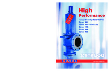
Wang et al. BMC Chemistry(2019) 13:94separations were carried out on a Zorbax SB-Aq column (250 mm 4.6 mm, 5 μm; Agilent, USA), maintained at 35 C. The mobile phases were acetonitrile (A)and 0.1% (v/v) phosphoric acid (B) (with a gradient elution program of 0–15 min, 12% A; 15–25 min, 13–14%A; 25–30 min, 14% A; 30–40 min, 14%–12% A), at a flowrate of 0.8 mL/min. The injection volume was 5–20 μLeach time. The detection wavelength was set to 340 nm.Results and discussionIdentification of chemical compoundsThe extract of F. fimbriata was analyzed by HPLC–ESI–MSn and HPLC-DAD. The UV chromatogram at 340 nmis shown in Fig. 1a, and its total ion chromatograms(TICs) are shown in Fig. 1b. Flavonoids typically exhibittwo major absorption bands in the ultraviolet region:Band I in the 320–385 nm region, representing B-ringPage 4 of 20absorption, and Band II in the 250–285 nm range, representing A-ring absorption [25]. These UV data are inaccordance with C-glycosyl apigenin and chrysoeriol,respectively [11, 26]. The majority of flavonoids with cinnamoyl acid have a UV spectrum with an intense Band Iat approximately 330 nm and a small Band II at approximately 270 nm (Fig. 1c), as a result of the overlapped UVspectra [27].C-glycosyl flavones, with the characteristic saccharides substitution directly attached to aglycone in ringA through a C–C bond, all show substituents in position 6 (C-6) and/or 8 (C-8) of the aglycone moiety [8].Apart from glucose, monosaccharides including xylose,arabinose, and rhamnose are ubiquitous in plants [28].Due to the cross-ring cleavages of the flavonoid saccharide residue, characteristic ions [Ag-H 42] and[Ag-H 72] were observed in the M S4 spectra for theFig. 1 HPLC–ESI–MSn analysis in the extracts of Flickingeria fimbriata: a HPLC–ESI–MS chromatogram at 340 nm; b HPLC–ESI–MS total ion current(TIC) profile in negative mode; c HPLC-DAD profile (340 nm) and UV spectra of compound 3, 10, 19, 21,22 and 26
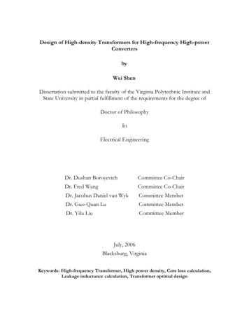
Wang et al. BMC Chemistry(2019) 13:94mono-C-glycosyl flavones, and characteristic ions [AgH 84] and [Ag-H 114] were observed in the MS4spectra for the 6,8-di-C-glycosyl flavones (Ag 270 forapigenin, Ag 300 for chrysoeriol) [24, 29]. In F. fimbriata, combining the loss of mass with the previouslyreported results, and considering the high contents ofvicenin-II, vicenin-I, schaftoside, isoschaftoside and vicenin-III, the xylose, arabinose, and glucose moieties werefound to be involved in glycosylation. The major fragmentation pathways concern the cross-ring cleavages ofthe saccharide residue and the loss of water molecules. Innegative mode, the characteristic ions of sugars in C-glycosyl flavones lost 120 Da and 90 Da in the hexose substituents, and 90 Da and 60 Da in pentose substituents bycrossing cleavages, respectively [30].In di-C-glycosyl flavones, sugar residues of differentmasses can be located, since the 6-C-sugar residue showsgreater fragmentation than the 8-C-sugar residue [8].From these findings, we deduced the types and positionof sugars. For the acylated-C-glycosyl flavones, and if thebase peak ions are made of [M-H-120] or [M-H-90] in MS2 spectra, there can be no acyl on the 6-C-sugar.If the base peak ions in M S2 spectra are constitutive ofPage 5 of 20the loss in acyl-relative ([M-H-Acyl] ) or acid-relative([M-H-Acid] ) neutral moiety, this suggests an acylation on the 6-C-sugar. When the position of acylationon the hydroxyl in position 2″ in sugar (2″-O), ions [(AgH 42)-18] were observed in the MS3 spectra for themono-C-glycosyl flavones, ions [(Ag-H 84)-18] and[(Ag-H 114)-18] were observed for the di-C-glycosylflavones, which differ from the position set to 6″ (6″-O).When hydroxyl in position 6″ in sugar (6″-O), ions [AgH 84] and [Ag-H 114] were obtained in the MS3spectra from di-C-glycosyl flavones (Fig. 2).The acyl group types were identified by neutral losses,which are characteristics of the acyl group or the acylatedglycosyl residue. The acylation of p-coumaroyl andferuloyl in the hydroxyl of the C-glucosylation sugarshowed higher polarity when compared to the acylationof 3,4-dimethoxycinnamoyl and 3,4,5-trimethoxycinnamoyl. These four types of acyl groups all belong to thederivatives of trans-cinnamoyl, but differ in the numberof hydroxy and methoxy substituents. Characteristicacyl-related product ions [M-H-Acyl] and acid-relatedproduct ions [M-H-Acid] were observed in the formertwo types in the CID MS2 spectra, whereas in the latterFig. 2 The proposed fragmentation pathways of acylated- C-glycosyl flavones when the position of acylation was on the hydroxyl in position 2″and 6″ in sugar (2″-O and 6″-O), a β-d-xyloside (14) and b xycinnamoyl)-β-d-glucoside (26)
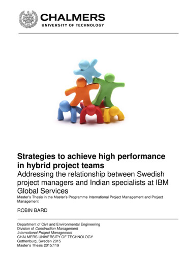
Wang et al. BMC Chemistry(2019) 13:94two, only [M-H-Acid] was be detected. That is to say,the lower polarity acylated-C-glycosyl flavones are without the loss of the radical acyl group neutral fragments.In the CID M S2 spectra, the neutral fragment losses are146 Da and 164 Da for p-coumaroyl (Fig. 3), and 176 Daand 194 Da for feruloyl (Fig. 4) in the hydroxyl of theC-glucosylation sugar, respectively [31], however, theyare only 208 Da for 3,4-dimethoxycinnamoyl (Fig. 5)and only 238 Da for 3,4,5-trimethoxycinnamoyl (Fig. 6).Finally, the acylated C-glycosyl flavones we found are allacylated with p-coumaroyl, feruloyl, 3,4-dimethoxycinnamoyl or 3,4,5-trimethoxycinnamoyl on the hydroxyl inthis work, and the majority of them are isomers.The position of the acyl group in the hydroxylof glucosyl flavones can also be identified by thePage 6 of 20characteristic of aglycone relative ions and the neutralfragments. When an acyl is in the 6″-O position onC-glucosylation glucose, simultaneous losses of acyland of hexose residue, here called acylhexose, wereobtained in fragment patterns, and the losses were266 Da, 296 Da, 310 Da and 340 Da for p-coumaroylhexose, feruloyl hexose, 3,4-dimethoxycinnamoylhexose, and 3,4,5-trimethoxycinnamoylhexose, respectively. To the best of our knowledge, this is the firstreport of 30 di-C-glycosyl flavones (Table 2) includingten non-acylated di-C-glycosyl flavones (Table 3), twoacylated-Mono-C-glycosyl flavones (Table 4), twelve6″-O -acylated-di-C-glucosyl flavones (Table 5) and six2″-O -acylated-di-C-glucosyl flavones (Table 6) on systematic MSn analysis in F. fimbriata.Fig. 3 CID MS2 spectra fragmentations of two pairs of isomeric C-glycosyl flavones, acylated with ρ-coumaroyl from the extracts ofFlickingeria fimbriata in negative ionization mode: a 11), b Apigenin-6-C-(2″-O-ρ-coumaroyl)-β-d-glucoside (12), c 8-C-β-d-xyloside (13), and d royl)-β-d-glucoside (18)
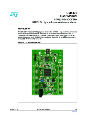
Wang et al. BMC Chemistry(2019) 13:94Page 7 of 20Fig. 4 CID MS2 spectra fragmentations of four C-glycosyl flavones, acylated with feruloyl from the extracts of Flickingeria fimbriata in negativeionization mode: a β-d-xyloside (14), b β-d-glucoside(16), c β-d-xyloside (17), and d )-β-d-glucoside (20)HPLC–ESI–MSn analysis of non‑acylated‑di‑C‑glycosylflavonesFor compounds 1 and 2, the base peak of fragment ions[(M-H)-120] in the CID MS2 spectra and the base peak ions [(M-H-120)-120] in the M S3 spectra suggested that the simultaneous glucosylation of glucoseoccurred in positions 6 and 8 [32]. A pair of fragmentsat m/z 353 [Ag(270)-H 84] and 383 [Ag(270)-H 114] in the MS3 spectra indicated that the nucleus of flavonoids is apigenin [33]. The coinstantaneous presence offragments at m/z 383 for [Ag(300)-H 84] and 411 for[Ag(300)-H 114] in the M S3 spectra indicated that thenucleus of flavonoids is chrysoeriol [29]. In both nucleuses of the flavones above, the presence of [Ag-H 56] and [Ag-H 28] were easily detected by the loss of28 Da (CO) and 56 Da (2CO) at the position of 6-C or/and 8-C, respectively. However, [Ag-H 28-15] couldonly be detected in the fragments of chrysoeriol-diC-glycosyl flavone by the loss of 15 Da (CH3) [5]. Fromall the results above, the compounds were found to beapigenin-6,8-di-C-β-d-glucoside (1, vicenin-II) andchrysoeriol-6,8-di-C-β-d-glucoside (2). In the compounds 3, 5, 6, and 8, di-C-glycosyl flavone was alsotypical due to [Ag-H 84] and [Ag-H 114] . Basepeak ions [(M-H)-90] and ions [(M-H)-60] in MS2 aswell as base peak ions [(M-H-90)-120] ([Ag-H 84] )in MS3 indicated the structure of Ag-6-C-pentose-8-Chexose. In the compounds 4, 7, 9, and 10. Additionally,the fragment ions [(M-H-90)-90] and [(M-H-60)-120] in MS2 from the 8-C-arabinoside flavone were producedmore extensively than 8-C-xyloside. The compoundswere found to be apigenin-6-C-β-d-xyloside-8-Cβ-d-glucoside (3, vicenin-I) and isoschaftoside),
Wang et al. BMC Chemistry(2019) 13:94Page 8 of 20Fig. 5 CID MS2 spectra fragmentations of five C-glycosyl flavones, acylated with 3,4-dimethoxycinnamoyl from the extracts of Flickingeria fimbriatain negative ionization mode: a glucoside-8-C-β-d-glucoside (19), b glucoside-8-C-β-d-xyloside (21), c oxycinnamoyl)-β-d-glucoside (22), d xycinnamoyl)-β-d-glucoside (26), and e thoxycinnamoyl)-β-d-glucoside lucoside(8). In compounds 4, 7, 9, and 10, the base peak ions[(M-H)-120] and ions [(M-H)-90] in MS2, and basepeak ions [(M-H-120)-90] ([Ag-H 84] ), apparentin MS3, indicated the structure of Ag-6-C-hexose-8C-pentose. Fragment ions [(M-H-120)-90] and [(MH-90)-90] in the M S2 spectra were produced more
Wang et al. BMC Chemistry(2019) 13:94Page 9 of 20Fig. 6 CID MS2 spectra fragmentations of five C-glycosyl flavones, acylated with 3,4,5-trimethoxycinnamoyl from the extracts of Flickingeriafimbriata in negative ionization mode: a -d-glucoside-8-C-β-d-xyloside (15), b ethoxycinnamoyl)-β-d-glucoside (23), c -d-glucoside-8-C-α-l-arabinoside (24), d thoxycinnamoyl)-β-d-glucoside (28), and e imethoxycinnamoyl)-β-d-glucoside (30)extensively for the 8-C-arabinoside flavone as opposedto 8-C-xyloside. Considering the results obtainedand the reported identification [33], we describethese compounds as apigenin-6-C-β-d-glucoside8-C-α-l-arabinoside (4, schaftoside), arabinoside oside(10) (Table 3). Compounds 1, 3, 4, 5, and 9 were confirmed by the reference standards using the eluted orderand retention time.HPLC–ESI–MSn analysis of acylated‑C‑glycosyl flavonesFor compounds 11 and 12, due to the higher relativeintensity produced by ions at m/z 293 [(M-H-164)-120] in MS2 in compound 12 relative to compound 11, togetherwith the base peak ions [(Ag-H 42)-18] obtained in the MS3 spectra, the characteristic of an acylation in the 2″-Oposition on mono-C-glycosyl flavones was confirmed(Table 4, Fig. 3a, b). 6-C-2″-O-coumaroyl-isomers caneliminate the acid between the 2″-O-coumaroyl groupof sugars and the 5- and 7-hydroxyl groups of aglycone, but 8-C-2″-O-coumaroyl-isomers only respond to
Wang et al. BMC Chemistry(2019) 13:94Page 10 of 20Table 2 C-glycosyl flavones structures based on the aglycones of apigenin and chrysoeriol from Flickingeria ylH30AraGlu-d2Ha: ρ-coumaroyl; b: feruloyl; c: 3,4-dimethoxycinnamoyl; d: 3,4,5-trimethoxycinnamoyl1The position of acylation is set to 2″-O in sugar2The position of acylation is set to 6″-O in sugarthe 7-hydroxyl group [27], which results in 6-C-2″-Ocoumaroyl isomers producing more acid-related ions.8-C-glycoside compounds, eluted before their 6-C isomer, also help distinguish the isomers [8]. Consequently,the compounds were deduced to be apigenin-8-C-(2″-Ocoumaroyl)-β-d-glucoside (11) and apigenin-6-C-(2″-Ocoumaroyl)-β-d-glucoside (12).For compounds 16, 18, and 20, base peak ions [M-H120] or [M-H-90] and ions [M-H-266/296] in the MS2 spectra showed the characteristic of a l)-henose flavone(Figs. 3d, 4b, d). Ions [Ag-H 84] and [Ag-H 114] were obtained in the MS3 spectra, which further confirmed the acylation at the 6″-O position (Table 5). Thecompound was determined to be �-d-glucoside (16). Ions[(M-H-90)-Acylhexose]-(or [Ag-H 84] ), found to beproduced at the base peak in MS3, prove the glucosylation of arabinose. It is likely that the compounds aroyl)β-d-glucoside (18) and -β-d-glucoside (20).For compounds 22, 23, 25–30, besides the aglycone ions [Ag-H 84] and [Ag-H 114] in the MS4 spectra (Table 5), base peak ions [M-H-120] together with ions [(M-H-120)-310/340] or [M-H90] with [(M-H-90)-310/340] in the M S2 spectra(Figs. 5c–e and 6b, d, e) characterize oyl/3,4,5trimethoxycinnamoyl)-henose flavones. The compoundswere determined to be xycinnamoyl)-β-d-glucoside(22) and thoxycinnamoyl)-β-d-glucoside (23). For compounds 26 and 29, when arabinose was substituted at6-C, 353[(M-H-90)-310] was abundant in the M S2spectra (Fig. 5d, e), whereas these ions showed a lowrelative intensity in M S2 for xylose substitution. Aswell, for compounds 28 and 30, 353[(M-H-90)-340] even became the base peak in the M S3 spectra for
ys-6-C-α- C27H30O15l-ara-8-C-βd-glu593593Chrys-6-C-β- C27H30O15d-glu-8-C-αl-araChrys-6-C-β- C27H30O15d-glu-8-C-βd-xyl593Chrys-6-C-β- H] (m/z)473 (77)baMS2 and MS3 in bracket means the ions were obtained in MS2 and MS3, respectivelyAg: aglycone, apigenin (Ag 270); chrysoeriol (Ag 300)Chrys chrysoeriol, glu glucose, ara arabinoside, xyl xyloside503 (5)353 (74)503 (74)383 (68)353 (46)533 (8)383 (100)383 (85)533 (28) 383 (71)533 (26) 383 (87)–503 (3)503 (100) 533 (5)473 (100) 503 (36)473 (100) 503 (41)503 (100) 533 (18)473 (90)353 (60)473 (100) 503 (69) 353 (93)443 (100) 473 (61)443 (84)–473 (100) 503 (60) 353 (83)443 (100) 473 (65)443 (61)473 (100) 503 (31)[AgH 84] 413 (47)413 (36)413 (55)413 (50)413 (30)383 (32)383 (97)383 (45)383 (56)383 (31)[AgH 114] 383 (100, MS2)383 (93)383 (100)383 (100)383 (100)353 (100)353 (100)353 (100)353 (100)353 (100)[AgH 84] 60 120 90MS3 (m/z)MS2 (m/z)aMain observed fragments. Other ions were found, but they have not been 7113MolecularformulaPeak No. Rt (min) CompoundsTable 3 [M-H] , MS2, MS3, and MS4 data of non-acylated di-C-glycosyl flavones in Flickingeria fimbriata355 (15)355 (32)355 (51)355 (29)325 (97)325(58)325 (81)325 (70)325 (75)[AgH 56] 413 (45, MS2) 355 (3, MS2)413 (100)413 (46)413 (44)413 (28)383 (13)383 (19)383 (20)383 (9)383 (12)[AgH 114] MS4 (m/z)312 (100)312 (100)312 (100)312 (100)–––––[Ag-H 2815] 327 (4, MS3) 312 (100, MS3)327 (7)327 (4)327 (2)327 (8)297 (100)297 (100)297 (100)297 (100)297 (100)[AgH 28] Wang et al. BMC Chemistry(2019) 13:94Page 11 of 20
ig-8-C-(2″-O-ρ-coum)-β-d-gluCompoundsba-Acid: ρ-coumaric acid ( 164 D
by high performance liquid chromatography electrospray ionization mass spectrometry and quanti cation of ve main C-glycosyl avones in Flickingeriambriata Yawen Wang †, Zhiyun Liang†, Xian Liao2, Chujuan Zhou1, Zhenshan Xie1, Sha Zhu1, Gang Wei 3,4 and Yuechun H