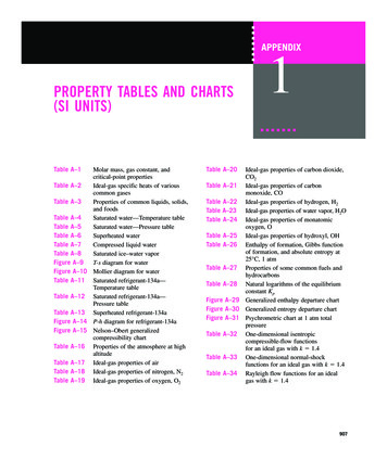
Transcription
Table of ContentsOverview of Advanced Cardiovascular Life Support . 2Updates to ACLS in 2015. 3Chain of Survival .4Adult BLS Chain of Survival. 4BLS and ACLS Surveys .5The BLS Survey . 5Solo Provider Adult BLS . 6Team Adult BLS . 7Cardiac Arrest.8Ventricular Fibrillation and Pulseless Ventricular Tachycardia . 9Ventricular Fibrillation and Pulseless Ventricular Tachycardia Algorithm . 10Pulseless Electrical Activity and Asystole . 11PEA and Asystole Algorithm . 12Respiratory Arrest . 13Airway Management .14Return of Spontaneous Circulation (ROSC) and Post Arrest Care . 16Rapid Differential Diagnosis of Cardiac Arrest . 17Bradycardia . 18Bradycardia Algorithm . 18Tachycardia . 19Tachycardia Algorithm . 20Atrioventricular (Heart) Block . 21Acute Coronary Syndrome. 22Acute Coronary Syndrome Algorithm. 23Acute Stroke . 24Time is Brain! Stroke Time Goals for Evaluation and Therapy . 25Team Dynamics . 26Resuscitation and Life Support Medications . 27
Overview of Advanced Cardiovascular Life SupportAdvanced Cardiac Life Support, or ACLS, is a system of algorithms and best practicerecommendations intended to provide the best outcome for patients in cardiopulmonary crisis.ACLS protocols are based on basic and clinical research, patient case studies, clinical studies, andreflect the consensus opinion of experts in the field. While the term Advanced Cardiovascular LifeSupport was coined by the American Heart Association, the content contained in this manual isbased on the most recent guidelines published by the American Heart Association, the AmericanCollege of Cardiology, the American Red Cross, and The European Society of Cardiology.Prior to taking ACLS, it is assumed that you are proficient and currently certified in Basic LifeSupport (BLS). Once you become certified in ACLS, the certification is valid for two years. However,we encourage you to regularly login back in to your account to check for updates on resuscitationscience advances.This Advanced Life Support provider manual includes: Updates to ACLS in 2015 Solo and Team BLS The ACLS Survey Cardiac and Respiratory Arrest Return of Spontaneous Circulation (ROSC) and Post Arrest Care Bradycardia and Tachycardia Management ECG Rhythm Recognition Atrioventricular Blocks Acute Coronary Syndrome and Acute Stroke Management Resuscitation Medications Team Dynamics and Systems of Care
Updates to ACLS in 2015As we learn more about resuscitation science and medicine, physicians and researchers realizewhat works best and what works fastest in a critical, life-saving situation. Therefore, it isnecessary to periodically update life-support techniques and algorithms. If you have previouslycertified in advanced cardiovascular life support, then you will probably be most interested inwhat has changed since the latest update in 2010. The table below also includes changesproposed since the last AHA manual was published. These changes will likely appear in futureeditions of the provider manual.
Chain of SurvivalAdvanced Cardiovascular Life Support continues to emphasize the Chain of Survival. The Chainof Survival is a sequence of steps or links that, when followed to its completion, increases thelikelihood that a victim of a life-threatening event will survive. The adult and pediatric chains ofsurvival are slightly different. The person who is providing BLS is only responsible for the earlylinks, that is, making sure the person is cared for by emergency personnel. The emphasis onearly care is to reinforce that time is a critical factor in life support care. The 2015 standardsinclude the concept of out of hospital care versus in-hospital care.Adult BLSChain ofSurvival (Out of Hospital)The links of the Adult ACLS Chain of Survival are: EMS – Early activation of EMS CPR – Early administration of High-quality CPR AED – Early use of an AED ACLS – Early Advanced Cardiovascular Life Support Post-Arrest Care – Transport victim to the hospitalAdult BLS Chain of Survival (In Hospital)This new chain should include: Primary Providers/ Code Team/ Cath Lab/ Post Arrest Care
BLS and ACLS SurveysACLS draws heavily on Basic Life Support (BLS). In fact, it is assumed that all people who arepursuing ACLS will be competent in the techniques of BLS—so much so that it is considered aprerequisite to ACLS.The first step in any resuscitation is to make sure therescuers (you!) and the victim are safe. Therefore, ifyour victim is in the middle of the highway or in aburning building, the first step is to move the victimto safety.Assuming you and the victim are in a safe location,the next step is to assess whether the patient isresponsiveIf patient is not responsive, move to BLS surveyIf patient is responsive, move to ACLS surveyThe BLS SurveyThe BLS Survey1. Responsive?Shake and Shout! Don’t be afraid to make noise.Check for effective breathing for 5 to 10 seconds.2. Activate EMS/Call CodeIn the hospital, you can call a “code” and send someone to get adefibrillator. In the community, call 911 and send for an AED3. CirculationSimultaneously, check the carotid pulse for no more than 10seconds while checking for respiratory effort. If no pulse, beginhigh quality CPR.If there is a shockable rhythm, pulseless ventricular tachycardia orventricular fibrillation, provide a shock4. DefibrillationAdult BLS is slightly different if there is one provider (solo) or more than one provider (team)present. The difference between solo provider BLS and team BLS is that responsibilities areshared when more than one person is present. These will be detailed in Solo and Team AdultBLS.For healthcare providers, the difference between a witnessed cardiac arrest and a victim who isfound down is the order of the initial steps. If you are alone and witness a victim suddenly collapse: Assume cardiac arrest witha shockable rhythm. If you can get an AED quickly, you may activate EMS, leave thevictim to get an AED, provide CPR for 2 minutes, and use the AED. If you are alone and find an unresponsive adult: Tailor response to the prospectivecause of injury. If you suspect cardiac arrest: Activate EMS, get AED, 2 min of CPR, use AED If you suspect asphyxia: 2 min of CPR, Activate EMS, get AED, use AED
Always make sure that you are safe and thevictim is safe before you start BLS. Check to see if the victim is responsive. Shakeand shout! Is the victim breathing effectively?Does the victim have a pulse in the carotidartery? If you witnessed the victim suddenly collapse,assume cardiac arrest with a shockablerhythm. If you can get an AED quickly, youmay activate EMS, leave the victim to get anAED, CPR for 2 minutes, and use AED. If you find an unresponsive adult, tailorresponse to the presumed cause of injury. If you suspect cardiac arrest: ActivateEMS, get AED, 2 min of CPR, use AED If you suspect asphyxia: 2 min of CPR,Activate EMS, get AED, use AED High Quality CPR includes Fast and deep compressions, 100to 120 compressions per minute Two inches deep, complete rebound If you can provide respiration, 2breaths for 30 comps If you cannot provide respiration,just give chest comps Check for a pulse and cardiac rhythm every two minutes. Follow directions on the AED.After providing a shock, immediately resume CPR. Keep going until EMS arrives or thevictim regains circulation.
Always make sure that your team is safe and thevictim is safe before you start BLS. Check to see if the victim is responsive. Shake andshout! Is the victim breathing effectively? Doesthe victim have a pulse in the carotid artery? One provider activates EMS and retrieves an AED.The other provider(s) stays with the victim. Provide High Quality CPR includes Fast and deep compressions, 100to 120 compressions per minute Two inches deep, complete rebound If you can provide breaths, 2 breathsfor 30 comps If you cannot provide breaths, justgive chest comps The provider who retrieved the AED applies theAED and follows directions given by the device.The provider that stayed with the victim providesCPR until the AED is ready. Check for a pulse and cardiac rhythm every twominutes. Follow directions on the AED. Ifa shock is indicated, clear everyone and administer a shock. After providing a shock,immediately resume Team CPR. In Team CPR, the provider giving chest compressions changes every 2 minutes Keep going until EMS arrives or the victim regains spontaneous circulation.
Cardiac ArrestCardiac arrest is the sudden sensation cessation of blood flow to the tissues in brain the resultsfrom a heart that is not pumping effectively. Four rhythms may occur during cardiac arrest:ventricular fibrillation, pulseless ventricular tachycardia, pulseless electrical activity, andasystole. The primary intervention for ventricular fibrillation and pulseless ventricular tachycardiais unsynchronized cardioversion, more commonly known as a “shock.” The primary interventionfor pulseless electrical activity and asystole is pharmacological, beginning with the administrationof epinephrine.While ACLS provides algorithms for each of these cardiac arrest rhythms, in the real world apatient may move between these rhythms during a single instance of cardiac arrest. Therefore,the provider must be able to accurately assess and adapt to changing circumstances. After every2 minutes of CPR, check for a pulse and check the cardiac rhythm. If the rhythm has switchedfrom shockable or to shockable, then switch algorithms.
Ventricular Fibrillation and Pulseless Ventricular TachycardiaIn ventricular fibrillation or pulseless ventricular tachycardia, the heart's conduction systemexhibits a disordered rhythm that can sometimes be corrected by applying energy to it. Thisenergy may come in the form of an automated external defibrillator (AED) defibrillator paddles,or defibrillator pads. VFib and VTach are treated with unsynchronized cardioversion, since thereis no way for the defibrillator to decipher the disordered waveform. In fact, it is important not toprovide synchronized shock for these rhythms.Ventricular fibrillation is recognized by a disordered waveform, appearing as rapid peaks andvalleys as shown in this ECG rhythm strip:Ventricular tachycardia may provide waveform similar to any other tachycardia; however, thebiggest difference in cardiac arrest is that the patient will not have a pulse and, consequently,will be unconscious and unresponsive. Two examples of ventricular tachycardia are shown in thisECG rhythm strips. The first is narrow complex tachycardia and the second is wide complextachycardia:
Ventricular Fibrillation and Pulseless Ventricular Tachycardia Algorithm Once you have determined thata patient has a shockablerhythm, immediately provide anunsynchronized shock. If you are using biphasic energy,use recommended settings onthe device. If you do not knowwhat that setting is, use thehighest available setting, (120 to200 J). If you are using a monophasicenergy source, administer 360 J. Resume CPR immediately after ashock. Minimize interruptions ofchest compressions. Provide 2rescue breaths for each 30compressions. Epinephrine (1 mg IV/IO) isgiven every 3 to 5 minutes (two2 minute cycles of CPR) Vasopressin (40 units IV/IO) canbe used instead of the first orsecond dose of epinephrine Amiodarone (IV/IO)o First dose 300 mgo Second dose 150 mgLidocaine may replace amiodarone when amiodarone is not available. o First dose: 1-1.5 mg/kg IVo Second dose: 0.5-0.75 mg/kg IV every 5 to 10 minIf the arrest rhythm is no longer shockable, move to PEA/Asystole algorithm If the patient regains consciousness, move to ROSC algorithm for him and him
Pulseless Electrical Activity and AsystolePulseless electrical activity or PEA is a cardiac rhythm that does not create a palpable pulse is even thoughit should. A PEA rhythm can be almost any rhythm except ventricular fibrillation (incl. torsade de pointes)or pulseless ventricular tachycardia.Asystole is the “flatline” on the ECG monitor. It represents a lack of electrical activity in the heart. It iscritically important not to confuse true asystole with disconnected leads or an inappropriate gain setting onan in-hospital defibrillator. Asystole may also masquerade as a very fine ventricular fibrillation. If the ECGdevice is optimized and is functioning properly, a flatline rhythm is diagnosed as asystole. Note thatasystole is also the rhythm one would expect from a person who has died. Consider halting ACLS efforts inpeople who have had prolonged asystole.It is inappropriate to provide a shock to pulseless electrical activity or asystole. Cardiac function can onlybe recovered in PEA or asystole through the administration of medications.
As long as the patient is in PEAor asystole, the rhythm is notshockable. Chest compressions/high-qualityCPR should be interrupted aslittleaspossibleduringresuscitation. After 2 min. of high-quality CPR,give 1 mg of epinephrine IV/IO.Consider advanced airway andwaveform capnography. Remember, chest compressionsareameansofartificialcirculation, which should deliverthe epinephrine to the heart.Without chest compressions,epinephrine is not likely to beeffective. Chest compressions should becontinued while epinephrine isadministered. Rhythm checks every 2 min. Epinephrine (1 mg IV/IO) is given every 3 to 5 minutes (after two, 2 minute cycles of CPR) Vasopressin (40 units IV/IO) can be used instead of the first or second dose of epinephrine If the arrest rhythm becomes shockable, move to VFib/Pulseless VTach algorithm If the patient regains circulation, move to ROSC algorithm
Respiratory ArrestWhile cardiac arrest is more common in adults than respiratory arrest, there are times whenpatients will have a pulse but are not breathing or not breathing effectively (e.g., agonalbreathing). A person who has a pulse but is not breathing effectively is in respiratory arrest.When you encounter a patient in need, you will not know he or she is in respiratory arrest, soperform a BLS assessment:
Airway ManagementIn ACLS, the term airway is used to refer both to the pathway between the lungs and the outside world and victim in thedevices that help keep that airway open. The simplest way to “manage an airway” is the head tilt-chin lift, which creates thestraightest passageway for air to flow into the lungs. As if the victim may have experienced head or neck trauma, airwaymanagement should include a jaw thrust, which leaves the head and neck unmoved, but which opens up the airway. Currentguidelines recommend that ventilations for a victim with pulses should be delivered every 5 to 6 seconds. It is critical that theprovider avoid delivering excessive ventilation which can lead to less effective circulation and cerebral vasoconstriction.
If one is to use a pocket mask or a bag mask to perform ventilations, it is important to make a tight seal with a maskon the victim's face. Proper use of these masks may require the rescuer to use one or even two hands to secure themask to the victim's face.A nasopharyngeal airway, which extends from the nose to the pharynx, can be used in both conscious and unconsciouspatients. An oropharyngeal airway can only be used in unconscious patients because it may stimulate the gag reflex.Advanced airways such as endotracheal tubes (ET tubes) and laryngeal mask airways (LMAs) usually require specializedtraining, but are useful in-hospital resuscitations (especially LMAs).
While nasopharyngeal and oropharyngeal airways are basic airways, they do require a bit ofpreparation and skill to use correctly.Inserting anOropharyngealAirway Select an airway that is thecorrect size for the patient Too big and it will damage thethroat Too small and it will press thetongue into the airway Place the device at the side ofthe patient’s face. Choose thedevice that extends from thecorner of the mouth to theearlobeInserting anNasopharyngealAirway Select an airway that is thecorrect size for the patient Place the device at the side ofthe patient’s face. Choose thedevice that extends from the tipof the nose to the earlobe. Usethe largest diameter device thatwill fit. Lubricate the airway with awater-soluble lubricant Clear the mouth or blood orsecretions with suction, ifpossible. Insert the device slowly, straightinto the face (not toward thebrain!) Insert the device so that thepoint is toward the roof of themouth or parallel to the teeth Do not press the tongue backinto the throat It should feel snug; do not forcethe device. If it feels stuck,remove it and try the othernostril. Once the device is almost fullyinserted, turn it so that thetongue is cupped by the interiorcurve of the device.Tips onSuctioning Adequate suctioning usuallyrequires negative pressures of 80 to -120 mmHg. Wallmountedsuction can deliver this, butportable devices may not. When suctioning the oropharynx,do not insert the catheter toodeeply. Extend the catheter tothe maximum safe depth andsuction as you withdraw. When suctioning an endotrachealtube, remember that the tube iswithin the trachea and you maybe suctioning near thebronchi/lung. Therefore steriletechnique should be used. Each suction attempt should befor no longer than 10 seconds.Prior to suctioning, give a briefperiod of 100% oxygen—remember that the patient will getno oxygen during suctioning. Monitor vital signs duringsuctioning and stop suctioningimmediately if the patientexperiences hypoxemia (O2 sats 94%), has a new arrhythmia, orbecomes cyanotic.
Return of Spontaneous Circulation (ROSC) and Post Arrest CareThe patient who has been successfully resuscitatedwill regain spontaneous circulation. You can detect spontaneous circulation by feeling apalpable pulse at the carotid artery. Even after Return of Spontaneous Circulation (ROSC),the patient still needs close attention and support. Thepatient is at risk for reentering cardiac arrest at anytime. Therefore, the patient should be moved to anintensive care unit. Titrate the patient's blood oxygen levels to 94% Does the person need an advanced airway? If so, itshould be placed. Add quantitative waveform capnography Titrate the patient's systolic blood pressure to at least 90 mmHg. this may require the addition of fluidsand/or vasopressors oEpinephrine IV 0.1-0.5mcg/kg/min oDopamine IV 5-10 mcg/kg/min oNorepinephrine IV 0.1-0.5 mcg/kg/min o 1-2 liters offluid resuscitation Does the person follow verbal commands? If not, theremay be neurological compromise. Consider inducingtherapeutic hypothermia with 4 C fluids during fluidresuscitation. Does the person have signs of myocardial infarction byECG? Move to ACS algorithm.
Rapid Differential Diagnosis of Cardiac ArrestMany different disease processes and traumatic events can cause cardiac arrest, but in anemergency, it is important to be able to rapidly consider and eliminate or treat the most typicalcauses of cardiac arrest. To facilitate remembering the main, reversible causes of cardiac arrest,they can be organized as the Hs and the Ts.The HsHypovolemiaHypoxiaSymptoms/Signs/TestsRapid heart rate, narrow QRS complex,Decreased heart rateInterventionFluid resuscitationAirway management, oxygenHydrogen Ion(Acidosis)Low amplitude QRS complexHypoglycemiaFingerstick glucose testingIV DextroseHypokalemiaFlat T waves, pathological U waveIV MagnesiumHyperkalemiaPeaked T waves, wide QRS complexHypothermiaThe on, sodium bicarbCalcium chloride, sodium bicarb,insulin/glucose, hemodialysisHistory of cold exposureRewarming w heart rate, narrow QRS complex, acute dyspnea, history ofchest traumaRapid heart rate and narrow QRS complexVariable, prolonged QT interval, neuro deficitsRapid heart rate, narrow QRS complexST segment elevation/depression, abnormal T wavesThoracotomy, venom (toxin-specific)Fibrinolytics, embolectomyFibrinolytics, Percutaneousintervention
BradycardiaBradycardia is any heart rate less than 60bpm.Inpractice,however,bradycardia is only a concern if it isunusual or abnormal for the patient orcausing symptoms.New cases of bradycardia should beevaluated, but most will not requirespecific treatment. Evaluation of bradycardia includescardiacandblood oxygenmonitoring and a 12 lead ECG ifavailable. Bradycardia may be treated by providingsupplemental oxygen and supporting thepatient's airway if needed. Unstable bradycardia (i.e., anabnormally slow heart rate that causesaltered mental status, hypotension,symptoms of shock, cardiac chest pain,or new signs and symptoms of heartfailure) should be treated immediately. Unstable bradycardia is first treatedwith intravenous atropine at a dose of 0.5 mg. Additional doses can be givenevery 3 to 5 min. up to a maximum of 3mg. Pulseless bradycardia is consideredPEA.If atropine is unsuccessful in treatingsymptomatic, unstable bradycardia, consider transcutaneous pacing, dopamine or epinephrine infusion, or transvenous pacing. Anintensive or cardiologist may need to be consulted for these interventions and the patientmay need to be moved to the intensive care unit
TachycardiaAtrial fibrillation is the most common arrhythmia. It is diagnosed by electrocardiogram,specifically the RR intervals follow no repetitive pattern. Some leads may show P waves whilemost leads do not. Atrial contraction rates may exceed 300 bpm. The ventricular rate oftenrange is between 100 to 180 bpm. The pulse may be “irregularly irregular.”Atrial flutter is a cardiac arrhythmia that generates rapid, regular atrial depolarizations at a rateof about 300 bpm. This often translates to a regular ventricular rate of 150 bpm, but may be farless if there is a 3:1 or 4:1 conduction. By electrocardiogram, or atrial flutter is recognized by asawtooth pattern sometimes called F waves. These waves are most notable in leads II, III, andaVF.Narrow QRS complex tachycardias include several different tachyarrhythmias. A narrow QRScomplex tachycardia is distinguished by a QRS complex of less than 120 ms. One of the morecommon narrow complex tachycardias is supraventricular tachycardia, shown below.Wide complex tachycardias are difficult to distinguish from ventricular tachycardia. Ventriculartachycardia leading to cardiac arrest should be treated using the ventriculartachycardia algorithm. A wide complex tachycardia in a conscious person should be treated usingthe tachycardia algorithm.
AlgorithmTachycardia is any heart rategreater than 100 bpm. In practice,however, tachycardia is usuallyonly a concern if it is 150 bpm orcausing symptoms.New cases of tachycardia should beevaluated with cardiac and bloodoxygen monitoring and a 12 leadECG if available. Tachycardia may be treated byproviding supplemental oxygen,supporting the patient's airway ifneeded, vagal maneuvers, and IVadenosine. Adenosine IV rapid push o First dose: 6 mgo Second dose: 12 mgUnstable tachycardia (i.e., anabnormally slow heart rate thatcauses altered mental status,hypotension,symptoms ofshock, cardiac chest pain, or newsigns and symptoms of heartfailure) should be treated withsynchronized cardioversion oradenosine. Consider beta-blockeror calcium channel blocker. Wide QRS tachycardia may require antiarrhythmic drugs. ProcainamideAmiodaroneSotalol20-50 mg IV, stop if hypotensive or maxdose of 17 mg/kg.Maintenance infusion at 1-4 mg/min.150 mg IV over 10 minutes with second dose forrecurrent VT.Maintenance at 1 mg/min for 6 h100 mg (1.5 mg/kg)over 5 min.
Atrioventricular (Heart) BlockAtrioventricular block or heart block is a failure of the heart's electrical system to properly coordinate conduction.There are four main types of atrioventricular block: first degree, second degree type I, second degree type II, andthird degree heart block. The types of second degree heart block are referred to as Mobitz type I and Mobitz type II.Second degree heart block Mobitz type I is also known as the Wenckebach phenomenon.Atrioventricular blocks may be acute or chronic. Chronic heart block may be treated with pacemaker devices. From theperspective of ACLS assessment and intervention, heart block is important because it can cause hemodynamicinstability and can evolve into cardiac arrest. In ACLS, heart block is often treated as a bradyarrhythmia.The PR interval is aconsistent size, butlonger or larger than itshould be in first degreeheart block.The PR interval increasesin size until a QRScomplexes dropped,resulting in missed“beat.”A QRS wave willoccasionally drop,though the PRinterval is thesame size.Complete dissociationbetween P waves andthe QRS complex. Noatrial impulses reach theventricle.
Acute Coronary Syndrome Acute coronary syndrome or ACS is aspectrum of signs and symptoms rangingfrom angina to myocardial infarction. ACSincludes ST segment elevation myocardialinfarction (STEMI) nonST segmentelevation myocardial infarction (NSTEMI),and unstable angina. Cardiac chest pain(any newchestdiscomfort) should be evaluated promptly.This includes high degree of suspicion byindividuals in the community, promptactionbyEMSpersonnel,rapidassessment in the emergency department,and definitive treatment. People with symptoms of cardiac ischemiashould be given oxygen, aspirin(if notallergic),nitroglycerin,andpossiblymorphine. Obtain a 12 lead ECG ASAP. The patient should be assessed in the EDwithin 10 min. of arrival. Draw and sendlabs (e.g., cardiac enzymes, coags),Obtain IV access.Give statin (if notcontraindicated). Obtain chest Xray.The results of the ECG will be the primaryguidance for how the patient with possiblecardiac chest pain is managed. The ECGdiagnosis of acute coronary syndrome can becomplex. STEMI is recognized by ST segmentelevation with/without pathological Q waves. NSTEMI can be a more challenging electrocardiographic diagnosis. It may result in ST segment depression, “flipped”T waves (T wave flattening or inversion), peaked T waves, U wave inversion, and bundle branch block. Theelectrocardiographic of diagnosis of an NSTEMI is beyond the scope of ACLS. Unstable angina is new onset cardiac chest pain without ECG changes, angina that occurs at rest and lasts for morethan 20 min., and/or angina that has become rapidly and progressively worse. STEMI and NSTEMI patients will have elevated cardiac markers in the blood (e.g. troponins) several hours after theacute event. People with unstable angina will not have elevated cardiac markers.
Acute Coronary Syndrome AlgorithmSTEMI patients should be treated per hospital protocol. This may include anti-plateletdrug(s), anticoagulation, a beta-blocker, an ACE inhibitor, a statin, and either PCI or afibrinolytic. NSTEMI is treated with medical therapy as above without a PCI or fibrinolytic, unless they donot improve with medical therapy. Patients with unstable angina are admitted and monitored for evidence of MI. Patients who do not “rule in” (develop MI) can undergo cardiac stress testing the next day or as an outpatient.
Acute StrokeThe Cincinnati Prehospital Stroke ScaleTaskAsk the patient to smileAsk the patient to say“you can't teach an old dognew tricks”AbnormalFace droops on one sideSpeech slursAsk patient to close eyesOne arm drifts lower and turns andextend both arms inward (pronator drift) straight out, palms upIf one of these tasks is abnormal, stroke probability is 72% If allthree tasks are abnormal, stroke probability is 85%The EMS team should take patients with suspectedstroke to a stroke center. While in transit, the
Support was coined by the American Heart Association, the content contained in this manual is based on the most recent guidelines published by the American Heart Association, the American College of Cardiology, the A