Transcription
Sox4 Is Required for the Survival of Pro-BCellsThis information is current asof March 13, 2013.ReferencesSubscriptionsPermissionsEmail AlertsJ Immunol 2013; 190:2080-2089; Prepublished online 23January 2013;doi: ppl/2013/01/24/jimmunol.1202736.DC1.htmlThis article cites 59 articles, 28 of which you can access for free #ref-list-1Information about subscribing to The Journal of Immunology is online at:http://jimmunol.org/subscriptionsSubmit copyright permission requests at:http://www.aai.org/ji/copyright.htmlReceive free email-alerts when new articles cite this article. Sign up at:http://jimmunol.org/cgi/alerts/etocThe Journal of Immunology is published twice each month byThe American Association of Immunologists, Inc.,9650 Rockville Pike, Bethesda, MD 20814-3994.Copyright 2013 by The American Association ofImmunologists, Inc. All rights reserved.Print ISSN: 0022-1767 Online ISSN: 1550-6606.Downloaded from http://jimmunol.org/ at Cleveland Health Sciences Library on March 13, 2013SupplementaryMaterialBaohua Sun, Saradhi Mallampati, Yun Gong, DonghaiWang, Véronique Lefebvre and Xiaoping Sun
The Journal of ImmunologySox4 Is Required for the Survival of Pro-B CellsBaohua Sun,* Saradhi Mallampati,* Yun Gong,† Donghai Wang,‡ Véronique Lefebvre,x,{ andXiaoping Sun*Bcells play pivotal roles in humoral immunity and are a keycomponent of the immune system. Like other blood celltypes, mature B cells arise from self-renewing pluripotenthematopoietic stem cells (HSCs) through a stepwise process involving coordinated cell proliferation along with progressive lineage commitment and differentiation. In the B cell lineage, HSCsfirst develop into lymphoid-primed multipotent progenitors, whichhave lost their self-renewal ability but remain multipotent, and theninto common lymphocyte progenitors (CLPs), which in turn develop into B cells, T cells, NK cells, and dendritic cells (1). The firstB cell–specific progenitors arising from CLPs are prepro-B cells,which sequentially develop into pro-B, pre-B, immature, and ultimately mature B cells.B cell development requires appropriate orchestration of a network of regulatory genes involved in cell survival, proliferation, and*Department of Laboratory Medicine, The University of Texas MD Anderson CancerCenter, Houston, TX 77030; †Department of Pathology, The University of Texas MDAnderson Cancer Center, Houston, TX 77030; ‡Division of Infectious Disease andImmunology, Department of Medicine, University of Massachusetts Medical School,Worcester, MA 01655; xDepartment of Cell Biology, Lerner Research Institute,Cleveland Clinic, Cleveland, OH 44195; and {Orthopedic and Rheumatologic Research Center, Lerner Research Institute, Cleveland Clinic, Cleveland, OH 44195Received for publication October 1, 2012. Accepted for publication December 18,2012.This work was supported by National Institutes of Health Grant 5R03AI079779-02(to X.S.), the MD Anderson Cancer Center Physician Scientist Program Award (toX.S.), American Cancer Society Research Scholar Grant 119645-RSG-10-131-01DDC (to X.S.), and MD Anderson Cancer Center Support Grant CA016672.Address correspondence and reprint requests to Dr. Xiaoping Sun or Dr. DonghaiWang, Department of Laboratory Medicine, The University of Texas MD AndersonCancer Center, 1515 Holcombe Boulevard, Houston, TX 77030 (X.S.) or Division ofInfectious Disease and Immunology, Department of Medicine, University of Massachusetts Medical School, 55 Lake Avenue North, Worcester, MA 01655 (D.W.).E-mail addresses: xsun@mdanderson.org (X.S.) and Donghai.Wang@umassmed.edu(D.W.)The online version of this article contains supplemental material.Abbreviations used in this article: CLP, common lymphocyte progenitor; EYFP,enhanced yellow fluorescent protein; HSC, hematopoietic stem cell; MFI, medianfluorescence intensity; pDC, plasmacytoid dendritic cell; Tg, transgenic.Copyright Ó 2013 by The American Association of Immunologists, Inc. 0022-1767/13/ 2736differentiation (2, 3). Particularly in early B cell development,several key transcription factors act in a hierarchical order toprecisely control the expression of critical genes (4, 5). Pu.1 isinvolved in the hematopoietic lineage fate decision at the branchpoint of myeloerythroid and myelolymphoid progenitor populations (6). Ikaros is a key factor in B lineage specification andcommitment. Ikaros-deficient or hypomorphic mutant mice havesevere defects in the development of the lymphoid system (7, 8).Ebf1 and Pu.1 activate IL-7R, the first gene that distinguishes CLPsfrom lymphoid-primed multipotent progenitors (9–11). E2Afunctions upstream of EBF, and the two transcription factors collaborate to activate Pax5 expression (12, 13). Pax5 is indispensablein B cell lineage commitment in that it activates B lineage–specificgenes and represses lineage-inappropriate genes (14, 15).Programmed cell death frequently occurs during B cell development, but despite intensive studies, the molecular mechanismsthat control this process are still elusive. Accumulating evidencehas demonstrated that members of the Bcl2 protein family, including Bcl2 (16), Bcl-xL (17, 18), Mcl1 (19), Bax (20), Bik (21),and Bim (22), are critical for B cell survival in vivo and in vitro.Other factors, such as Stat5 and Dicer1, are also involved inmaintaining B cell survival. Stat5 has been shown to mediatethe survival function of IL-7 signaling during B cell development(23, 24). Conditional deletion of Dicer1 in the earliest stage ofB cell development results in an almost complete absence of preB cells, owing to a massive increase in apoptosis in pre-B cells (25,26). In adult mice, signaling by stem cell factor and its receptorc-Kit is critical in pro-B and pro-T cell development (27). Thedownstream signaling pathways activated by c-Kit include activation of PI3K and Src kinase. However, c-Kit–mediated Src kinaseactivation, but not PI3K activation, is important for adult B celldevelopment, likely through signaling events related to cell proliferation and cell survival (28).Sox4 is a member of the SOX (SRY-related HMG box) transcription factor family and has been shown to play important rolesin embryonic development and differentiation (29). Germlinedeletion of Sox4 is embryonically lethal in mice (30). Embryoswith this deletion died at day 14 of development owing to circu-Downloaded from http://jimmunol.org/ at Cleveland Health Sciences Library on March 13, 2013The development of mature B cells from hematopoietic stem cells is a strictly orchestrated process involving multiple regulatorygenes. The transcription factor Sox4 is required for this process, but its role has not been systematically studied, and the underlyingmechanisms remain unknown. To determine when and how Sox4 functions in the stepwise process of B cell development, we usedmice harboring conditional null alleles for Sox4 and a Cre transgene. Sox4 deletion in hematopoietic stem cells almost entirelyeliminated pro-B cells in both fetal livers and adult bone marrow, resulting in a severe deficiency in later stage B cells, includingcirculating mature B cells. Sox4-deficient pro-B cells, particularly those expressing the stem cell factor receptor c-Kit, readilyunderwent apoptosis, and even more so when c-Kit activity was inhibited by imatinib. C-Kit–expressing pro-B cells showeddecreased activation of the c-Kit downstream protein Src upon Sox4 deletion. Likewise, the level of the anti-apoptotic Bcl2protein was decreased in residual pro-B cells, and its restoration using a Bcl2 transgene allowed not only partial rescue ofpro-B cell survival but also B cell maturation in the absence of Sox4. Our findings indicate that Sox4 is required for the survivalof pro-B cells and may functionally interact with c-Kit and Bcl2. The Journal of Immunology, 2013, 190: 2080–2089.
The Journal of ImmunologyMaterials and MethodsSignaling Technology, Danvers, MA), cells were incubated with surface Absas indicated. After being washed with staining solution, cells were incubatedin fixation solution (00-8222-49; eBioscience) for 20 min and subsequentlywashed twice with permeabilization buffer (00-8333-56; eBioscience). Cellswere then incubated with specific primary Abs for 30 min at room temperature and then incubated for 20 min on ice with PE-, allophycocyanin-,or FITC-conjugated anti-rabbit second Abs.Semiquantitative RT-PCRTotal RNA was isolated from freshly sorted bone marrow prepro-B or proB cells with TRIzol (Invitrogen, Carlsbad, CA). Next, 50 ng total RNA wasapplied for first-strand cDNA synthesis using the SuperScript II First-StrandSynthesis Kit (Invitrogen). Serially diluted cDNA was used for 28–32 cyclesof PCR with gene-specific primer pairs (Supplemental Table 1). PCRproducts were then separated in 1.5% agarose gel and visualized by aDyversity imaging system (Syngene, Frederick, MD).Statistical analysisThe Student t test, assuming unequal variances between the two samples,was applied to determine the significance of difference between experimental mice and their littermates. The p values were considered significantwhen the probability of a difference was ,0.05.MiceMice with loxP sites flanking the entire coding region of the Sox4 genewere described previously (36). Vav-Cre mice were provided by Dr.Dimitris Kioussis at the National Institute for Medical Research, TheRidgeway, London (37). H2K-Bcl2 transgenic (Tg) mice were provided byDr. Irving Weissman at Stanford University, Stanford, CA (38). Genotyping was performed by PCR, using genomic DNA extracted from mousetails. All mice were bred and maintained in a specific pathogen–free animal facility at The University of Texas MD Anderson Cancer Center,Houston, TX. All mouse experiments were performed in accordance withfederal laws as well as guidelines of the National Institutes of Health,and protocols were approved by the MD Anderson Animal Care and UseCommittee.Imatinib treatmentTo inhibit the c-Kit signaling pathway, 4- to 5-wk-old mice were given i.p.injections of 100 mg/kg imatinib (LC Laboratories, Woburn, MA) twicedaily in a volume of 100 ml PBS for 2, 3, or 7 consecutive days, as indicated. Mice were euthanized the day after the last injection, and sampleswere harvested for flow cytometric analysis.Cell surface staining, flow cytometry, and cell sortingSingle-cell suspensions were prepared at the time of autopsy from fetal liver,bone marrow (femurs and tibias), and spleen. All samples were incubatedwith ice-cold RBC lysis buffer with ammonium chloride for 4 min in thedark before staining with a combination of mouse-specific Abs at 4 C for 15min in staining solution (PBS supplemented with 1% BSA). Blood wascollected from the hearts of the mice in 15-ml Falcon tubes (Becton-Dickinson Biosciences, San Jose, CA) containing 50 ml 0.5 M EDTA in PBS.After two rounds of RBC lysis, the WBCs were subjected to staining.Abs used for flow cytometric analysis included the following: FITC–antiIgM (Jackson ImmunoResearch, West Grove, PA); FITC–anti-CD21(8D9), PE–anti-CD11c (N418), PE–anti-CD25 (PC61,5), PE–anti-NK1.1(PK136), allophycocyanin–anti-AA4.1 (AA4.1), PerCP–Cy5.5–anti-B220(RA3-6B2), biotin–anti-BP1(FG35.4), and eFluor 450–Streptavidin, allfrom eBioscience (San Diego, CA); FITC–anti-CD24 (M1-69), FITC–antiIgD (11-26c.2a), PE–anti-CD43 (S7), PE–anti-Ter119 (Ter-119), PE–antic-kit (2B8), allophycocyanin–anti-CD19 (1D3), biotin–anti-Cxcr4 (2B11),and biotin–anti–IL-7R (B12-1), all from Becton-Dickinson Biosciences.Biotinylated Abs were detected with FITC-, allophycocyanin-, PerCPCy5.5–, eFluor 450–, or PE-conjugated streptavidin (Becton-DickinsonBiosciences). Samples were acquired on a FACSCalibur or Influx highspeed sorter (BD), and data were analyzed with FlowJo software (TreeStar, Ashland, OR). The absolute number of each B cell population wasderived from the total number of bone marrow cells per mouse and thepercentage of the population in question.Annexin V and intracellular stainingFor cell apoptosis analysis, the staining buffers containing surface Abs werecarefully removed before biotin-conjugated Annexin V was applied accordingto the manufacturer’s instructions (Becton-Dickinson Biosciences). For intracellular detection of Bcl2 (2870), total Src (2109), or p-Src (2101; CellResultsSox4 is required for pro-B cell developmentTo determine at which step in normal B cell development Sox4 isfirst required, we generated mouse strains harboring the Sox4conditional null allele and the Vav-Cre transgene. The Vav-Cretransgene is under the control of the Vav1 gene promoter and isexpressed in HSCs (39, 40). Sox4fl/flVav-Cre mice were present atnormal mendelian ratios and externally indistinguishable fromtheir littermate controls (Sox4fl/ Vav-Cre and Sox4 / Vav-Cre)at birth and until at least 1 y of age.Flow cytometric analysis revealed that the number of pro-B cells,including fraction B (B220 CD43 CD24 BP1–) and fraction C–C9(B220 CD43 CD24 BP1 ), was much lower in the bone marrowof adult Sox4fl/flVav-Cre mice [Hardy’s nomenclature for subpopulations of developing B cells (41)]. Whereas control littermates had 0.55 million pro-B cells, the Sox4fl/flVav-Cre mice hadonly 0.04 million (p 0.0019; Fig. 1A). The number of fraction Acells (i.e., prepro-B, B220 CD43 CD24–BP1–) was slightly decreased (0.14 million in control mice versus 0.07 million in Sox4fl/flVav-Cre mice; p 0.064). Fraction A cells are heterogeneous andcan give rise to, in addition to B cells, NK and plasmacytoiddendritic cells (pDCs) (42–44). The AA4.1 subset of fraction Acells contains B lineage precursors (45), the NK1.1 subset produces NK cells, and the CD11c subset generates pDCs (46, 47).The numbers of AA4.1 , NK1.1 , and CD11c fraction A cells inSox4fl/flVav-Cre bone marrow were comparable to those in thecorresponding subsets of control mice (Fig. 1B). These data indicate that deletion of Sox4 significantly affected pro-B cells butonly minimally affected prepro-B cells. Moreover, a moderatedecrease in the number of Lin2c-Kit Sca-1 primitive HSCs(36 3 103 in control mice versus 12 3 103 in Sox4fl/flVav-Cre mice;p 0.03; Fig. 1C) and a minimal increase in the number of CLPs(Lin2IL-7R c-Kit Sca-1 ; 65 3 103 in control mice versus 78 3103 in Sox4fl/flVav-Cre mice; p 0.18; Fig. 1D) suggested that themarked reduction in pro-B cells was unlikely to be caused bychanges in earlier stage cells (HSCs, CLPs, and prepro-B cells).As a consequence of the pro-B cell depletion, bone marrow fraction D (pre-B, IgM–B220 CD25 ), fraction E (immature B, IgM B220low), and fraction F (mature B, IgM B220high) cells were almost completely eliminated (Fig. 1E). In the spleen, the percentagesof immature (B220 AA4.1 , including transitional T1, T2, and T3)and mature (B220 AA4.1–, including marginal zone and follicular)B cells were severely reduced (7.12% and 61.3% in control miceDownloaded from http://jimmunol.org/ at Cleveland Health Sciences Library on March 13, 2013lation failure as a result of malformation of the semilunar valvesand ventricular septum. In vitro culture of liver cells from Sox42/2embryos failed to generate B cells in the presence of IL-7. Reconstitution of lethally irradiated adult mice with the Sox42/2 fetalliver cells showed a stringent arrest of donor B cell development atthe pro-B cell stage. These findings indicated that Sox4 is indispensable for B cell development in the fetal liver. However, howSox4 deficiency causes the fetal B cell developmental arrest andwhat role Sox4 plays in adult B cell development remain unknown.Sox4 has since been shown to be critical for cell survival anddifferentiation in many cell lineages other than B cells in embryonic development and postnatal life, and to act largely in redundancy with its close relatives Sox11 and Sox12 (31–35). In thisstudy, we used Sox4fl/fl mice and Vav-Cre recombinase to investigate the effect of Sox4 deletion in HSCs on B cell development.Our results showed that Sox4 was essential for pro-B cell survivaland might functionally interact with c-Kit and Bcl2.2081
2082Sox4 IN REGULATION OF B CELL DEVELOPMENTDownloaded from http://jimmunol.org/ at Cleveland Health Sciences Library on March 13, 2013FIGURE 1. B cell development in the bone marrow of Sox4fl/flVav-Cre mice. Flow cytometric analyses were performed to assess B cells at differentdevelopmental stages in Sox4fl/ Vav-Cre (fl/ Cre) and Sox4fl/flVav-Cre (fl/fl Cre) mice. The numbers in the plots are the percentages of the indicated cellgroups. Bar graphs show the total number of cells per mouse, presented as mean SD with p values indicated. (A) Bone marrow B220 CD43 lymphocyteswere analyzed for fraction (Fr.) A (prepro-B; B220 CD43 Cd24–), fraction B (early pro-B; B220 CD43 Cd24 BP1–), and fraction C–C9 (late pro-B; B220 CD43 CD24 BP1 ). Note that the percentage of fraction A cells was 24.3% in Sox4fl/ Vav-Cre mice and 79.3% in Sox4fl/flVav-Cre mice (flow cytometricplot). This difference was mostly due to the decrease in fraction B–C9 cells; the total number of fraction A cells was 0.14 million in Sox4fl/ Vav-Cre miceand 0.07 million in Sox4fl/flVav-Cre mice (bar graph). (B) Bone marrow fraction A cells were analyzed for the expression of NK1.1, AA4.1, and CD11c. (C)Bone marrow Lin2 cells were analyzed for the expression of c-Kit and Sca-1 to assess the number of hematopoietic stem cells. Lin, lineage markers,including CD11b, B220, CD4, CD8, Ter119, NK1.1, Gr-1, and CD19. (D) Bone marrow Lin2 and IL-7R cells were analyzed for the expression of c-Kitand Sca-1 to assess the number of common lymphocyte progenitors. (E) Bone marrow lymphocytes were analyzed for fractions D (pre-B; IgM2B220 CD25 ), E (immature B; IgM B220low), and F (mature B; IgM B220high) B cells (n 3–5 mice per group).
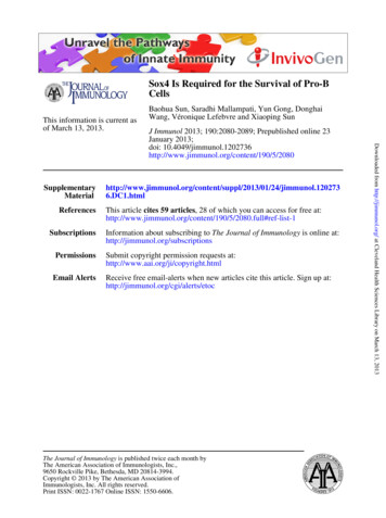
The Journal of Immunology2083versus 0.56% and 5.31% in Sox4fl/flVav-Cre mice, respectively; Fig.2A, 2B). As shown in Fig. 2C and 2D, B220 CD19 peripheralblood B cells were essentially absent. However, the peripheral bloodresidual B cells in Sox4fl/flVav-Cre mice expressed IgM and IgD onthe surface, indicating that these cells were able to develop intomature B cells.It was shown previously that Sox42/2 fetal liver hematopoieticcells fail to develop into mature B cells in recipient adult miceafter transplantation (30). However, it was not known which earlystage B cells were absent in the fetal livers of the Sox4-null embryos. We found that pro-B cells (B220 CD43 CD24 CD19 )were almost undetectable in the fetal livers of day 13.5 Sox4fl/flFIGURE 3. Sox4 deletion in B cells at various stages in Sox4fl/flVav-Cre mice. (A and B) Total RNA prepared from bone marrow FACS-sorted prepro-Bcells (A) and pro-B cells (B) was subjected to RT-PCR to assess Sox4 expression. The cDNA of pro-B cells was 53 serially diluted for semiquantitative RTPCR (B). (C) Quantitative RT-PCR analysis of Sox4 mRNA expression in pro-B cells of Sox4fl/ Vav-Cre and Sox4fl/flVav-Cre mice, with or without Bcl2 Tgoverexpression. (D and E) Fractions A–F (D) and peripheral blood B cells [B220 CD19 ; (E)] from Sox4f/ Vav-CreRosa26EYFP (blue line) and Sox4fl/flVavCreRosa26EYFP (red line) mice were subjected to flow cytometric analysis to assess EYFP expression, which serves as an indicator for Sox4 deletion.Downloaded from http://jimmunol.org/ at Cleveland Health Sciences Library on March 13, 2013FIGURE 2. B cell development in the spleen, peripheral blood, and fetal liver of Sox4fl/flVav-Cre mice. (A) Splenocytes were analyzed for immature(I, B220 AA4.1 ) and mature (M, B220 AA4.1–) B cells. The immature B cells were further analyzed for transitional T1 (B220 AA4.1 IgM CD23–), T2(B220 AA4.1 IgM CD23 ), and T3 (B220 AA4.1 IgM–D23 ) cells, and the mature B cells were further analyzed for marginal zone (MZ, B220 AA4.1–IgM CD23–) and follicular (FO, B220 AA4.1–IgM CD23 ) B cells. (B) The number of individual cells per million spleen lymphocytes. (C) Peripheralblood B cells (B220 CD19 ) were analyzed for the expression of IgM and IgD. (D) Percentage of B cells in peripheral blood. (E) Fetal liver cells from dayE13.5 embryos were analyzed for B220 CD43 CD24 CD19 pro-B cells (data are representative of two independent experiments). Gating strategies andabbreviations are the same as in Fig. 1, and n 3–5 mice per group unless otherwise specified.
2084Sox4 IN REGULATION OF B CELL DEVELOPMENTDownloaded from http://jimmunol.org/ at Cleveland Health Sciences Library on March 13, 2013FIGURE 4. Effect of Sox4 deletion and c-Kit inhibition on the apoptosis of B cells. (A) Apoptotic frequency measured as the percentage of Annexin V–positive cells in fractions A–F in Sox4fl/ Vav-Cre (blue line) and Sox4fl/flVav-Cre (red line) mice. The histogram shows one of the representative experiments, whereas the bar graph shows results (mean SD with p values indicated) from five experiments. (B) The expression of critical B cell developmentalgenes in pro-B cells was determined by semiquantitative RT-PCR. Also included were two antiapoptotic genes, Bcl2 and Mcl1. The actin cDNA was usedas control. Three cDNA concentrations with 53 serial dilution were used for each gene. The size of the PCR products is in- (Figure legend continues)
The Journal of ImmunologySox4 is vital for the survival of pro-B and pre-B cellsTo measure apoptosis in minute populations of B cells, particularlyin mice with B cell deficiency caused by Sox4 deletion, we usedhighly sensitive multicolor flow cytometry with Annexin V staining(49, 50). Annexin V staining may vary in B cells with a differentstatus of activation (51), but is a reliable marker for apoptosiswhen comparing cells at similar stages in development (52, 53).As shown in Fig. 4A, a minimal increase in the frequency ofapoptosis in prepro-B (fraction A) cells was detected in Sox4fl/flVav-Cre mice (3.55% versus 6.44%; p 0.39), whereas the frequency of apoptosis in pro-B (fraction B–C9) and pre-B (fractionD) cells increased dramatically (respectively, 5.61% versus 33.6%,p 0.011; and 1.76% versus 10.1%, p 0.014). In contrast, bonemarrow immature and mature (fractions E–F) B cells of Sox4fl/flVav-Cre mice had a frequency of apoptosis comparable to that incontrols. These results suggested that Sox4 was crucial for maintaining the survival of pro-B and pre-B cells but not required forthe survival of later stage B cells.Sox4 cooperates with the c-Kit signaling pathway to regulatepro-B cell survivalTo find out whether Sox4 exerts its function by regulating the genesknown to be critical in early B cell development, we sorted out thesmall number of residual pro-B cells and characterized the expression of these genes by semiquantitative RT-PCR. Of the genesstudied, including IL-7r, Cxcr4, E2a, Ebf1, Pax5, Foxp1, Stat5a,Ezh2, and Rag1, none showed a significant difference in expression between Sox4fl/flVav-Cre mice and their control littermates(Fig. 4B). Because Sox4 deficiency caused pro-B cell apoptosis,we also determined the mRNA levels of two antiapoptotic genes—Bcl2 and Mcl1, which were also known to be involved in earlyB cell development—and found no significant difference in them,either (Fig. 4B). However, the possibility still existed that Sox4regulates some of these gene products at the posttranscriptional level.We analyzed apoptosis in residual c-Kit pro-B cells from thebone marrow of the Sox4fl/flVav-Cre mice. Lack of Sox4 expression significantly increased cell death in c-Kit pro-B cells (IgM2B220 c-Kit CD19 ; 6.95% versus 36.2%; p , 0.001) but not inc-Kit non–pro-B cells (IgM2B220 c-Kit CD192; 11.5% versus11.1%; p 0.9; Fig. 4C). Although deletion of one copy of theSox4 gene (Sox4fl/ Vav-Cre) resulted in only minimal apoptosis ofc-Kit pro-B cells (Fig. 4D, left), it caused nearly three times thefrequency of apoptosis in pre-B cells compared with control(3.36% versus 9.65%; Fig. 4D, right), which did not express c-Kit.We hypothesized that c-Kit compensates for the deleterious effectof Sox4 deletion in pro-B cells. We administered 100 mg of thec-Kit inhibitor imatinib by i.p. injection twice daily for 2, 3, or 7 d.Whereas the frequency of apoptosis in c-Kit pro-B cells wascomparable between Sox4 fl/ Vav-Cre and Sox4 / Vav-Cre miceafter 0 and 2 d of injection, the difference in the frequency ofapoptosis was 3-fold (29.1% versus 10.9%) and 1.5-fold (46.9%versus 30.2%) after 3 and 7 d of injection, respectively (Fig. 4E).Because Src kinase is downstream of c-Kit and plays a critical rolein pro-B cell development (28, 54), we determined the levels oftotal Src and phosphorylated Src (p-Y416) in pro-B cells fromSox4fl/flVav-Cre and Sox4 fl/ Vav-Cre mice by flow cytometricanalysis. We found no overt difference in total Src, but phosphorylated Src (p-Y416) was significantly reduced upon Sox4deletion (median fluorescence intensity [MFI] 29.4 versus 13.2;Fig. 4F).B cell development in Sox4fl/flVav-Cre mice can be partiallyrescued by Bcl2Because the frequency of apoptosis in Sox4-deficient pro-B cellswas significantly increased and lack of the antiapoptosis proteinBcl2 was reported to increase the susceptibility of pro-B cells toproapoptotic agents (55, 56), we analyzed the level of Bcl2 proteinin Sox4-deficient pro-B cells. Although Bcl2 mRNA did not showsignificant change (Fig. 4B), Bcl2 protein was reduced in proB cells (MFI 261.3 versus 96.7), but not in prepro-B cells (MFI509.1 versus 633.3; Fig. 5A, left) in the Sox4fl/flVav-Cre mice,according to flow cytometric analysis. A reduction by 66% in Bcl2protein was detected by Western blot in B220 bone marrowB cells (Fig. 5A, right). These data indicated that the proapoptoticeffect of Sox4 deficiency in pro-B cells was associated with reduction of Bcl2 at the protein level.Forced expression of Bcl2 in Bcl2-Tg mice has been shown torestore hematopoietic cell development in a number of gene-dicated in bp. (C) The frequency of apoptosis in c-Kit pro-B cells (IgM–B220 c-Kit CD19 ) was measured by the percentage of Annexin V–positive cellsin Sox4fl/ Vav-Cre and Sox4fl/flVav-Cre mice. The apoptotic frequencies of c-Kit non–pro-B cells (IgM–B220 c-Kit CD192) are shown for comparison.The histogram shows one of the representative experiments, whereas the bar graph shows results (mean SD with p values indicated) from fiveexperiments. (D) Comparison of apoptosis in c-Kit pro-B cells and CD25 pre-B cells between Sox4 / Vav-Cre and Sox4fl/ Vav-Cre mice. (E) Frequency ofapoptosis in bone marrow pro-B cells (IgM2B220 CD19 c-Kit ) after imatinib treatment. Percentages of Annexin V–positive cells from mice that hadreceived imatinib injections for the indicated number of days were determined by flow cytometry. Data are representative of two mice per group. (F) Flowcytometric analysis of total Src and phosphorylated Src (p-Y416) in c-Kit pro-B cells from Sox4fl/ Vav-Cre and Sox4fl/flVav-Cre mice. The numbersrepresent MFI. Gating strategies and abbreviations are the same as in Figs. 1 and 2. The bar graph shows the percentage of p-Src in Sox4fl/flVav-Cre pro-Bcells compared with that in Sox4fl/ Vav-Cre pro-B cells. n 3–5 mice per group unless otherwise specified.Downloaded from http://jimmunol.org/ at Cleveland Health Sciences Library on March 13, 2013Vav-Cre embryos (Fig. 2E). This result suggested that deletion ofthe Sox4 gene in embryonic HSCs resulted in the abrogation offetal liver pro-B cells and that this abrogation was responsible forthe B cell deficiency in fetal liver cell–reconstituted adult bonemarrow and in vitro culture.To exclude the possibility of incomplete or escape of Sox4deletion in the residual B cells in Sox4fl/flVav-Cre mice, we sortedout the minute populations of prepro-B cells and pro-B cells andanalyzed the Sox4 mRNA levels. Sox4 mRNA was not detectablein bone marrow fraction A cells (Fig. 3A) or residual pro-B cells(Fig. 3B) in Sox4fl/flVav-Cre mice by RT-PCR. Real-time RT-PCRshowed that Sox4 mRNA in pro-B cells was decreased 133-fold inSox4fl/flVav-Cre mice (Fig. 3C, left). We also crossed the Sox4fl/flVav-Cre mice with Rosa26–enhanced yellow fluorescent protein(EYFP) Cre reporter mice to assess Cre activity in residual B cells.It was found that .90% of bone marrow prepro-B (fraction A)cells and .95% of peripheral blood residual B220 CD19 B cellswere EYFP positive (Fig. 3D, 3E), implying that the floxed Sox4allele had been deleted in these cells. The relative low percentagesof EYFP cells in pro-B (fractions B–C9) and pre-B (fraction D)cells of Sox4fl/flVav-Cre mice compared with control mice wereexpected because of the high frequency of apoptosis in these cells(see below); EYFP (or GFP ) cells become EYFP2 (or GFP2 )cells when undergoing apoptosis (48). The presence of EYFP B cells in peripheral blood suggested that a minor portion of proB cells overcame the effect of Sox4 deficiency and continued todevelop into mature B cells.2085
2086Downloaded from http://jimmunol.org/ at Cleveland Health Sciences Library on March 13, 2013FIGURE 5. Effect of Bcl2 on B cellapoptosis induced by Sox4 deletion. (A)Left panel, Flow cytometric analysis ofBcl2 expression in prepro-B (B220 CD43 CD242CD192) and pro-B cells (B220 CD43 CD24 CD19 ) from Sox4fl/ VavCre (blue line) and Sox4fl/flVav-Cre (redline) mice. The numbers represent MFI.Data represent three independent experiments. Right panel, Western blot results ofthe B220 bone marrow cells from thesemice. (B–D) Frequency of apoptosis measured as the percentage of Annexin V–positive cells in bone marrow c-Kit pro-Bcells (B), pre-B cells (C), and IgM (immature and mature) B cells (D), with orwithout forced Bcl2 expression. Data represent three mice per group. Gating strategies and abbreviations are the same as inFigs. 1 and 2. (E) Semiquantitative RTPCR of Sox4 expression in prepro-B(B220 CD43 CD242 CD192 ) and pro-B(
The Journal of Immunology Sox4 Is Required for the Survival of Pro-B Cells Baohua Sun,* Saradhi Mallampati,* Yun Gong,† Donghai Wang,‡ Ve ronique Lefebvre,x,{and Xiaoping Sun* The development of mature B cells from hematopoietic stem cells is a st