Transcription
0-Frontmatter-Preface-Foreword-RL.qxd4/5/20052:14 PMPage iRabbit and RodentDENTISTRYH A N D B O O KVITTORIO CAPELLO, DVMDipl ECZM-Small Mammals, Dipl ABVP-ECMWith Margherita Gracis, DVM, Dipl AVDC and EVDCEdited by Angela M. Lennox, DVM, Dipl ABVP-Avian, ECMZoological Education Network
0-Frontmatter-Preface-Foreword-RL.qxd4/5/20052:14 PMPage iiiiPREFACEAlthough great care has been taken to provide accurate and current information, neither the authors nor the publisher nor the editor shall be liable for any loss, damage orliability directly or indirectly caused or alleged to be caused by this book. The materialcontained herein is not intended to provide specific advice or recommendation for anyspecific situation.Library of CongressA catalog record of this book is available from the Library of Congress(http://catalog.loc.gov)ISBN: 0-9706395-1-1Zoological Education Network, Inc.PO Box 541749, Lake Worth, Florida 33454-1749 USAwww.exoticdvm.comCreative and Project Director: Richard Larson 2005 Zoological Education Network, Inc. All rights reserved.Neither this book nor any part may be reproduced or transmitted in any form or by anymeans, electronic or mechanical, including photocopying, microfilming and recording,or by any information storage and retrieval system, without written permission from thepublisher.Current printing (last digit):10 9 8 7 6 5 4 3 2 1Printed in Canada
0-Frontmatter-Preface-Foreword-RL.qxd4/5/20052:14 PMPage iiiiiiPREFACEThe authors thank:Akiteru Amimoto, DVM, PhDAmica Pet Clinic, UbeYamaguchi, JapanCristiano Colombo, DVMBusto Arsizio (Va), ItalyDavid A. Crossley, DVMAnimal Medical Centre Referral ServicesManchester, UKGabriele Ghisleni, DVMMilano, ItalySonia Giola, DVMMilano, ItalyCristina Pieralisi, DVMCentro Diagnostico VeterinarioMilano, ItalyAlexander Reiter, DVM, Dipl Tzt, Dipl AVDC, Dipl EVDCDepartment of Clinical StudiesVeterinary Hospital of the University of PennsylvaniaPhiladelphia, USAAyako Okuda, DVM, PhD, Dipl AVDCVettec Dentistry, Sumida-kuTokyo, JapanGiuseppe Ripamonti, DVMVarese, ItalyGermana Scerbanenco, DVMMilano, ItalyYosuke Sugiura, DVMDepartment of Anatomy, Faculty of Veterinary MedicineAzabu University, SagamiharaKanagawa, Japanfor their contributions to this handbook.(contributors are listed alphabetically)
0-Frontmatter-Preface-Foreword-RL.qxd4/5/20052:14 PMPage vvPREFACECONTENTSForewordPrefaceDavid CrossleyVittorio Capello1Introduction to Dentistryin Exotics and Exotic Mammals13Vittorio Capello and Margherita GracisLAGOMORPHSTHE RABBITAnatomy of the Skull and TeethDental FormulaSuperficial MusclesDeep Muscles, Arteries, Veins and NervesMedial MusclesNasolacrimal DuctRODENTSPorcupine-like (Hystrychomorph) RodentsRat-like (Myomorph) RodentsSquirrel-like (Sciuromorph) RodentsTHE GUINEA PIGAnatomy of the Skull and TeethDental FormulaSuperficial and Deep MusclesTHE CHINCHILLAAnatomy of the Skull and TeethDental FormulaTHE GOLDEN HAMSTERAnatomy of the Skull and TeethDental FormulaTHE RUSSIAN HAMSTERAnatomy of the Skull and TeethDental FormulaTHE RATAnatomy of the Skull and TeethDental FormulaSuperficial and Deep Muscles3Oral Physiology3737384143Vittorio CapelloVittorio Capello2Anatomy of the Skull and TeethTHE PRAIRIE DOGAnatomy of the Skull and TeethDental FormulaSIZE COMPARISON OF THE 23334343536LAGOMORPHSJaw at RestJaw RetractionJaw ProtrusionChewing MovementsRODENTS4The Clinical Exam43444445464749Vittorio CapelloTHE RABBITCommon Presenting SymptomsAssociated with Dental DiseaseCommon Presenting SignsAssociated with Dental DiseaseCommon Presenting PrimaryDental DiseaseCommon Presenting DiseasesSecondary to Dental DiseaseFacial and Oral Findings Associatedwith Oro-dental DiseaseNormal Incisor TeethTHE GUINEA PIGNormal Incisor TeethTHE CHINCHILLANormal Incisor TeethTHE HAMSTERNormal Incisor TeethTHE PRAIRIE DOGNormal Incisor TeethDENTAL RECORDS49495050505152555556565959606060
0-Frontmatter-Preface-Foreword-RL.qxd4/5/20052:14 PMPage viviPREFACE5Radiology of the Skull and Teeth65Vittorio Capello and Margherita GracisEQUIPMENT AND INSTRUMENTSRADIOGRAPHIC PROJECTIONS6567Skull RadiographsUse of Non-screen Dental FilmsTHE RABBITSkull RadiographsContrast Radiography of the Nasolacrimal DuctDental RadiographsTHE GUINEA PIGSkull RadiographsTHE CHINCHILLASkull RadiographsTHE DEGUSkull RadiographsTHE GOLDEN HAMSTERSkull RadiographsTHE RUSSIAN HAMSTERSkull RadiographsTHE RATSkull RadiographsTHE PRAIRIE DOGSkull RadiographsDental RadiographsTHE 98SIZE COMPARISONOF RADIOGRAPHIC IMAGES6Endoscopy98101Vittorio Capello and Margherita GracisINSTRUMENTSTHE RABBITTHE GUINEA PIGTHE CHINCHILLATHE DEGUTHE GOLDEN HAMSTERTHE PRAIRIE DOG7Other Diagnostics101102104105106106107109Vittorio CapelloHEMATOLOGY AND BIOCHEMISTRYBACTERIAL CULTURE AND SENSITIVITY109109HISTOPATHOLOGYCOMPUTERIZED TOMOGRAPHICSCANNING8Dental Diseases110110113Vittorio CapelloTHE RABBITDENTAL DISEASES OF INCISOR TEETHCongenitalTraumatic InjuriesMandibular Prognathism and MaxillaryBrachygnathismMalocclusion of Cheek TeethMetabolic Bone DiseaseStages of Incisor MalocclusionPATHOPHYSIOLOGY OF DENTALDISEASEImproper and/or Insufficient WearingMetabolic Bone DiseaseDENTAL DISEASES OF CHEEK TEETHStages of Cheek Teeth Malocclusionand Acquired Dental DiseaseSpurs and Malocclusion of MandibularCheek TeethLongitudinal Fractures of Cheek TeethSpurs and Malocclusion of MaxillaryCheek TeethEnd Stage Acquired Dental Diseaseof Cheek TeethTHE GUINEA 3135139Dental Diseases of Incisor TeethDental Diseases of Cheek TeethTHE CHINCHILLA139141145Dental Diseases of Incisor TeethDental Diseases of Cheek TeethTHE GOLDEN HAMSTER145146152Dental Diseases of Incisor TeethDental Diseases of Cheek TeethTHE RUSSIAN HAMSTER152154155Dental Diseases of Incisor TeethDental Diseases of Cheek TeethTHE GERBILTHE RATTHE PRAIRIE DOGDental Diseases of Incisor TeethPseudo-odontomaDental Diseases of Cheek TeethTHE CHIPMUNK155156156157158158159161163
0-Frontmatter-Preface-Foreword-RL.qxd4/5/20052:14 PMPage viiviiPREFACE9Secondary Diseases165Vittorio CapelloTHE DENTAL DISEASE “SYNDROME”THE RABBITSystemic DiseaseSkin DiseaseOcular DiseaseGastrointestinal DiseaseLesions of the Tongue and Oral MucosaFacial Abscesses and OsteomyelitisTHE GUINEA PIGGastrointestinal DiseasePeriapical Abscesses and OsteomyelitisTHE CHINCHILLATHE HAMSTER10Medical 718719019119211Dental Instrumentsand Equipment213Vittorio Capello and Margherita GracisVittorio CapelloANTIBIOTIC THERAPYANALGESIC THERAPYSUPPORTIVE THERAPYOTHER MEDICAL THERAPIES12Dental ProceduresREDUCTION OF INCISOR HEIGHTIN RABBITSPARTIAL PULPECTOMY ANDDIRECT PULP CAPPINGREDUCTION OF INCISOR HEIGHTIN RODENTSOCCLUSAL ADJUSTMENT:Cheek Teeth in RabbitsCheek Teeth in Guinea PigsCheek Teeth in ChinchillasEXTRACTION OF ARADICULARHYPSODONT TEETH:Mandibular Incisor TeethMaxillary Incisor TeethComplicationsCheek Teeth, IntraoralCheek Teeth, ExtraoralEXTRACTION OF ARADICULARHYPSODONT TEETH INPORCUPINE-LIKE rgical Treatmentof Periapical Abscessations249Vittorio Capello and Margherita Gracis193Vittorio Capello and Margherita GracisDIAGNOSISOTHER INSTRUMENTSMECHANICAL TRIMMINGMANUAL TRIMMINGDENTAL EXTRACTION193201202205207SURGICAL TREATMENT OFABSCESSES OF DENTAL ORIGIN211PATIENT SELECTION ANDDECISION MAKING249MARSUPIALIZATIONPostoperative DebridementPostoperative Follow-upLong-term Follow-up250254255258INTRODUCTION OF PMMA BEADS259CALCIUM HYDROXIDE PACKING261DIFFUSE OSTEOMYELITIS263DEBRIDEMENT OF MAXILLARYABSCESSATION264ABSCESSATION OF THENASOLACRIMAL DUCT266CHEEK TEETH ABSCESSATION:In the Guinea PigIn the Chinchilla268271References274
0-Frontmatter-Preface-Foreword-RL.qxd4/5/20052:14 PMPage viiiviiiPREFACEFOREWORDThere are many books available on rabbitand rodent medicine but few include aserious look at dental disease, and with acouple of exceptions, those that do tend to bepoorly illustrated. It is therefore very nice to seethis book which brings together basic scientificknowledge and presents it along with graphicalillustration of the range of pathology and practical treatment methods.Owners now expect to have their animals, ofwhatever species, treated appropriately placingincreasing pressures on their veterinarians.Luckily an increasing proportion of these owners realize that it costs money to obtain the necessary specialized equipment and skills to treatthe less frequently seen species, and that theyhave to pay a professional fee for veterinarytreatment. Long gone are the days when thelimit to what an owner would pay was the cost ofa new animal.Whilst there were some dedicated “rabbit” surgical and dental instruments available at thetime I qualified (27 years ago) the selection wasvery limited: a mouth gag, a cheek dilator and atotally inappropriate rasp for grinding overlongcheek teeth. Without the right equipment we areseverely limited in the quality of work we canoffer. This has changed dramatically over thelast 10 years. An increasing number of dedicated instruments are being produced as the resultof work done by a small number of pioneers ofgood “exotic” animal dentistry. At the last countthere were 11 different commercial designs ofrabbit and rodent incisor elevators and luxatorson the market, one new design per year sincethe first was introduced.Medicine advances rapidly, the amount ofknowledge doubling every 5-10 years. Generaltechnological advances occur at a similar rateand with each advance the cost of equipmentfalls in real terms. Air driven dental units, ultrasound scanners and video-endoscopic instrumentation are now affordable by most veterinaryclinics, opening up the possibility of their use inall species, not just cats and dogs. Endoscopesare particularly useful for examining inside smallopenings, such as the mouths of rabbits andsmall rodents. As can be seen from the illustrations in Chapter 6, these are very useful both fordiagnostic examination and for observation during treatment.David Crossley
0-Frontmatter-Preface-Foreword-RL.qxd4/5/20052:14 PMPage ixixPREFACEP R E FA C Exotic or non-traditional pet species areincreasing in popularity worldwide. As aresult, veterinarians interested in themedicine and surgery of these special speciesare faced with challenges to provide the highestquality care.with a useful tool to aid in the diagnosis andMore and more continuing education seminarsand publications describe advanced exotic medicine and surgery topics such as endoscopy,orthopedics, surgery and others. Recently, thesubject of rabbit and rodent dentistry hasreceived considerable attention.Another chapter describes dental instrumenta-In the past, dental disease of rabbits wasdescribed simply as “slobbers,” “lumps,” or simply “malocclusion” with little thought to underlying etiology. Today, the many differing clinicalsigns, symptoms, and etiologies are well recognized and more properly grouped as a syndrome,including primary dental disease, and dental disease secondary to other underlying conditions.The handbook utilizes a modern visual styleEIn addition, dental disease is being recognizedin rodent species as well, and it is clear thesespecies with their unique anatomical and physiological differences often require a slightly different diagnostic and treatment approach.The primary goal of the Rabbit and RodentDentistry Handbook is to provide the practitionertreatment of dental disease in these variousspecies. Veterinarians new to exotic mammalmedicine will appreciate sections focusing ongross and radiographic anatomy, physiology, andvisual and endoscopic examination techniques.tion and equipment. Of primary importance arechapters on the manifestations of dental diseaseand treatment recommendations. Many procedures are described in step-by-step detail.with over 1000 full color photos and accompanying text, a style well recognized by those familiar with Exotic DVM magazine and the variousexcellent publications produced by ZoologicalEducation Network.We owe a special acknowledgement to Dr. DavidCrossley, our friend and mentor, who has kindlyprovided many charts, illustrations and photographs for this handbook. We would also like tothank Dr. Ayako Okuda, who provided additional much needed support for this project.Vittorio Capello
0-Frontmatter-Preface-Foreword-RL.qxd4/5/20052:16 PMPage xxPREFACEANGELA M. LENNOXVITTORIO CAPELLOMARGHERITA GRACISDVM, Dipl ECZM-SM, Dipl ABVP-ECMDVM, Dipl AVDC, Dipl EVDCDVM, Dipl ABVP-Avian, ECMDr. Vittorio Capello graduated in1989 from the School of VeterinaryMedicine of the University of Milano,Italy. He has practiced exotic animalmedicine exclusively since 1993,providing professional services fortwo veterinary clinics in Milano. Dr.Capello’s focus has been the medicine and surgery of small exoticpets, in particular rabbits, rodentsand ferrets.Dr. Capello has lectured, publishedand taught exotic animal coursesand practical laboratories throughout Italy and other parts of Europe,and is a frequent guest lecturer atthe International Conference onExotics, where he was voted mostappreciated speaker two years in arow. He has written articles forExotic DVM Magazine and theJournal of Exotic Mammal Medicineand Surgery. Other works includethe Small Rodent Surgeries sectionin “The Exotic Guidebook” (ZoologicalEducation Network) and an educational CD for Italian veterinarianson the medicine and surgery of thepet hamster. Dr. Capello is a memberof the advisory board of Exotic DVMmagazine.Dr. Margherita Gracis graduated in1993 from the Veterinary School ofthe University of Milano, Italy. Afterworking in private practice for a fewyears, she completed a Residency inVeterinary Dentistry at the Veterinary School of the University ofPennsylvania, Philadelphia (USA).From 1998 until 2000 she worked atthe same institution as a lecturer inVeterinary Dentistry. Since 2000, Dr.Gracis has been working at tworeferral clinics in Milano (Italy) andin Monza (Milano, Italy), dedicated todentistry and oral surgery. She is aDiplomate of both the American(AVDC) and the European (EVDC)Veterinary Dental Colleges. Dr. Gracisis the current Past President of theEuropean Veterinary Dental Society(EVDS) and the Italian VeterinaryDental Society (SIODOV).Dr. Angela Lennox graduated in1989 from Purdue University Schoolof Veterinary Medicine, and has practiced avian and exotic animal medicine exclusively since 1991. She isthe owner of the Avian and ExoticAnimal Clinic of Indianapolis. Dr.Lennox was awarded board certification in avian medicine by theAmerican Board of Veterinary Practitioners in 2004, and is an adjunctprofessor at Purdue University whereshe teaches courses in exotic petmedicine to both veterinary and veterinary technician students. She currently serves as the President of theAssociation of Exotic Mammal Veterinarians. Dr. Lennox is a frequentguest lecturer at the InternationalConference on Exotics and theConference of the Association ofAvian Veterinarians, and has servedas editor for numerous publications,including Seminars in Exotic PetMedicine, Veterinary Clinics of NorthAmerica, Exotic DVM magazine andthe Journal of Exotic MammalMedicine and Surgery. She is a member of the advisory board of ExoticDVM magazine.
2-Anatomy of the Skull and Teeth-RLnew.qxd4/5/20051:08 PMPage 3CHAPTER 2Anatomy of theSkull and TeethVITTORIO CAPELLOMARGHERITA GRACISThe distinctive anatomy of rabbits and rodents must be thoroughly appreciated in order to successfully diagnose and treat dental disease. A discussion of dental anatomy is more meaningful with an understandingof precise terms used in dentistry.Table 2.1. Dental and Periodontal Anatomic Glossary (Figures 2.1, 2.2)AlveolarcrestThe most coronal portion of the alveolarboneFurcationIn a brachyodont tooth with multiple roots,the area where roots divergeApexThe termination or end of a tooth root. Thisterm is also used to define the termination ofaradicular (see elodont) teeth. It is normally“open” (with a single large foramen) inaradicular hypsodont teeth and immaturebrachyodont teeth, “closed” (with an apicaldelta) in adult brachyodont teethGingivalmarginThe most coronal portion of the gingivaGingivalsulcusThe shallow space between the gingiva andthe tooth, measured from the gingival marginto the gingival attachment to the toothsurface (junctional epithelium)A group of fine channels at the apex of abrachyodont tooth root through which thepulp blood vessels and nerves passLaminaduraThe wall of the dental alveolus or socket,visible radiographically as a radiopaque lineNeckThe portion of the brachyodont toothbetween the crown and the rootApicaldeltaCementoenameljunctionWhere enamel and cementum meet(corresponds to the neck of brachyodontteeth)Crown oranatomicalcrownPortion of the tooth covered by enamelCrown,clinicalExposed portion of the tooth within themouth, above the gingival marginCrown,reserveIn a hypsodont tooth, the part of the crownlocated below the gingival marginPeriodontal The space between the tooth and thespacealveolar bone occupied by periodontalligament fibersPulp cavityThe pulp chamber and root canal in a tooth,containing pulp tissueRootPortion of the brachyodont tooth covered bycementum. The reserve crown of aradicularhypsodont teeth is often improperly referredto as “root”
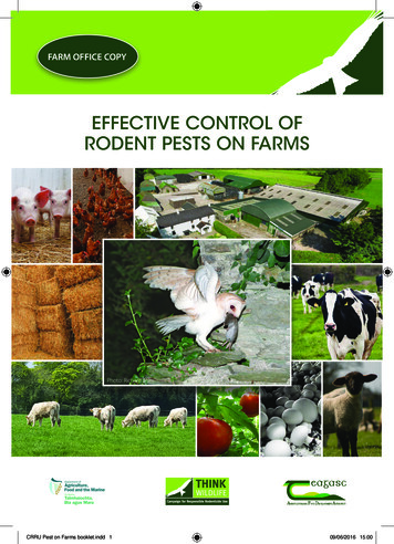
2-Anatomy of the Skull and Teeth-RLnew.qxd4/5/20051:10 PMPage 88CHAPTER 2RABBIT AND RODENT DENTISTRY HANDBOOKLAGOMORPHSTHE RABBITTable 2.6. Dentition of LagomorphsDiphyodontDeciduous and permanent sets of teethHeterodontDifferently shaped incisor and cheek teethElodontContinuously growing and erupting teeththat do not develop anatomical roots (alsocalled aradicular)HypsodontLong crowned teethDuplicidentataDouble row of maxillary incisor teethAnatomy of the Skull and TeethFigure 2.9. Gross anatomy of the skull of the European rabbit (Oryctolagus cuniculus), left lateralview. The mouth is long, with a narrow opening. Incisor and cheek teeth are separated by an edentulous gap called diastema. Lagomorphs, as well as rodents, lack canine teeth. The rabbit temporomandibular joint is dorsal to the dental occlusal plane. This skull morphology is typical of true herbivores, and is similar to larger species such as horses, cattle, sheep and goats. Also, rabbits havetypical herbivorous dentition, with continuously growing incisor and cheek teeth that do not formanatomical roots. The embedded portion of the teeth, commonly referred to as the root, is in factthe so-called reserve crown (see Figure 2.2).When the lower jaw is at rest, mandibular incisor teeth occlude between the first and second maxillary incisor teeth, and the cheek teeth are slightly separated. Cheek teeth come into direct contactonly during chewing, through the movement of the mandible caudally, and shifting of the condyloidprocess into a step of the temporal joint surface (see Figure 3.2).Incisor teeth of lagomorphs are anatomically and functionally similar to those of rodents, and forthis reason were once classified as a suborder of rodents. However, unlike rodents with only one pairof maxillary incisor teeth, rabbits and hares usually have two pairs of teeth, namely two larger labialfirst incisor teeth and two smaller palatal second incisor teeth (or “peg” teeth) (Figures 2.10, 2.11).Peg teeth are cylindrical in shape, and may be absent in some individuals. The maxillary first and themandibular incisor teeth are typically chisel-shaped. Peg teeth may protect the palate from the sharpincisal edge of the mandibular incisor teeth.
2-Anatomy of the Skull and Teeth-RLnew.qxd4/5/20051:11 PMPage 99ANATOMY OF THE SKULL AND TEETHCHAPTER 2THE RABBITFigure 2.10. Close up of incisor teeth, gross specimen, lateral view.Figure 2.11. Close up of incisor teeth on live animal, lateralview.Figure 2.12. Dorsal view of the skull.Figure 2.13. Ventral view of the skull, mandible removed.Figure 2.14. Ventral view of the skull, with the mandible inplace.
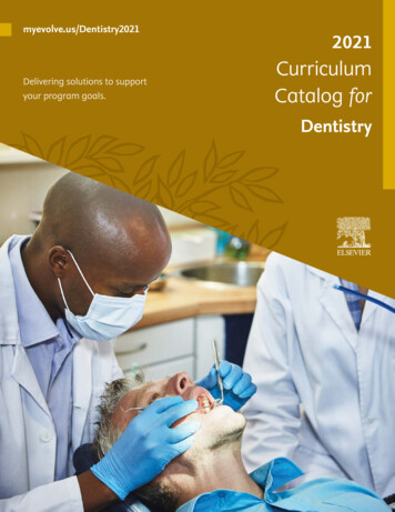
2-Anatomy of the Skull and Teeth-RLnew.qxd4/5/20051:12 PMPage 1010CHAPTER 2RABBIT AND RODENT DENTISTRY HANDBOOKTHE RABBITFigure 2.15. Cheek teeth. Rabbits have three premolar andthree molar teeth on each maxillary quadrant, and two premolar and three molar teeth on each mandibular quadrant.Premolar and molar teeth are visually indistinguishable, andare therefore simply called “cheek teeth.” Longitudinal groovesare present on the buccal surface of the cheek teeth. Transverse enamel ridges on the occlusal surface interlock with theopposite teeth during chewing, providing an efficient roughsurface for grinding and crushing of fibrous food material.As maxillary and mandibular cheek teeth differ in number,each mandibular tooth occludes with two maxillary teeth. Thefirst and the sixth maxillary cheek teeth only occlude with thefirst and the fifth mandibular cheek teeth, respectively.Table 2.7. Dental Formula2I0C3P3M1I0C2P3M 28The permanent dental formula of lagomorphs includes sixincisor and 22 cheek teeth. Lagomorphs are diphyodont, withtwo sets of teeth. The deciduous teeth are lost just before orshortly after birth.Figure 2.16. Dorsal view of the skull, focusing on the leftorbital fossa. The apex of four overgrown cheek teeth can beseen protruding through the bony plate in the pterygopalatinefossa.
2-Anatomy of the Skull and Teeth-RLnew.qxd4/5/20051:16 PMPage 1515ANATOMY OF THE SKULL AND TEETHTHE RABBITDeep Muscles, Arteries, Veins and NervesFigure 2.28. Diagram of the deep muscles, arteries, veins and nerves of the head of the rabbit, lateral view.Reprinted from A Colour Atlas of Anatomy of Small Laboratory Animals, Vol. I, Popesko P, Rajtovà V, Horàk J, pp 32, 35, 39, 40, 1992, with permission from Elsevier.CHAPTER 2
2-Anatomy of the Skull and Teeth-RLnew.qxd4/5/20051:22 PMPage 2323ANATOMY OF THE SKULL AND TEETHCHAPTER 2THE GUINEA PIGabFigures 2.40a,b. Cheek teeth. Guinea pigs and other porcupine-like rodents have one premolar and three molar teeth on theleft and right maxilla and mandible. Premolar and molar teeth have no anatomical or physiological differences, and are therefore simply called “cheek teeth.” Therefore, guinea pigs have four mandibular and four maxillary cheek teeth on each side, fora total of 16 cheek teeth. Deep longitudinal grooves are present on the buccal surface of the cheek teeth (Figure 2.43). Eachmandibular cheek tooth occludes with one single opposing maxillary tooth. Mandibular and maxillary teeth of each arcade arealigned to form a straight line angled obliquely in a disto-mesial, bucco-palatal direction. In particular, right and left maxillaryarcades converge rostrally to the point that the first cheek teeth closely meet.abFigures 2.41a,b. a) Diagram of cheek teeth of guinea pigs, skyline view. b) Close-up of a rostral view of the cheek teeth, incisors removed. The oblique occlusal plane is clearly visible. Guinea pigs are anisognathic, with the mandible much wider thanthe maxilla. The maxillary dental arcades are therefore closer to each other than the mandibular arcades. The cheek teeth ofguinea pigs normally come in full occlusion when the jaw is at rest. The mandibular teeth are curved with a pronounced buccal convexity, and the maxillary teeth with a prominent palatal convexity. This results in a 30 degree oblique occlusal plane thatslopes from buccal to lingual, dorsal to ventral.Illustration modified from Crossley DA: Clinical aspects of rodent dental anatomy, J Vet Dent 1995, 12: 131-135 with permission.
5-Radiology of the skull and teeth-RL.qxd4/5/200512:31 PMPage 65CHAPTER 5Radiology of theSkull and TeethVITTORIO CAPELLOMARGHERITA GRACISEQUIPMENT AND INSTRUMENTSRadiographic examination of the skull and teeth isan essential diagnostic tool in cases of suspecteddental disease in lagomorphs and rodents. Multipleviews are necessary for a full evaluation; the diagnosis should not be based on any single radiographicimage. The radiographic series should alwaysinclude a lateral skull view, two lateral oblique skullviews, and a ventrodorsal or dorsoventral skull view.A rostral skull view and one or more intraoral dental views may also be useful. Deep sedation or general anesthesia is usually necessary for perfect positioning. For some anesthetic procedures, larger rabbits and rodents are intubated. However, for the purposes of skull radiography, an endotracheal tubemay interfere with the image.Figure 5.1. Good quality skull radiographs can be obtainedwith the use of standard radiographic equipment and screenfilms.Figure 5.2. High-resolution mammography x-ray films areparticularly advantageous.
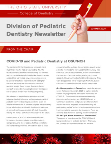
5-Radiology of the skull and teeth-RL.qxd4/5/200512:37 PMPage 7171RADIOLOGY OF THE SKULL AND TEETHTHE RABBITSkull Radiographs: Lateral View, Radiographic AnatomyabFigures 5.14a,b. The apex of the mandibular incisor teeth (a, red circle) normally extends tothe level of the ipsilateral first cheek tooth. Maxillary incisor teeth are more curved thanmandibular incisors, and their apex is normally located half the length of the diastema, at somedistance from the corresponding radiopaque hard palate (a, yellow circle). The tip of the radiolucent pulp system of normal incisor teeth usually extends to the level of the alveolar ridge,or just above it (green circles). The maxillary second incisor teeth (peg teeth) are short andsmall, with a slight curvature. The buccal alveolar margins of both maxillary first and mandibular incisor teeth are more apical than the palatal and lingual margins. Note the regular, smoothpalisade formed by the cheek teeth. The mandibular cheek teeth apexes are at some distancefrom the ventral cortex of the mandible.CHAPTER 5
8-Dental Diseases-RL.qxd4/5/20053:40 PMPage 126126CHAPTER 8RABBIT AND RODENT DENTISTRY HANDBOOKTHE RABBITDENTAL DISEASES OF CHEEK TEETHMany different patterns of abnormalities from mildto severe may be recognized in cases of acquireddental disease of cheek teeth (ADD). The severity ofthe pathologic changes can be staged. Both radiography and endoscopy are important for staging ofADD, and endoscopy in particular allows a moredetailed examination of the oral cavity. Thoroughevaluation and diagnosis are particularly impor-tant, as the patient’s clinical signs may be mild ormay be absent.Early diagnosis is the key to early treatment andresolution of lesions involving soft tissues of thegingiva, tongue and oral mucosa (see Chapter 12),which can be a source of constant pain in the petrabbit. Prompt treatment will also prevent the progression of dental disease.Stages of Cheek Teeth Malocclusion and Acquired Dental DiseaseFigure 8.35. The earliest stage of acquired dental disease(ADD) of cheek teeth in rabbits is elongation of the crowns.Because both the reserve crown and the clinical crown beginto take up more space, abnormalities related to increasedpressure begin to occur. The zig-zag pattern of the cheek teethocclusal plane is still normal (see Figure 5.13), but the radiolucent line is less visible when the mandible is at rest, even in aproper lateral projection. Pressure on the reserve crownsbegins to increase when the animal chews. Since there is notanother tooth cranial to the first premolars, they begin tocurve, with increasing mesial convexity (red arrow).In some early cases, slight deformation of the ventralmandibular cortical bone due to the increased pressure maybe visible (yellow arrow). Due to the abnormal convexity,interproximal space of mandibular cheek teeth begins towiden (blue arrows). Malocclusion of incisor teeth is usuallynot present at this stage.Figure 8.36. ADD of cheek teeth, later stage. Abnormalchanges to the occlusal plane due to excessive and irregularcrown elongation are clearly visible, with height differencesbetween adjacent molars of up to a few millimeters. An irregular zig-zag radiolucent line or the superimposition of two different zig-zag lines are present. This abnormal occlusal planeis called “wave mouth.” Mandibular cheek teeth root deformities are also visible. Malocclusion of incisor teeth is not stillpresent.
12-Dental procedures-RL.qxd4/5/200511:20 AMPage 213CHAPTER 12Dental ProceduresVITTORIO CAPELLOMARGHERITA GRACISREDUCTION OF INCISOR HEIGHT IN RABBITSElongated incisor teeth may require reduction toallow normal food prehension and in many cases,to help restore the normal occlusion of the cheekteeth. However, aradicular hypsodont teeth oftengrow very quickly, at an average rate of 2 mm perweek for maxillary incisor teeth and 2.4 mm perweek for mandibular incisor teeth. Therefore, thisprocedure may need to be repeated frequently.Incisor reduction is indicated when malocclusion ismild and can be readily corrected. Extraction maybe more appropriate in case of severe malocclusion.TCERRINCOFigure 12.1. Simple amputation of overgrown incisor teethwith nail clippers or other similar instruments must be discouraged, as this technique is associated with patient discomfort and a high rate of potentially severe complications,including vertical fractures. Fractures and the application ofthese types of forces to the teeth can lead to damage of theapical germinal tissues. Clipping does not allow the restoration of a normal incisal edge, and creates rough surfaces thatcan produce secondary injuries to the tongue and lips.Figure 12.2. The pulp system of elongated incisor teethoften extends beyond the gingival margin and may be seenas a pink discoloration of the clinical crown.
12-Dental procedures-RL.qxd4/5/200511:39 AMPage 229229DENTAL PROCEDURESCHAPTER 12EXTRACTION OF ARADICULAR HYPSODONT TEETHFeeding behavior is not negatively impacted by theextraction of incisor teeth. Lagomorphs and rodentswithout incisors use their lips and tongue for foodprehension.Indications for extraction of incisor teeth includecongenital or acquired severe malocclusion, dentalfractures, and endodontic and periapical disease.The following
DENTISTRY HANDBOOK VITTORIO CAPELLO, DVM Dipl ECZM-Small Mammals, Dipl ABVP-ECM With Margherita Gracis, DVM, Dipl AVDC and EVDC Edited by Angela M. Lennox, DVM, Dipl ABVP-Avian, ECM Zoological Education Network 0-Fr