Transcription
Gunadi et al. BMC Pediatrics (2018) EARCH ARTICLEOpen AccessNRG1 variant effects in patients withHirschsprung diseaseGunadi1* , Nova Yuli Prasetyo Budi1, Raman Sethi2,3, Aditya Rifqi Fauzi1, Alvin Santoso Kalim1, Taufik Indrawan1,Kristy Iskandar4, Akhmad Makhmudi1, Indra Adrianto5 and Lai Poh San2,3AbstractBackground: Hirschsprung disease (HSCR) is a heterogeneous genetic disorder characterized by absence of ganglioncells along the intestines resulting in functional bowel obstruction. Mutations in neuregulin 1 (NRG1) gene have beenimplicated in some cases of intestinal aganglionosis. This study aims to investigate the contribution of the NRG1 geneto HSCR development in an Indonesian population.Methods: We analyzed the entire coding region of the NRG1 gene in 54 histopathologically diagnosed HSCR patients.Results: All patients were sporadic non-syndromic HSCR with 53/54 (98%) short-segment and 1/54 (2%) long-segmentpatients. NRG1 gene analysis identified one rare variant, c.397G C (p.V133 L), and three common variants, rs7834206,rs3735774, and rs75155858. The p.V133 L variant was predicted to reside within a region of high mammalianconservation, overlapping with the promoter and enhancer histone marks of relevant tissues such as digestiveand smooth muscle tissues and potentially altering the AP-4 2, BDP1 disc3, Egr-1 known1, Egr-1 known4, HEN1 2transcription factor binding motifs. This p.V133 L variant was absent in 92 non-HSCR controls. Furthermore, the rs7834206polymorphism was associated with HSCR by case–control analysis (p 0.037).Conclusions: This study is the first report of a NRG1 rare variant associated with HSCR patients of South-East Asianancestry and provides further insights into the contribution of NRG1 in the molecular genetic pathogenesis of HSCR.Keywords: Hirschsprung disease, Indonesia, NRG1 variant, Transcription factor binding motifBackgroundHirschsprung disease (HSCR), a heterogeneous geneticdisorder, is characterized by the lack of ganglion cellsalong varying lengths of the intestines resulting in functional obstruction among children [1]. According to thelength of aganglionosis, HSCR is categorized into threemajor types: short-segment (aganglionosis up to theupper sigmoid colon), long-segment (aganglionosis up tothe splenic flexure and beyond) and total colonic aganglionosis (TCA) [1, 2].The incidence of HSCR varies among populations with15, 21 and 28 cases per 100,000 live births in Caucasians,Africans and Asians, respectively [1, 2]. These differencesmight be influenced by susceptibility factors such as theREarranged during Transfection (RET) rs2435357 risk* Correspondence: drgunadi@ugm.ac.id1Pediatric Surgery Division, Department of Surgery, Faculty of Medicine,Public Health and Nursing, Universitas Gadjah Mada/Dr. Sardjito Hospital,Yogyakarta 55281, IndonesiaFull list of author information is available at the end of the articleallele frequency [3]. Our recent studies clearly demonstratedthe presence of a higher frequency of RET rs2435357susceptibility allele in Indonesian ancestry (50%) [4, 5].Nevertheless, we observed that semaphorin 3 rs11766001common variant had different effects on HSCR dependingon the ethnic background [6].HSCR is a complex genetic disorder and at least 15 geneshave been implicated in the development of HSCR, withthe gene encoding the receptor tyrosine kinase RETaccounting for up to 21% of sporadic cases [7]. These genesencode proteins that are important for the enteric gangliadevelopment and are classified into three groups: 1) thosethat are associated with RET pathways (RET, GFRα1,GDNF, NTN, PSPN); 2) those implicated in endothelintype B receptor/EDNRB (EDNRB, EDN3, ECE-1) pathways;and 3) transcription factors that influence both RET and/orEDNRB pathways (SOX10, ZFXH1B, PHOX2B) [7, 8].Neuregulin 1 (NRG1) has been implicated as a diseasesusceptibility gene in Chinese [9, 10]. Both common (single The Author(s). 2018 Open Access This article is distributed under the terms of the Creative Commons Attribution 4.0International License (http://creativecommons.org/licenses/by/4.0/), which permits unrestricted use, distribution, andreproduction in any medium, provided you give appropriate credit to the original author(s) and the source, provide a link tothe Creative Commons license, and indicate if changes were made. The Creative Commons Public Domain Dedication o/1.0/) applies to the data made available in this article, unless otherwise stated.
Gunadi et al. BMC Pediatrics (2018) 18:292nucleotide polymorphisms, SNPs) and rare variants ofthis gene are reported to confer disease risk withover-representation in patient cohorts. However, in the firststudy on Caucasian, Luzón-Toro et al. [11] did not find anysignificant association of NRG1 SNPs in Spanish HSCRsuggesting that the association of such common polymorphisms to the disease may be restricted to specific populations. Furthermore, they found three novel NRG1 rarevariants that were hypothesized to have functional consequences during embryonic development of HSCR whichcould lead to HSCR in their patients. It is believed thatNRG1 plays an essential role in the signaling pathway ofthe enteric nervous system (ENS) [9–12]. In addition, thedevelopment of ENS requires a balance of neurogenesisand gliogenesis by RET/GDNF and ERBB2/NRG1 pathways, respectively. Nrg1 suppresses Gdnf-induced neuronaldifferentiation and Gdnf negatively controls Nrg1-signalingby reducing the expression of its receptor, ErbB2 [13].Recently, we studied two previously reported NRG1SNPs and demonstrated that that only one commonvariant rs7835688 as a genetic risk factor for Indonesian HSCR [4]. Therefore, we aimed to perform a comprehensive study of the entire NRG1 gene to investigatefully if there is any link between NRG1 variants withIndonesian HSCR patients.MethodsPatientsWe recruited 54 patients with HSCR, of whom 38 weremales and 16 females, corresponding to a gender ratio of2.4:1. Diagnosis of HSCR at Dr. Sardjito Hospital,Yogyakarta, Indonesia was based on clinical findings, contrastenema and histopathology examinations. Hematoxillin-eosinstaining and S100 immunohistochemistry were utilizedfor the histopathology assessment [4–6, 14–16]. Forcontrols, 92 ethnically-matched individuals with nodiagnosis of HSCR were enrolled.The study was reviewed and approved by the InstitutionalReview Board (IRB) of the Faculty of Medicine, UniversitasGadjah Mada/Dr. Sardjito Hospital, Indonesia (KE/FK/787/EC/2015). Written informed consent was obtained from allparents of the subjects for this study.Genomic DNA extraction and polymerase chain reaction(PCR)Genomic DNA was extracted from whole blood fromeach individual by using QIAamp DNA Extraction Kit(QIAGEN, Hilden, Germany), according to the manufacturer’s instructions. The extracted DNA sampleswere stored at 20 C until analysis.PCR was conducted by using a Swift Maxi thermal cycler(Esco Micro Pte. Ltd., Singapore). The primer sequencesfor NRG1 gene analysis were designed according to a previous study [10].Page 2 of 9DNA sequencingTo identify mutations, all 16 exons of the NRG1 thathad been PCR amplified in 20 fragments were screenedusing Sanger sequencing analysis with a BigDye TerminatorV3.1 Cycle Sequencing Kit (Applied Biosystems, FosterCity, CA). The products were separated and analyzed on a3730xl Genetic Analyzer (Applied Biosystems, Foster City,CA) using DNA Sequencing Analysis Software (AppliedBiosystems, Foster City, CA) [17, 18].Bioinformatics analysesData from the 1000 Genomes Project (http://www.internationalgenome.org) and ExAC (http://exac.broadinstitute.org) ancestry controls were utilized for comparison of variant frequencies among population. The in silico tools usedto predict coding variant effects on protein function wereSIFT (http://sift.jcvi.org/), PolyPhen-2 (http://genetics.bwh.harvard.edu/pph2/), LRT 100/), Mutation Taster (http://www.mutationtaster.org), Mutation Assessor (http://mutationassessor.org/r3/), FATHMM (http://fathmm.biocompute.org.uk), CADD (http://cadd.gs.washington.edu) algorithmand DANN (https://cbcl.ics.uci.edu/public data/DANN/)(Additional file 1).SIFT predicts whether a change of amino acid changein a protein will have an impact on phenotype. This calculation is based on the premise that protein evolutionis correlated with protein function, the more importantpositions of amino acids will correspondingly be moreconserved in an alignment of the protein family. A SIFTscore ranges from 0 to 1 and is classified as two groups:1) predicted damaging if score 0.05; and 2) tolerated ifscore 0.05 (http://sift.jcvi.org/). PolyPhen-2 score calculates the possible effect of a change of amino acid onthe human protein structure and function. Its scoreranges from 0 to 1.0 and consists of three interpretation:1) benign ( 0.452 in PolyPhen2 HDiv and 0.446 inPolyPhen2 HVar); 2) possibly damaging (0.453 to 0.956in PolyPhen2 HDiv and 0.447 to 0.909 in PolyPhen2HVar); and 3) probably damaging ( 0.957 in PolyPhen2HDiv and 0.909 in PolyPhen2 HVar) (http://genetics.bwh.harvard.edu/pph2/). Mutation Taster determinesthe disease potential of an alteration of amino acid usingthe Bayes classifier, with four possible analysis: 1) diseasecausing (i.e. probably deleterious); 2) disease causingautomatic (i.e. known to be deleterious); 3) polymorphism(i.e. probably harmless); and 4) polymorphism automatic(i.e. known to be harmless) (http://www.mutationtaster.org).DANN uses deep neural network (DNN) algorithm topredict pathogenicity of variants and scores from 0.98to 1 are considered to be protein disrupting (Additionalfile 1). All these tools predict whether a mutation leadsto damaging function that may be potentially diseasecausing but it is well acknowledged that such in silico
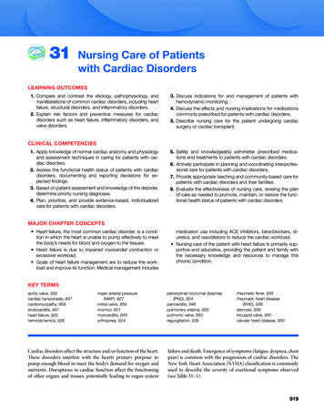
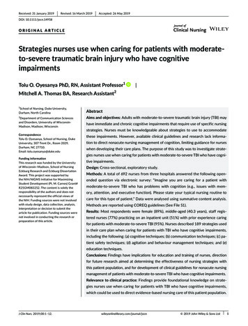
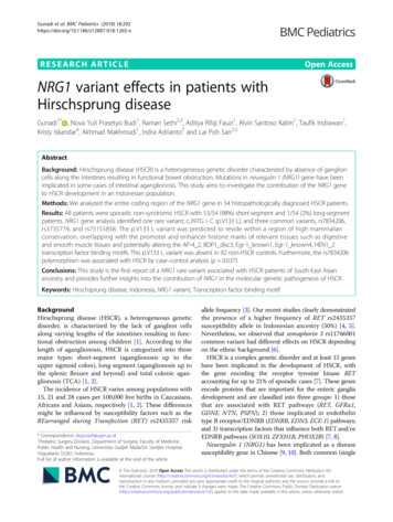
Gunadi et al. BMC Pediatrics (2018) 18:292Page 3 of 9Table 1 Clinical characteristics of Indonesian HSCR patientsCharacteristicsN (%); monthsGenderMaleFemaleAge at diagnosis38 (70)16 (30)34.6 44.5broadinstitute.org/genome bio/siphy/index.html) tests. Theclinical significance of variants was analyzed using ClinVartool (https://www.ncbi.nlm.nih.gov/clinvar/). The HaploRegdatabase haploreg.php) was utilized to evaluate the impact of the variant by annotation of epigenomic, conservation and regulatory motif information (Additional file 1).Degree of aganglionosisShort segment53 (98)Statistical genetic analysisLong segment1 (2)Chi-square test was used to establish p-values for thecase–control association analysis for NRG1 variants(rs7834206, rs3735774, and rs75155858). A p-value lessthan 0.05 was considered significant.Age at definitive surgery38.7 43.9Type of surgical approachTransanal endorectal pull-through21 (43)Duhamel procedure12 (25)Soave procedure11 (22)Others5 (10)HSCR Hirschsprung diseasetools are only used as predictive guides and may notnecessarily agree with each other as they can yield conflicting results [19–21].The predicted conservation scores of variants were determined using GERP p/index.html), PhyloP l), and SiPhy (http://portals.ResultsAll fifty-four patients (n 54) were sporadic non-syndromic HSCR. Neither familial nor syndromic HSCRwere included in this study. According to the degree ofaganglionosis, the distribution of the cases was 53/54(98%) short-segment, 1/54 (2%) long-segment and nonewere TCA. The mean age at diagnosis and age at definitiveoperation was 34.6 44.5 months (range, 1–174 months)and 38.7 43.9 months (range, 1–175 months), respectively (Table 1).DNA analysis covering coding regions of all 16 exonsof NRG1 gene in our cohort of 54 Indonesian HSCRFig. 1 Sanger sequencing of NRG1 exon 7 showed a missense variant, c.397G C, which led to substitution of valine with leucine at amino acid133 (p.V133 L) in the NRG1 protein. The arrow indicates the mutation
Gunadi et al. BMC Pediatrics (2018) 18:292Page 4 of 9Table 2 NRG1 variants found in Indonesian HSCR patientsVariantsReferenceNucleotideAmino Acidc.-97C A5’-UTRc.136G Ac.2298G Tc.397G G613 Vrs35641374αp.V133 LCases/Allele (54/108)Control/Allele (92/184)GenotypeGenotypeCC: 26CC: 59CA: 24CA: 31AA: 4AA: 2AlleleAlleleC: 76C: 149A: 32A: 35GenotypeGenotypeGG: 47GG: 75GA: 6GA: 17AA: 1AA: 0AlleleAlleleG: 100G: 167A: 8A: 17GenotypeGenotypeGG: 26GG: 39GT: 18GT: 42TT: 10TT: 11AlleleAlleleG: 70G: 120T: 38T: 64GenotypeGenotypeGG: 53GG: 92GC: 1GC: 0CC: 0CC: 0AlleleAlleleG: 107G: 184C: 1C: 0Odds ratio (95% CI)p-value1.79 (1.03–3.11)0.037*0.79 (0.33–1.89)0.591.02 (0.62–1.67)0.94, a p-value of 0.05 was considered significant; α, NP 039253; μ, NP 039251*patients demonstrated the presence of four variants. Thesevariants were: one rare missense variant, c.397G C,in exon 7, which led to a substitution of valine withleucine (p.V133 L) in one HSCR patient (Fig. 1), andthree common variants, rs7834206, rs3735774, andrs75155858 (Table 2).We compared the observed NRG1 rare variant allelefrequency in Indonesian HSCR patients with those reported for the 1000 Genomes Project and ExAC ancestrycontrols. The frequency of c.397G C (p.V133 L) rarevariant in our HSCR patients (0.9%) was higher thanthose reported for the 1000 Genomes and ExACTable 3 NRG1 variants frequency in Indonesian HSCR and population databasesVariantHSCR patientsIndonesian control1000 GenomesμExACμp-valuevs. 1000 Genomesvs. ExACc.397G C (p.V133 L)0.009000.0007 0.0001 0.0001rs78342060.300.190.18N/A 0.350.350.3511*, a p-value of 0.05 was considered significant; μ, East Asian ancestries; HSCR, Hirschsprung disease; N/A, not available
0.22 (tolerated)00.36 (tolerated)0c.397G C(p.
Age at diagnosis 34.6 44.5 Degree of aganglionosis Short segment 53 (98) Long segment 1 (2) Age at definitive surgery 38.7 43.9 Type of surgical approach Transanal endorectal pull-through 21 (43) Duhamel procedure 12 (25) Soave procedure 11 (22) Others 5 (10) HSCR Hirschsprung disease