Transcription
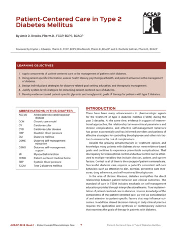
Vol. 4(4), pp. 46-57, September, 2013DOI 10.5897/JPAP2013.0001ISSN 2I41-260X 2013 Academic l of Physiology andPathophysiologyReviewThe pathogenesis and pathophysiology of type 1 andtype 2 diabetes mellitusOzougwu, J. C.1*, Obimba, K. C.2, Belonwu, C. D.3, and Unakalamba, C. B.4Physiology and Biomedical Research Unit, Department of Biological Sciences, College of Basic and Applied Sciences,Rhema University Aba, Abia State, Nigeria.2Department of Biochemistry, School of Science, Federal University of Technology Owerri. Imo State. Nigeria.3Department of Biochemistry, Faculty of Chemical Sciences, University of Portharcourt. Rivers State. Nigeria.4Department of Zoology and Environmental Biology, University of Nigeria, Nsukka, Enugu state, Nigeria.Accepted 17 June, 2013The aim of this paper is to review the information on type 1 and type 2 diabetes with emphasis on itsetiology, pathogenesis and pathophysiology via literature review. Diabetes is a group of metabolicdisorders characterized by a chronic hyperglycemic condition resulting from defects in insulinsecretion, insulin action or both. Type 1 diabetes is the result of an autoimmune reaction to proteins ofthe islets cells of the pancreas while type 2 diabetes is caused by a combination of genetic factorsrelated to impaired insulin secretion, insulin resistance and environmental factors such as obesity,overeating, lack of exercise and stress, as well as aging. The pathogenesis of selective β-celldestruction within the islet in type 1 diabetes mellitus is difficult to follow due to marked heterogeneityof the pancreatic lesions. At the onset of overt hyperglycemia, a mixture of pseudoatrophic islets withcells producing glycogen, somatostatin and pancreatic polypeptide, normal islets and islets containingboth β-cells and infiltrating lymphocytes and monocytes may be seen. The autoimmune destruction ofpancreatic β cells leads to a deficiency of insulin secretion that leads to the metabolic derangementsassociated with type 1 diabetes. The main pathophysiological features of type 2 diabetes are impairedinsulin secretion and increased insulin resistance. The impairment of pancreatic β cell function notablyshows progression overtime in type 2 diabetes although aging, obesity, insufficient energyconsumption, alcohol drinking, smoking, etc are independent risk factors of pathogenesis of type 2diabetes mellitus.Key words: Diabetes Mellitus, Pathophysiology, Pathogenesis, Etiology.INTRODUCTIONDiabetes mellitus (DM) is a group of metabolic disorderscharacterized by a chronic hyperglycemic conditionresulting from defects in insulin secretion, insulin actionor both. Permanent neonatal diabetes is caused byglucokinase deficiency, and is an inborn error of theglucose-insulin signaling pathway (Njolstad et al., 2003).The prevalence of diabetes is increasing rapidlyworldwide and the World Health Organization (2003) haspredicted that by 2030 the number of adults with diabeteswould have almost doubled worldwide, from 177 million in2000 to 370 million. Experts project that the incidence ofdiabetes is set to soar by 64% by 2025‚ meaning that a*Corresponding author. E-mail: jevaschubby@yahoo.com. Tel: 2348034006816
Ozougwu et al.47Table 1. Some causes of insulin resistance.S/No.Causes1Obesity/overweight (especially excess visceral adiposity)2Excess glucorticoids (cushing’s syndrome or steroid therapy)3Excess growth hormone (acromegaly)4Pregnancy, gestational diabetes5Polycystic ovary disease6Lipodystrophy (acquired or genetic, associated with lipid accumulation in liver)7Autoantibodies to the insulin receptor8Mutations of insulin receptor9Mutations of the peroxisome proliferators’ activator receptor γ (PPAR γ)10Mutations that cause genetic obesity (e.g., melanocortin receptor mutations)11Hemochromatosis (a hereditary disease that causes tissue iron accumulation).Source: Guyton and Hall (2006).Table 2. Clinical characteristics of patients with Type 1 and Type 2 diabetes mellitus.FeaturesType 1Type 2Age of onsetUsually less than 20 yearsUsually greater than 30 yearsBody massLow (wasted) to normalObesePlasma insulinLow or absentNormal to high initiallyPlasma glucagonHigh, can be suppressedHigh, resistant to suppressionPlasma glucoseincreasedIncreasedInsulin sensitivityNormalReducedTherapyinsulinWeight loss, thiazolidinediones, metformin, sulfonylureas, insulinSource: Guyton and Hall (2006).staggering 53.1 million citizens will be affected by thedisease (Rowley and Bezold, 2012). The estimatedworldwide prevalence of diabetes among adults in 2010was 285 million (6.4%) and this value is predicted to riseto around 439 million (7.7%) by 2030 (Shaw et al., 2010).There are two main types of diabetes mellitus: i. Type 1diabetes, also called insulin dependent diabetes mellitus(IDDM), is caused by lack of insulin secretion by betacells of the pancreas.ii. Type 2 diabetes, also called non-insulin dependentdiabetes mellitus (NIDDM), is caused by decreasedsensitivity of target tissues to insulin.The reduced sensitivity to insulin is often called insulinresistance and its causes are shown in Table 1. In bothtypes of diabetes mellitus, metabolism of all the mainfoodstuffs is altered. The basic effect of insulin lack orinsulin resistance on glucose metabolism is to preventthe efficient uptake and utilization of glucose by mostcells of the body, except those of the brain (Guyton andHall, 2006). As a result of this, blood glucoseconcentration increases, cell utilization of glucose fallsincreasingly lower and utilization of fats and proteinsincreases. The clinical characteristics of patients withtype 1 and type 2 diabetes mellitus are shown in Table 2.EPIDEMIOLOGY ANDDIABETES (IDDM)ETIOLOGYOFTYPE1Type 1 diabetes represents around 10% of all cases ofdiabetes, affecting approximately 20 million peopleworldwide (American Diabetes Association, 2001).Although type 1 diabetes affects all age groups, themajority of individuals are diagnosed either at around theage of 4 to 5 years, or in their teens and early adulthood(Blood et al., 1975). The incidence of type 1 diabetes isrising. Across Europe, the average annual increase in theincidence in children under 15 years is 3.4% (EURODIAB
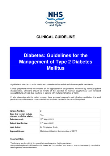
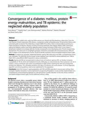
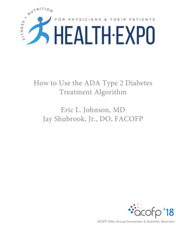
48J. Physiol. Pathophysiol.ACE study Group, 2000), with the steepest rise in thoseunder 5 years old (Karvonen et al., 1999). Type 1diabetes is the result of an autoimmune reaction toproteins of the islets cells of the pancreas (Holt, 2004).There is a strong association between IDDM and otherendocrine autoimmunity (for example, Addison disease)and an increased incidence of autoimmune diseases areseen in family members of IDDM patients. The threetypes of autoantibodies known are:i) Islet cell cytoplasmic antibodies (ICCA): The primaryantibodies found in 90% of type 1 diabetics are againstislet cell cytoplasmic proteins. The presence of ICCA is ahighly accurate predictor of future development of IDDM.ii) Islet cell surface antibodies (ICSA): Autoantibodiesdirected against islets cell surface antigens (ICSA) havealso been described in as many as 80% of type 1diabetics. Some patients with type 2 diabetes have beenidentified, which are ICSA positive.iii). Specific antigenic targets of islet cells: Antibodies toglutamic acid decarboxylase (GAD) have been identifiedin over 80% of patients newly diagnosed with IDDM. AntiGAD antibodies decline over time in type 1 diabetics. Thepresence of anti GAD antibodies is a strong predictor ofthe future development of IDDM in high risk populations.Anti insulin antibodies (IAAs) have been identified inIDDM patients and in relatives at risk to developingIDDM. These IAAs are detectable even before the onsetof insulin therapy in type 1 diabetics. IAA is detectable inaround 40% of young children with IDDM (Raju and Raju,2010).Pathogenesis of type 1 diabetes mellitusType 1 diabetes mellitus is a chronic autoimmunedisease associated with selective destruction of insulinproducing pancreatic β-cells (Figure 1). The onset ofclinical disease represents the end stage of β-celldestruction leading to type 1 diabetes mellitus. Al Homsiand Lukic (1992) explained that several featurescharacterize type 1 diabetes mellitus as an autoimmunedisease:1. Presence of immuno-competent and accessory cells ininfiltrated pancreatic islets;2. Association of susceptibility to disease with the class II(immune response) genes of the major histocompatibilitycomplex (MHC; human leucocyte antigens HLA);3. Presence of islet cell specific autoantibodies;4. Alterations of T cell mediated immunoregulation, inparticular in CD4 T cell compartment;5. The involvement of monokines and TH1 cellsproducing interleukins in the disease process;6. Response to immunotherapy and;7. Frequent occurrence of other organ specific auto-immune diseases in affected individuals or in their familymembers.The pathogenesis of selective β-cell destruction withinthe islet in type 1 DM is difficult to follow due to markedheterogeneity of the pancreatic lesions. At the onset ofovert hyperglycemia, a mixture of pseudoatrophic isletswith cells producing glycogen (a cells), somatostatin (dcells) and pancreatic poly-peptide (PP cells), normalislets, and islets containing both b-cells and infiltratinglymphocytes and monocytes may be seen (Al-Homsi andLukic, 1992). Lymphocytic infiltration is found only in theislet containing residual β-cells and is likely that thechronicity with which type 1 DM develops reflects thisheterogeneity of islet lesions (Al-Homsi and Lukic, 1992).In contrast to this chronicity in the natural history of thedisease, β-cells are rapidly destroyed when pancreas istransplanted from identical twin donors into their longterm diabetic twin mates in the absence ofimmunosuppression. In these cases, massive insulitisdevelops rapidly with infiltrating T lympocytes indicatingan anamnestic autoimmune reaction (Al Homsi andLukic, 1992). In addition, this observation also indicatesthat the chronic time course in type 1 DM (but not in atransplanted pancreas) is a consequence of downregulatory phenomena taking part in immunopathogenesis of the disease (Al Homsi and Lukic, 1992).Activation of islet antigen - specific CD4 T cells appearto be absolute prerequisite for the development ofdiabetes in all animal models of type 1 DM (Gill andHaskins, 1993). CD4 islet specific T-cell clones derivedfrom diabetic NOD mice, when injected into prediabetic ornon diabetes prone Fl mice, induce insulitis and diabetes.It was also reported that CD4 T cells are sufficient toinduce insulitis while CD8 T cells contribute to theseverity of the damage (Yagi et al., 1992). These findingstogether with the evidence that insulitis in chronic graftversus host disease may occur in the absence of CD8 Tcells suggest that CD4 T cells may be the onlyimmunocompetent cells required in the disease process.However, it seems that only one subset of CD4 T cellsare responsible for disease induction.CD4 T cell bearing alloantigen RT6 are absent indiabetes prone BB rats and appear to protect AO ratsfrom MLD-STZ induced diabetes (Greineh et al., 1987).Down-regulation of diabetogenic autoimmune responseby the spleen cells derived from animals treated withadjuvants could also be explained by CD4 T cellsubsets interplay (Ulaeto et al., 1992). High level of THItype cytokines IL-2 and interferon g are found to correlateor/and to enhance induction of autoimmune diabetes inexperimental models (Fowell et al., 1991; Campbell et al.,1991). The TH-1 type cells, and in particular their productIFN-g, activate macrophages. In animal, models of type 1DM electron microscopic studies of pancreata showed
Ozougwu et al.49Genetic SusceptibilityIneffective DefenseBarriersIneffective ImmuneResponseInsulating Agent eg.Pancreotropic Virus[Typea quote sT- Lymphocytefrom thedocument orthe summaryof aninterestingpoint. You canCD4position thetext boxanywhere inthe document.Use the TextBox Tools tabINF-γto change theformatting ofthe pull quotetext box.]Autoimmune ProcessBeginsMacrophages (Ag PresentingCell)AgTNF-αPeptidesInterleukin 1B- Cell Destruction 90% DestructionPrecipitating FactorsOr StressorsType 1 Diabetes MellitusFigure 1. Pathogenesis of type 1 diabetes mellitus.Figure 1: Pathogenesis of Type 1 Diabetes Mellitusthat macrophages are the first cell type invading theislets(Kolb-Bachofen et al., 1988). In vitro studies andstudies on perfused pancreas suggest that Interleukin 1(IL-1) and tumor necrosis factor (TNFa), two cytokinesmainly produced by macrophages, induce structuralchanges of β-cells and suppression of their insulinreleasing capacity (Mandrup-Poulsen et al., 1987).However, it seems that IL-1 and TNF do not contribute
50J. Physiol. Pathophysiol.appreciably to the cytotoxic activity of macrophages(Kroncke et al., 1991). Interferon g is also a powerfulactivator of macrophages for nitric oxide synthesis.Recently, evidence has been provided indicating that NOsynthase activity is involved in diabetes development(Lukic et al., 1991). These data indicated, for the firsttime, that nitric oxide may be a pathogenic factor inautoimmunity and suggested a possibility that a newclass of immunopharmacological agents, capable ofmodulating nitric oxide secretion may be tested in theprevention of type 1 DM development (Kolb and KolbBachofen, 1992).Pathophysiology of type 1 diabetes (IDDM)The autoimmune destruction of pancreatic β-cells, leadsto a deficiency of insulin secretion which results in themetabolic derangements associated with IDDM. Inaddition to the loss of insulin secretion, the function ofpancreatic α-cells is also abnormal and there is excessivesecretion of glucagons in IDDM patients. Normally,hyperglycemia leads to reduced glucagons secretion,however, in patients with IDDM, glucagons secretion isnot suppressed by hyperglycemia (Raju and Raju, 2010).The resultant inappropriately elevated glucagons levelsexacerbate the metabolic defects due to insulin deficiency. The most pronounced example of this metabolicdisruption is that patients with IDDM rapidly developdiabetic ketoacidosis in the absence of insulinadministration. Although insulin deficiency is the primarydefect in IDDM, there is also a defect in theadministration of insulin. There are multiple biochemicalmechanisms that account for impairment of tissue’sresponse to insulin. Deficiency in insulin leads touncontrolled lipolysis and elevated levels of free fattyacids in the plasma, which suppresses glucosemetabolism in peripheral tissues such as skeletal muscle(Raju and Raju, 2010). This impairs glucose utilizationand insulin deficiency also decreases the expression of anumber of genes necessary for target tissues to respondnormally to insulin such as glucokinase in liver and theGLUT 4 class of glucose transporters in adipose tissue.Raju and Raju (2010) explained that the major metabolicderangements, which result from insulin deficiency inIDDM are impaired glucose, lipid and protein metabolismwhich are explained in details as follows:Effects on glucose metabolismUncontrolled IDDM leads to increased hepatic glucoseoutput. First, liver glycogen stores are mobilized thenhepatic gluconeogenesis is used to produce glucose.Insulin deficiency also impairs non hepatic tissue utiliza-tion of glucose. In particular in adipose tissue andskeletal muscle, insulin stimulates glucose uptake. This isaccomplished by insulin mediated movement of glucosetransporters proteins to the plasma membrane of thesetissues. Reduced glucose uptake by peripheral tissues inturn leads to a reduced rate of glucose metabolism. Inaddition, the level of hepatic glucokinase is regulated byinsulin. Therefore, a reduced rate of glucose phosphorrylation in hepatocytes leads to increased delivery to theblood. Other enzymes involved in anabolic metabolicmetabolism of glucose are affected by insulin.The combination of increased hepatic glucoseproduction and reduced peripheral tissues metabolismleads to elevated plasma glucose levels. When thecapacity of the kidneys to absorb glucose is surpressed,glucosuria ensues. Glucose is an osmotic diuretic and anincrease in renal loss of glucose is accompanied by lossof water and electrolyte. The result of the loss of water(and overall volume) leads to the activation of the thirstmechanism (polydipsia). The negative caloric balance,which results from the glucosuria and tissue catabolismleads to an increase in appetite and food intake that ispolyphagia (Raju and Raju, 2010).Effect on lipid metabolismOne major role of insulin is to stimulate the storage offood energy in the form of glycogen in hepatocytes andskeletal muscle, following the consumption of a meal. Inaddition, insulin stimulates hepatocytes to synthesize andstore triglycerides in adipose tissue. In uncontrolled IDDMthere is a rapid mobilization of triglycerides leading toincreased levels of plasma free fatty acids. The free fattyacids are taken up by numerous tissue (except the brain)and metabolized to provide energy. In the absence ofinsulin, malonyl COA levels fall, and transport of fattyacyl-COA into the mitochondria increases. Mitochondrialoxidation of fatty acids generates acetyl COA that can befurther oxidized in the TCA cycle. However, in heaptocytes the majority of the acetyl COA is not oxidized bythe TCA cycle but is metabolized into the ketone bodies(acetoacetate and b-hydroxybutyrate). These ketonebodies are used for energy production by the brain, heartand skeletal muscle. In IDDM, the increased availabilityof free fatty acids and ketone bodies exacerbates thereduced utilization of glucose, furthering the ensuinghyperglycaemia. Production of ketone bodies in excess ofthe body’s ability to utilize them leads to ketoacidosis. Aspontaneous breakdown product of acetoacetate is theacetone that is exhaled by the lungs, which gives adistinctive odor to the breath. Normally, plasmatriglycerides are acted upon by lipoprotein lipase (LPL)that requires insulin. LPL is a membrane bound enzymeon the surface of the endothelial cells lining the vessels,
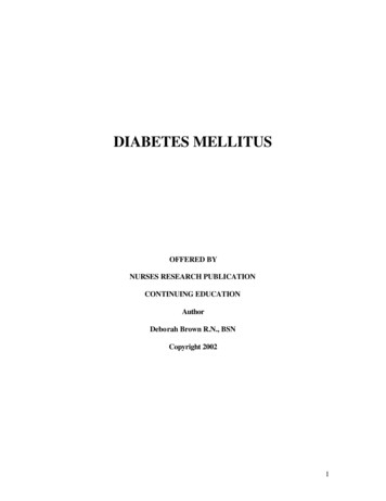
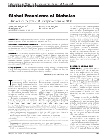
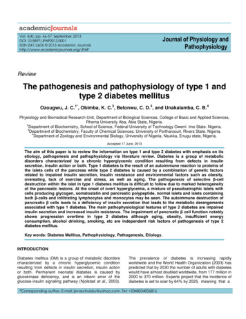
Ozougwu et al.which allows fatty acids to be taken from circulatingtriglycerides for storage in adipocytes (Raju and ceridemia.Effects on proteinInsulin regulates the synthesis of many genes, eitherpositively or negatively, which affect overall metabolism.Insulin has an overall effect on protein metabolism,increasing the rate of protein synthesis and decreasingthe rate of protein degradation. Thus insulin deficiencywill lead to increased catabolism of protein. Theincreased rate of proteolysis leads to elevated concentration of amino acids in plasma (Raju and Raju, 2010).Glucogenic amino acids serve as precursors for hepaticand renal glyconeogenesis, which further contributes tothe hyperglycaemia seen in IDDM.EPIDEMIOLOGY ANDDIABETES (NIDDM)ETIOLOGYOFTYPE2Type 2 diabetes is the predominant form of diabetes andaccounts for at least 90% of all cases of diabetes mellitus(Gonzalez et al., 2009). The rise in prevalence ispredicted to be much greater in developing than indeveloped countries (69 versus 20%) (Shaw et al., 2010).In developing countries, people aged 40 to 60 years (thatis, working age) are affected most, compared with thoseolder than 60 years in developed countries (Shaw et al.,2010). This increase in type 2 diabetes is inextricablylinked to changes towards a Western lifestyle (high dietwith reduced physical activity) in developing countriesand the rise in prevalence of overweight and obesity(Chan et al., 2009; Colagiuri, 2010). There areapproximately 1.4 million people with diagnosed type 2diabetes in the UK (Bennett et al., 1995). The incidenceof diabetes increases with age, with most cases beingdiagnosed after the age of 40 years. This equates to alifetime risk of developing diabetes of 1 in 10 (Neil et al.,1987). Type 2 diabetes is a heterogenous disordercaused by a combination of genetic factors related toimpaired insulin secretion, insulin resistance andenvironmental factors such as obesity, over eating, lackof exercise, and stress as well as aging (Kaku, 2010). It istypically a multifactorial disease involving multiple genesand environmental factors to varying extents (Holt, 2004).Type 2 diabetes is the common form of idiopathicdiabetes and is characterized by a lack of the need forinsulin to prevent ketoacidosis. It is not an autoimmunedisorder and the susceptible genes that predispose toNIDDM have not been identified in most patients. Thiscould be due to the heterogeneity of the genes respon-51sible for the susceptibility to NIDDM.Pathogenesis of type 2 diabetesUnder normal physiological conditions, plasma glucoseconcentrations are maintained within a narrow range,despite wide fluctuations in supply and demand, througha tightly regulated and dynamic interaction betweentissue sensitivity to insulin (especially in liver) and insulinsecretion (DeFronzo, 1988). In type 2 diabetes thesemechanisms break down, with the consequence that thetwo main pathological defects in type 2 diabetes areimpaired insulin secretion through a dysfunction of thepancreatic β-cell, and impaired insulin action throughinsulin resistance (Holt, 2004). Type 2 diabetes mellitushas a greater genetic association than type 1 DM, thepathogenesis of type 2 diabetes mellitus is characterizedby impaired insulin secretion and insulin resistance asshown in Figure 2. The 100% concordance rate inidentical twins is thought to be over-estimated, due to aselection or reporting bias. A population based twin studyin Finland has shown a concordance rate of 40%, andenvironmental effect may be a possible reason for thehigher concordance rate for type 2 diabetes mellitus thanfor type 1 diabetes mellitus (Kaprio et al., 1992). Type 2diabetes mellitus affects 1 to 2% of caucasians (Cook etal., 1993) but it is much higher in some ethnic groupssuch as Pima Indians (Knowler et al., 1990) and Arabs(Richens et al., 1988) and approaches 50% in SouthIndia. This indicates that genetic factors are moreimportant than environmental factors. Except for maturityonset diabetes of the young (MODY), the mode ofinheritance for type 2 diabetes mellitus is unclear. MODY,inherited as an autosomal dominant trait, may result frommutations in glucokinase gene on chromosome 7p.Glucokinase is a key enzyme of glucose metabolism inbeta cells and the liver (Froguel et al., 1993; Hattersley etal., 1992). MODY is defined as hyperglycemia diagnosedbefore the age of twenty-five years and treatable for overfive years without insulin in cases where islet cellantibodies (ICA) are negative and HLA-DR3 and DR4 areheterozygous. MODY is rare in Caucasians, less than1%, and more common in blacks and Indians, more than10% of diabetics. Chronic complications in MODY werethought to be uncommon but later were found to be morecommon, indicating its heterogeneity.Considering MODY as a separate entity maymasquerade its association with specific geneticdiseases; and without a definite genetic marker, it shouldbe treated as type 1 DM (Tattershall, 1991). Identificationof a nonsense mutation in the glucokinase gene and itslinkage with MODY was reported for the first time in aFrench family, implicating a mutation in a gene involvedin glucose metabolism in the pathogenesis of type 2
52J. Physiol. Pathophysiol.GENESLIFESTYLEIMPAIRED INSULINSECRETIONINSULIN RESISTANCEIGTTYPE 2 DIABETESPROGRESSIVE HYPERGLYCEMIAFigure 2. Pathogenesis of type 2 diabetes characterized by impaired insulin secretion and insulin resistancediabetes mellitus (Vionnet et al., 1992). Later, sixteenmutations were identified in 18 MODY families. Theyincluded 10 mutations that resulted in an amino acidsubstitution, 3 that resulted in the synthesis of truncatedprotein, and 3 that affected RNA processing.Hyperglycemia in these families was usually mild andbegan in childhood, whereas the hyperglycemia of aredafter puberty (Froguel et al., 1993). Moleculargenetic studies in type 2 diabetes mellitus, with theexception of MODY, have not been as successful as intype 1 diabetes mellitus. Mutations in the insulin genelead to the synthesis and secretion of abnormal geneproducts, leading to what are called insulinopathies(Gabbay, 1980). Most of the patients with insulinopathieshave hyperinsulinemia, inherited in autosomal fashion,heterozygous for normal and mutant alleles, and normallyrespond to exogenous insulin administration. Al Homsiand Lukic (1992) explained that most insulin genemutations lead to:(a) Abnormal insulins - Such as insulins Chicago andWakayama where the mutation leads to an amino acidreplacement at an important site for receptor interaction;or(b) The mutation may interfere in the proinsulinprocessing to insulin (Chan et al., 1987).The association of the polymorphic (hypervariable) 5'flanking region of the human insulin gene and type 2diabetes mellitus is lacking in some population groups,indicating that it may be one of many factors in amultifactorial disease. Even MODY patients have shownno association with this region. It was mentioned earlierthat there is a strong association between HLA-DR3/4and type 1 diabetes mellitus. It was also reported thatsuch an association is present with type 2 diabetesmellitus, rendering HLA-DR3/4 markers for beta celldestruction in these patients (Richens et al., 1988;Tattershall, 1991). Pancreatic abnormalities in isletsecretory cells in type 2 diabetes mellitus are noted inbeta, alpha and delta cells of the islets. Defects involvinginsulin secretion include relative decrease in basal
Ozougwu et al.secretion, decreased first and second phases of insulinresponse, glucose insensitivity and amino acidhypersensitivity of insulin release. The number andvolume of beta cells are usually decreased to half thenormal, and the alpha cell mass is increased leading tohyperglucagonemia. The islets exhibit hyalinization andamyloid deposition, containing islet amyloid polypeptide(IAPP) or amylin. This is a minor secretory peptide of thebeta cells released along with insulin and C-peptide, butits role in the pathogenesis of type 2 DM is not wellunderstood (Steiner et al., 1991). This amylin is thoughtto produce insulin resistance (Molina et al., 1990). IAAPis reduced with progression of type 2 DM (Enoki et al.,1992). Intimate contact between beta cells and amyloiddeposit in type 2 DM is noted by electron microscopy(Westermark, 1973). Away from the islets in the exocrinepancreas, fatty infiltration and diffuse fibrosis are evident.Defective islet cell function is the primary event whichmay be due to an autoimmune reaction producinghyperglycemia in type 2 DM (Zawala et al., 1992). Theinsulin receptor gene is located on chromosome 19 and itencodes a protein having alpha and beta subunitsincluding the transmembrane domain and the tyrosinekinase domain (Kahu and White, 1988). Mutationsaffecting the insulin receptor gene have been identifiedand their association with type 2 diabetes mellitus andtype A insulin resistance is recognized.Type A insulin resistance is hereditary and type B is anautoimmune disorder (Levy and Hug, 1993). Restrictionfragment length polymorphism (RFLP) analysis of theinsulin receptor gene (Ohagi et al., 1992), erythrocyteglucose transporter gene, and HLA genes, were notfound useful as genetic markers for type 2 DM. Insulinresistance is insufficient to cause overt glucoseintolerance, but may play a significant role in cases ofobesity where there is known impairment of insulinaction. Insulin resistance by itself may be a secondaryevent in type 2 DM, since it is also found in non-diabeticobese individuals. Insulin secretion defect may be theprimary event, presenting as impaired pulsatile secretionof insulin. Hence, hyperglycemia is an inducer as well asa consequence of impaired islet cell function and insulinresistance. Many factors contribute to the insulininsensitivity including obesity and its duration (Evephartet al., 1992), age, lack of exercise, increased dietary fatand decreased fibres and genetic factors.Fish oil is found to prevent insulin resistance inanimals, but not in humans. It has a protective effectagainst thrombosis and vasospasm in type 2 DM (McVeigh et al., 1993). Insulin resistance in type 2 DM is nottotally clear, it may involve reduced insulin receptornumber, it may be secondary to hyperinsulinemia andhyperglycemia, (Vuorinen-Markkola et al., 1992) or it mayresult from reduced tyrosine kinase activity (Comi et al.,1987; Bonadonna et al., 1993; Sten-linder et al., 1993) or53even abnormalities distal to the receptor involvingglucose transporter proteins through a family of glucosetransporter genes (Mueckler, 1990). The GLUT2 gene,expressed in liver and pancreatic beta cells, and GLUT4,expressed in skeletal muscle and adipocytes, are strongcandidate genes for the genetic susceptibility to type 2DM. Analysis of these two glucose transporter genes, inaddition to GLUT1, encoding for the brain/erythrocyteglucose transporter, has yielded, in Caucasians, noassociation of any RFLP marker on haplotype with eithertype 2 DM or obesity (Oelbaum, 1992).Obesity has genetic as well as environmental causes. Ithas a strong effect on the development of type 2 DM(Bjorntorp, 1992; Haffner et al., 1992) as it is found inWestern countries (NDDG, 1979; Wilson et al., 1981) andsome ethnic groups such as Pima Indians (Joffe et al.,1992; Knowler et al., 1993). Obesity is more than just arisk factor; it has a causal effect in the development oftype 2 DM against a genetic background. The evolutionfrom obesity to type DM results from a succession ofpathophysiological events:(a) Augmentation of the adipose tissue mass, leading toincreased lipid oxidation;(b) Insulin resistance noted early in obesity, revealed byeuglycemic clamp, as a resistance to insulin mediatedglucose storage and oxidation, blocking the function ofthe glycogen cycle;(c) Despite maintained insulin secreti
worldwide prevalence of diabetes among adults in 2010 was 285 million (6.4%) and this value is predicted to rise to around 439 million (7.7%) by 2030 (Shaw et al., 2010). There are two main types of diabetes mellitus: i. Type 1 diabetes, also called insulin dependent diabetes mellit