Transcription
Clinical StudyV L Wester and othersHCC as a screening tool for CS176:6695–703Scalp hair cortisol for diagnosis ofCushing’s syndromeVincent L Wester1, Martin Reincke4, Jan W Koper1, Erica L T van den Akker2,Laura Manenschijn1, Christina M Berr4, Julia Fazel4, Yolanda B de Rijke3,Richard A Feelders1 and Elisabeth F C van Rossum11Division of Endocrinology, Department of Internal Medicine, 2Department of Pediatrics, 3Departmentof Clinical Chemistry, Erasmus MC, University Medical Center Rotterdam, Rotterdam, the Netherlands,and 4Medizinische Klinik und Poliklinik IV, Klinikum der Ludwig-Maximilians-Universität München,Munich, GermanyCorrespondenceshould be addressedto E F C van RossumEmaile.vanrossum@erasmusmc.nlEuropean Journal of EndocrinologyAbstractObjective: Current first-line screening tests for Cushing’s syndrome (CS) only measure time-point or short-term cortisol.Hair cortisol content (HCC) offers a non-invasive way to measure long-term cortisol exposure over several months oftime. We aimed to evaluate HCC as a screening tool for CS.Design: Case-control study in two academic referral centers for CS.Methods: Between 2009 and 2016, we collected scalp hair from patients suspected of CS and healthy controls. HCCwas measured using ELISA. HCC was available in 43 confirmed CS patients, 35 patients in whom the diagnosis CSwas rejected during diagnostic work-up and follow-up (patient controls), and 174 healthy controls. Additionally, wecreated HCC timelines in two patients with ectopic CS.Results: CS patients had higher HCC than patient controls and healthy controls (geometric mean 106.9 vs 12.7 and8.4 pg/mg respectively, P 0.001). At a cut-off of 31.1 pg/mg, HCC could differentiate between CS patients and healthycontrols with a sensitivity of 93% and a specificity of 90%. With patient controls as a reference, specificity remainedthe same (91%). Within CS patients, HCC correlated significantly with urinary free cortisol (r 0.691, P 0.001). In twoectopic CS patients, HCC timelines indicated that cortisol was increased 3 and 6 months before CS became clinicallyapparent.Conclusions: Analysis of cortisol in a single scalp hair sample offers diagnostic accuracy for CS similar to currently usedfirst-line tests, and can be used to investigate cortisol exposure in CS patients months to years back in time, enablingthe estimation of disease onset.European Journal ofEndocrinology(2017) 176, 695–703IntroductionGlucocorticoids are a class of steroid hormones that areproduced under the influence of the hypothalamic–pituitary–adrenal (HPA) axis and have mediating effectsin metabolism, inflammation, circulation and behavior.Cushing’s syndrome (CS) occurs when there is an excess ofglucocorticoids, either from an exogenous source or from anexcessive endogenous production of cortisol. EndogenousCS is a rare disorder, most commonly caused by an ACTHproducing pituitary adenoma (Cushing’s disease), lesswww.eje-online.orgDOI: 10.1530/EJE-16-0873 2017 European Society of EndocrinologyPrinted in Great Britainfrequent are primary adrenal causes and ectopic CS (1).Ectopic CS results from secretion of adrenocorticotropichormone (ACTH) from a non-pituitary source, or veryrarely, from ectopic corticotropin-releasing hormone(CRH) secretion. Ectopic CS is often associated withhigher ACTH levels than in Cushing’s disease, leadingto more fulminant CS (2). While a number of signs andsymptoms are deemed highly suggestive for CS (e.g. facialplethora, proximal muscle weakness, purple striae andPublished by Bioscientifica Ltd.Downloaded from Bioscientifica.com at 04/10/2022 08:05:07PMvia free access
European Journal of EndocrinologyClinical StudyV L Wester and otherseasy bruising), many are non-specific and highly prevalentin the general population, such as metabolic syndromefeatures, osteoporosis and depression (3). This overlapwith common chronic conditions is likely to cause a delayin the diagnosis of CS in many cases.Diagnosis of CS is further complicated by the factthat no single biochemical test for cortisol exposure offersperfect diagnostic accuracy. First-line tests for CS that arerecommended by the Endocrine Society guideline areurinary free cortisol (UFC) in 24-h urine collections, latenight salivary cortisol (LNSC) and the 1 mg overnightdexamethasone suppression test (DST) (3). Cortisolsecretion can be variable in CS, illustrated by the fact thatthere is a high variability of UFC in patients with activeCS (4) and that many patients have at least one normalUFC (5). In general, multiple tests are needed to establish adiagnosis, and thus, both UFC and LNSC measurements areusually performed on at least two different occasions (3).Over the past few years there is an increasing use ofcortisol in scalp hair as a measure of long-term cortisolexposure (6). Hair cortisol content (HCC) has been usedto investigate long-term cortisol levels in association withcardiovascular disease (7, 8, 9), obesity (10, 11, 12) andmetabolic syndrome (13). We and others have previouslyreported increased HCC in a small number of CS patients,including cases of cyclic CS (14, 15). HCC has practicaladvantages over currently used diagnostic tests, sincesample collection can easily be performed in an outpatientsetting and is not dependent on patient adherence tosampling instructions. Furthermore, HCC measurementoffers retrospective information about cortisol levelsover months of time in a single measurement, therebypotentially circumventing the limitations posed by thevariability in cortisol secretion in endogenous CS (6).We aimed to establish the optimal cut-off value ofHCC for the diagnosis of endogenous CS. In order to dothis, we measured HCC in patients with confirmed CS,in healthy controls and in patients who were initiallysuspected to have CS but in whom CS was excludedduring work-up. Furthermore, we aimed to explore thepotential of HCC to retrospectively assess the onset ofhypercortisolism in severe (ectopic) CS.HCC as a screening tool for CS176:6696medical center (Erasmus MC, Rotterdam, the Netherlands)and were suspected of CS were requested to take part inthe present study. Within this period, we included allpatients who were evaluated for Cushing’s syndrome andconsented to take part in the present study, and in whomCushing’s syndrome could either be confirmed, or excludedbased on the diagnostic tests and clinical evaluation ofthe treating endocrinologist. Part of these patients wereincluded in a previously published case series (15). Weexcluded patients who used topical hydrocortisone, aswell as users of systemic corticosteroids. Patients wereclassified as having endogenous CS if hypercortisolismcould be biochemically confirmed in the three monthsbefore study inclusion (using UFC, DST and/or LNSC), andthe cause of hypercortisolism could be demonstrated. ForCushing’s disease, the combination of hypercortisolismwith an inferior petrosal sinus sampling showing acentral to peripheral ACTH gradient, or a pituitaryadenoma 6 mm on MRI was considered diagnostic. Foradrenal Cushing’s syndrome, histopathology of a cortisolproducing adenoma was considered diagnostic. Forectopic Cushing’s syndrome, histopathology of an ACTHor CRH producing tumor was considered diagnostic.Healthy individuals from our previously publishedvalidation study, which were included from August2009 through April 2010, served as controls (16). Fromall participants, written informed consent was obtained.This study was approved by the institutional review boardof Erasmus MC. Using questionnaires, information abouthair characteristics, which have been shown to influencehair cortisol, frequency of hair washing, the use of hairproducts and hair treatments like coloring and bleachingwere obtained from all participants. Furthermore, thisquestionnaire included a question about the recent use ofcorticosteroids, including topical corticosteroids (6).Patients: HCC timelines in ectopic Cushing’s syndromeSubjects and methodsTwo patients with ectopic CS and long hair were recruitedat the Klinikum der Ludwig-Maximilians-UniversitätMünchen (Munich, Germany) with the objective to createretrospective timelines of cortisol exposure. From both theparticipants, written informed consent was obtained. Thisstudy was approved by the institutional review board of theKlinikum der Ludwig-Maximilians-Universität München.Patients and controls: HCC for diagnosis of Cushing’ssyndromeMeasurement of hair cortisol concentrationsBetween 2009 and 2016, patients who visited theendocrinology outpatient clinic at a single academicIn all participants we cut a scalp hair sample ofapproximately 150 hairs at the posterior vertex, as closewww.eje-online.orgDownloaded from Bioscientifica.com at 04/10/2022 08:05:07PMvia free access
European Journal of EndocrinologyClinical StudyV L Wester and othersto the scalp as possible. Hair processing and analysis wasperformed as described previously (16). Depending onthe length of the hair, from the most proximal 1–3 cmat least 10 mg of hair was weighed. For the creation oftimelines in patients with ectopic CS, the entire length ofhair samples was divided into segments of 1 cm length,corresponding to cortisol exposure during periods of1 month (17). Depending on the quantity of hair, moredistal hair was divided in 2 cm segments, correspondingto 2-month periods.After weighing, the hair was finely cut using scissors.We extracted cortisol from the hair in 1 mL of methanolduring 16 h at 52 C. After extraction, the methanol wastransferred into clean glass tubes, evaporated undernitrogen stream and the residue was reconstitutedin 250 µL of phosphate buffered saline (pH 8.0). Wethen vortexed the samples and analyzed them usinga commercially available ELISA kit for cortisol in saliva(SLV-2930, DRG Instrument GmbH, Marburg, Germany).We previously determined the intra- and inter-assayvariations. For the intra-assay variation, coefficients ofvariance (CV) were 3.1% at 4.4 ng/mL, 2.3% at 21.3 ng/mL and 2.6% at 35.0 ng/mL. The inter-assay CVs were 7.0,2.3 and 8.2% respectively (18).Measurement of cortisol concentrations in urine,serum and salivaTwenty-four-hour UFC was measured on two consecutivedays as a part of the routine diagnostic procedure forCS. UFC was measured using either one of two in-housemethods: chemiluminescence immunoassay usingunextracted urine (Immulite XPi, Siemens AG) or liquidchromatography/tandem mass spectrometry (LC/MS–MS,Waters Xevo-TQ-S, Milford, MA, USA). The upper limitsof normal of these assays are 850 (validated for cortisolproduction rate) and 133 nmol/24 h respectively.Serum cortisol was measured using chemiluminescenceimmunoassay (Immulite XPi, Siemens AG). A morningcortisol after 1 mg dexamethasone overnight of more than50 nmol/L suggested hypercortisolism. Salivary cortisolwas measured using ELISA (SLV-2930, DRG InstrumentGmbH, Marburg, Germany, or DES6611, DemeditecDiagnostics GmbH, Kiel-Wellsee, Germany). For LNSC,a cut-off value of 9.3 nmol/L was used, as reportedpreviously (19).In the two patients with ectopic CS from theKlinikum der Ludwig-Maximilians-Universität München,serum cortisol was measured using solid phase antigenlinked technique (Liaison, DiaSorin Deutschland GmbH,HCC as a screening tool for CS176:6697Dietzenbach, Germany), and salivary cortisol wasmeasured using a luminescence immunoassay (CortisolLuminescence Immunoassay, IBL International GmbH,Hamburg, Germany). UFC was measured using twodifferent chemiluminescence immunoassays for patientA (DVIA Centaur, Siemens AG) and patient B (Liaison,DiaSorin Deutschland GmbH, Dietzenbach, Germany).Statistical analysisIBM SPSS Statistics version 21 (IBM) and GraphPad Prismversion 5.01 (GraphPad Software) were used for statisticalanalysis. Baseline characteristics were analyzed usingchi square, Mann–Whitney U and Kruskall–Wallis tests.Cortisol values were logarithmically transformed to obtaina normal distribution. HCC is expressed as a geometricmean and 95% confidence interval, in pg/mg hair. Wecompared HCC between CS patients, healthy controlsand non-CS patients with analysis of (co)variance. Todetermine cut-off values, sensitivity and specificity, wecreated receiver operating characteristic (ROC) curves.Correlations between HCC and first-line screening testsin CS patients were analyzed using Pearson’s correlation.ResultsDiagnosis accuracy of HCC in CSWe included 43 patients with confirmed endogenousCS, including patients from a previously publishedcase series (15). In addition, we identified 35 patientswho had suspected CS, but in whom CS diagnosis wasexcluded during diagnostic work-up (patient controls:median follow-up 3.5 months, range: 0.5–91.2). All35 patient controls had UFCs below the upper limit ofnormal. LNSC values were measured in 33 out of 35patient controls, and were all below the cut-off value of9.3 nmol/L. Patients with CS were on average older thanthe 174 healthy controls (median age 50 (range: 15–76)vs 32 years (range: 18–63), P 0.001) and had a higherBMI (29.4 (range: 18.3–81.6) vs 23.5 kg/m2 (range: 16.9–43.3), P 0.001, Table 1). CS patients had lower BMI thanpatient controls (29.4 (range: 18.3–81.6) vs 35.2 (range:21.4–46.6) kg/m2, P 0.011) and were significantly older(50 (range: 15–76) vs 38 (15–79 years), P 0.009). Thegroup of healthy controls consisted mostly of men,followed by CS patients and patient controls (43 vs30 vs 17%, P 0.011). The three groups were similar interms of hair characteristics (Table 1). HCC levels werehighest in CS patients (geometric mean: 106.9 pg/mg,www.eje-online.orgDownloaded from Bioscientifica.com at 04/10/2022 08:05:07PMvia free access
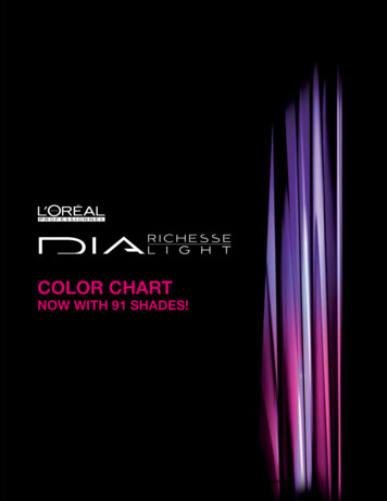
Clinical StudyV L Wester and othersHCC as a screening tool for CS698176:6Table 1 Baseline characteristics and hair cortisol content. Differences in baseline characteristics were analyzed using chi squareand Kruskal–Wallis tests. Differences in hair cortisol content were analyzed using analysis of (co)variance. Hair cortisol data forhealthy controls, and part of that of CS patients have been published previously (15, 16).Patient characteristicsMale (n, %)Age (years) (median, range)BMI (kg/m2) (median, range)Hair characteristicsHair washing 3 per week (yes/no, %)Use of hair products (yes/no, %)Hair coloring* (yes/no, %)Hair bleaching* (yes/no, %)Hair cortisol contentNon-adjusted (pg/mg hair) (geometric mean, 95% CI)Adjusted model 1† (pg/mg hair) (geometric mean, 95% CI)Adjusted model 2‡ (pg/mg hair) (geometric mean, 95% CI)First-line screening testsUrinary free cortisol (ULN) (geometric mean, 95% CI)Late-night salicary cortisol (nmol/L) (geometric mean,95% CI)DST cortisol (nmol/L) (geometric mean, 95% CI)74 (43%)32 (18–63)23.5 (16.9–43.3)127/45 (74%)84/89 (49%)36/138 (21%)13/161 (7%)Patient controls,n 35CS patients,n 43Pdiff6 (17%)38 (15–79)35.2 (21.4–46.6)13 (30%)50 (15–76)29.4 (18.3–81.6)0.011 0.001 0.00125/9 (74%)17/17 (50%)13/21 (38%)4/30 (12%)8.4 (7.0–10.0)8.3 (6.7–10.3)8.6 (6.9–10.6)24/16 (60%)12/28 (30%)10/33 (23%)1/42 (2%)0.2090.0910.0880.27012.7 (8.6–18.6)13.8 (8.6–21.8)15.2 (9.5–24.1)106.9 (77.1–147.9)95.8 (66.6–137.6)93.0 (63.7–135.6) 0.001 0.001 0.0010.42 (0.31–0.57)2.2 (1.4–3.5)3.81 (2.87–5.05)27.7 (18.1–42.4) 0.001 0.001476 (369–615) 0.00140 (30–54)*Hair coloring and bleaching in the 3 months prior to hair collection. †Model 1 was adjusted for age, sex and BMI. ‡Model 2 was adjusted for age, sex,BMI and hair characteristics.BMI, body mass index; CI, confidence interval; CS, Cushing’s syndrome; DST, dexamethasone suppression test; ULN, upper limit of normal.(geometric mean: 412.5 (95% CI: 176.7–961.1) pg/mg)compared to patients with Cushing’s disease andadrenal CS (82.6 (95% CI: 53.0–128.6) and 80.5(95% CI: 39.2–164.3) pg/mg respectively. F(2,40) 6.18,P 0.005, both pairwise comparisons vs ectopic ACTHsecretion P 0.01, Fig. 3). Within Cushing’s diseasepatients, the 6 patients with a macroadenoma had asignificantly higher HCC than the 20 patients withA 100B 1008080Sensitivity (%)95% CI: 77.1–147.9, F(2,249) 88.9, P 0.001), andsignificantly higher than in healthy controls (8.4 pg/mg, 95% CI: 7.0–10.0, P 0.001) and patients controls(12.7 pg/mg, 95% CI: 8.6–18.6, P 0.001). Adjustmentfor age, sex and BMI and hair characteristics did notchange these results (Table 1). In healthy controls, HCCwas not significantly influenced by sex (t(172) 1.05,P 0.293), age (Pearson’s r 0.65, P 0.395) or BMI(Pearson’s r 0.111, P 0.180).The ROC curve revealed an optimal cut-off for thediagnosis of CS of 31.1 pg/mg hair, when healthy controlswere used as a reference population. For this cut-off,sensitivity and specificity were 93 and 90% respectively(AUC 0.958, Fig. 1A). When we used patient controlsas a reference, the optimal cut-off for the diagnosiswas the same as with healthy controls. In this analysis,sensitivity and specificity remained similar at 93% and91% respectively (AUC 0.951, Fig. 1B).In CS patients, HCC levels significantly correlatedwith UFC (available in n 41, Pearson’s r 0.691, P 0.001,Fig. 2). HCC also correlated with serum cortisol after 1 mgDST (n 25, r 0.724, P 0.001) and with LNSC (n 33,r 0.761, P 0.001).Most CS patients had Cushing’s disease (n 26,60%), followed by adrenal CS (n 10, 23%) and ectopicACTH secretion (n 7, 16%). HCC was significantlyhigher in patients with ectopic ACTH secretionSensitivity (%)European Journal of EndocrinologyHealthy controls,n 1746040Sensitivity: 93%Specificity: 90%200AUC: 0.958020406080False positive rate (%)1006040Sensitivity: 93%Specificity: 91%200AUC: 0.951020406080100False positive rate (%)Figure 1ROC curves of HCC for the diagnosis of CS, with healthycontrols (A) or patient controls (B) as a reference population.The arrows indicate a cut-off value of 31.1 pg/mg. AUC, areaunder the curve. **P 0.01. Hair cortisol data for healthycontrols, and part of the CS patients have been publishedpreviously (15, 16).www.eje-online.orgDownloaded from Bioscientifica.com at 04/10/2022 08:05:07PMvia free access
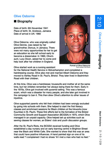
V L Wester and othersB100001000100r 0.691P 0.001n 411010.1110Hair cortisol (pg/mg)Hair cortisol (pg/mg)AHCC as a screening tool for CSC100001000100100r 0.724P 0.001n 2510110Urinary free cortisol (ULN)1001000DST cortisol (nmol/L)10000Hair cortisol (pg/mg)Clinical Study699176:6100001000100r 0.761P 0.001n 33101110100100010000LNSC (nmol/L)Figure 2Correlation between hair cortisol and first-line diagnostic tests in Cushing’s syndrome patients. Panel A, urinary free cortisol(UFC). Panel B, dexamethasone suppression test (DST). Panel C, late-night salivary cortisol (LNSC). Hair cortisol content isexpressed on a logarithmic scale, in pg/mg hair. UFC is expressed in times upper limit of normal (ULN) on a logarithmic scale. Thehorizontal dotted line represents the cut-off value for hair cortisol of 31.1 pg/mg. The vertical dotted line represents the upperlimit of normal for first-line diagnostic test. R, Pearson’s correlation. Hair cortisol data for healthy controls, and part of the CSEuropean Journal of Endocrinologypatients have been published previously (15, 16).a microadenoma (227.2 (98.2–524.2) vs 60.9 (44.8–82.6) pg/mg, t(24) 3.63, P 0.001).Hair cortisol timelines in patients with ectopic CSPatient A was a 54-year old woman, presented at theKlinikum der Ludwig-Maximilians-Universität in Munichin October 2014 with severe hypokalemic hypertension(minimum serum potassium 1.8 mmol/L) and edema, forevaluation of mineralocorticoid excess. However, plasmarenin concentration (2 U/L, normal: 4.4–46) and plasmaaldosterone ( 83 pmol/L, normal: 139–979 pmol/L) weresuppressed. Before October 2014, she was asymptomatic.Classical signs and symptoms of CS were absent. ACTHdependent hypercortisolism was found, with a UFC of44 358 nmol/24 h (normal: 414 nmol/24 h), and a baselineserum cortisol of 3862 nmol/L (normal: 138–690 nmol/L).The patient underwent CRH stimulation testing, lowand high-dose dexamethasone testing, and sinus petrosalinferior sampling suggesting an ectopic ACTH source, butintensive imaging studies including FDG PET, DOTATATEPET and DOPA PET scanning were negative. The patientunderwent emergency bilateral adrenalectomy aftercontrolling hypercortisolism by CYP11B2 blockade usingintravenous etomidate. The source of ACTH has not beendetected thus far (last follow-up June 2016).In the middle of November 2014, approximately4 weeks after the onset, a hair sample was obtained, witha length of 26 cm, allowing us to retrospectively assesscortisol levels for over 2 years back in time (26 months;Fig. 4A). The most proximal 12 cm of hair, whichcorrespond with the period from November 2013 toNovember 2014, were divided into 1 cm segments. Themost distal 14 cm of hair were divided into 2 cm longsegments, spanning the period between September 2012and November 2013. Between April 2013 and April 2014,HCC values in Patient A were around the previouslyestablished cut-off value ( 31.1 pg/mg). From April 2014onward, 6 months before clinical presentation, a gradualrise in HCC can be observed, with a maximum of 78.2 pg/mg. In the hair segments corresponding to the fall of2012, increased HCC can be observed as well (Fig. 4A).Patient A did not recall any symptoms of CS duringthis period.Patient B was a 59-year-old woman, presentedin September 2015 at the Klinikum der LudwigMaximilians-Universität in Munich with symptoms ofrapid weight gain (7 kg in 2 weeks), edema, abdominalobesity, plethora, hirsutism, hair loss, proximal muscleweakness, irritability and polydipsia. All symptoms hada recent onset of less than one month earlier. Clinicalexamination showed plethora, dorsal fat pad, proximalmuscle weakness, edema of the lower extremities, moonface, abdominal obesity, hirsutism, alopecia and dry skin.ACTH-dependent hypercortisolism was found, with aLNSC of 308 nmol/L (normal: 4.1 nmol/L), and a UFC of11 361 nmol/24 h (normal: 229 nmol/24 h). A CRH test,high-dose dexamethasone suppression test and inferiorsinus pretrosal sampling were all in accordance with anectopic Cushing’s syndrome. On PET-CT, a suspectedpancreatic lesion was seen, but this was not evidentfrom an abdominal MRI and endoscopic sonographywww.eje-online.orgDownloaded from Bioscientifica.com at 04/10/2022 08:05:07PMvia free access
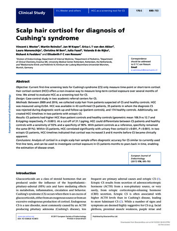
V L Wester and othersClinical StudyHair cortisol (pg/mg)10000**HCC as a screening tool for CS176:6700A**10001001010Healthyn 17 4Non-CSn 3 5Pituitaryn 2 6Adrenaln 10Ectopic ACTHn 7Figure 3BHair cortisol concentrations in healthy controls, patientswithout Cushing’s syndrome (Non-CS patients), and in patientswith Cushing’s syndrome. Hair cortisol values are presented ona semi-logarithmic scale. Values in patients with Cushing’ssyndrome are stratified by etiology. Solid horizontal linesEuropean Journal of Endocrinologyrepresent group medians, the dotted horizontal linerepresents the cut-off value of 31.1 pg/mg. Hair cortisol datafor healthy controls, and part of the CS patients have beenpublished previously (15, 16).Figure 4that followed. Based on a diagnosis of occult ectopicCushing’s syndrome, adrenostatic therapy was startedin December 2015, with ketoconazole, metyrapone andhydrocortisone substitution. After this, the patient becamenormocortisolemic. In June 2016, UFC was 151 nmol/24 h(normal: 229/24 h).In November 2015, slightly over 2 months after theonset of symptoms, a hair sample of 11 cm length wasobtained, allowing us to create a retrospective timelinespanning December 2014 to November 2015 (Fig. 4B). Themost proximal 7 cm of hair was divided into 1 cm longsegments, while the most distal 4 cm was divided into two2 cm long segments. In hair segments corresponding toDecember 2014 to June 2015, cortisol levels appear belowor just above the cut-off level ( 31.1 pg/mg). From June2015, 3 months before the onset of symptoms, cortisollevels raised sharply, up to 311.2 pg/mg.DiscussionIn the present study, we used HCC to distinguish betweenindividuals with CS, and those without. We found that acut-off value of 31.1 pg/mg discriminates well between CSand healthy controls. Additionally, we showed that thistest performs equally well when CS patients are comparedHair cortisol timelines in patients with a recent onset ofectopic Cushing’s syndrome. The horizontal dotted linerepresents the cut-off value of 31.1 pg/mg. The gray areacorresponds to period in which the patient had signs and/orsymptoms of Cushing’s syndrome.to patients who were suspected to have CS, but in whomthe diagnosis could be excluded (patient controls).The sensitivity of 93% and specificity of 90% (healthycontrols) and 91% (patient controls) we found are well inline with current first-line tests for CS as reported in recentmeta-analyses: UFC (mean sensitivity and specificity of 84and 92% respectively) (20), LNSC (95 and 92%) (21) and1 mg DST (99 and 88%) (20). However, it must be notedthat the performance of first-line test differs significantlybetween studies.In two patients with recent onset of ectopic CS, wecreated retrospective timelines of cortisol exposure. Inboth cases an increase can be seen months before theeffects of hypercortisolism became clinically evident. Inthe case of patient A, the hair sample surprisingly seemsto indicate hypercortisolism as much as 2 years before haircollection. This may indicate that she has suffered froma previous period of hypercortisolism and thus a cyclicalform of CS, although the patient did not recall symptomsof CS around that period. We previously showed that hairwww.eje-online.orgDownloaded from Bioscientifica.com at 04/10/2022 08:05:07PMvia free access
European Journal of EndocrinologyClinical StudyV L Wester and otherscortisol can well reveal cyclical forms of Cushing’s withmore than one period of symptomatic hypercortisolism inthe past (15). Care must be taken not to draw exaggeratedconclusions based on hair cortisol timelines, especiallyin the lack of supporting evidence. However, thesecases do illustrate the potential of HCC measurements,offering the unique possibility to retrospectively assesscortisol levels months up to years back in time (6). Theuse of HCC as a historical record of Cushing’s syndromewas first shown by Thompson et al. (14), and we havepublished additional examples in which HCC timelinescorresponded well with the clinical course in Cushing’s(15). Now, for the first time we showed that the onset ofhypercortisolism due to ectopic ACTH production couldbe estimated. From this, we may learn in the future howthe duration of the disease (before diagnosis) may affectthe somatic and neuropsychological outcomes of thedisease both in short-term and, importantly, in long-termafter curation.Besides the addition of retrospective long-termcortisol measurement, scalp hair cortisol offers significantadvantages over current first-line tests for hypercortisolism.First, hair sampling can easily be performed in an inpatientor outpatient setting. Unlike all of the other first-linetests, it does not depend on the adherence of patients toinstructions concerning sample collection or medicationuse. Obviously, complete baldness makes scalp hairsample collection impossible, but in our experience, evenin individuals with little scalp hair a useful sample canbe obtained successfully. Second, our results are based oncollecting just one hair sample. Currently, often multipleurine or saliva collections are required to establish thediagnosis of CS (3, 5). Recently, it has been shown thatHCC in the proximal 1 cm of scalp hair (representativeof the month before sample collection) correlates wellwith integrated daily salivary cortisol, based on threedaily saliva samples, which were collected for a 30-dayperiod (22). This is strong evidence that cortisol in hairtruly reflects the average long-term exposure to cortisol,which dampens the effects of the incidental fluctuations(such as stress at the moment of sample collection) thatseriously affect the concentrations that are measured inthe other tests.Although the measurement of scalp hair cortisolis more laborious than cortisol measurements in othermatrices, due to hair sample preparation and extraction,it requires no sophisticated techniques. Hair cortisolmeasurements have been performed for years by severalexpertise centers (14, 16, 23). A previously publishedround robin comparison between hair cortisol methodsHCC as a screening tool for CS176:6701from different labs found that different methodscorrelated well, with r2values between 0.88 and 0.97(24). Further standardization of analytical procedures,as well as automation of hair sample work-up may helproutine laboratories set up hair cortisol assays. Like anyanalytical method hair cortisol relies on proper training oflaboratory personnel, in order to limit operator variability.Furthermore, when using any immunoassay basedmethod for an extended period of time, there is a risk ofassay performance drift. In our lab, we have consistentlyused the same equipment and ELISA kits. However, we dorely on quality controls procedures as performed by themanufacturing company.Several limitations have to be considered wheninterpreting our data. We included patients in our study,who either had unequivocally confirmed CS, and patientsin whom it could unequivocally be excluded. Since thediagnosis of CS was based on current first-line tests, ourdata do not allow a comparison between the diagnosticaccuracy of HCC and these other tests. Additionally, wedid not investigate the added value of hair cortisol incases in which the diagnosis of CS remains unclear. Futureprospective studies may show the additional value of HCCin these cases.Experience with current first-line tests for CSillustrates the importance of investigating possibleconfounders of screening tests for CS. UFC may befalsely elevated due to high urine volumes and stress,and falsely low with impaired renal function. TheDST is subjected to the absorption and metabolism ofdexamethasone, which can be influenced by medicati
Methods: Between 2009 and 2016, we collected scalp hair from patients suspected of CS and healthy controls. HCC was measured using ELISA. HCC was available in 43 confirmed CS patients, 35 patients in whom the diagnosis CS was rejected during diagnostic work-up and follow-up (patient controls), an