Transcription
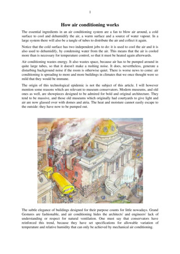
Review article 1Focus on abnormal air: diagnostic ultrasonography for theacute abdomenBeatrice Hoffmanna, Dieter Nürnbergc and Mary C. WestergaardbEmergency ultrasonography is a frequently used imagingtool in the bedside diagnosis of the acute abdomen.Classic indications include imaging for acute abdominalaneurysm, acute cholecystitis, hydronephrosis, and freeintra-abdominal fluid in patients with trauma or suspectedvascular or ectopic pregnancy rupture. Point-of-caresonographic imaging often emphasizes the diagnosticutility of fluid and edema, both as a significant finding andas a desirable adjunct for improved imaging. Conversely,the finding of sonographic intra-abdominal air is commonly‘tolerated’ as a necessary evil that can foil imageacquisition. This is in stark contrast to the accepteddiagnostic utility of air in other imaging modalities forthe acute abdomen, such as computed tomography andconventional radiography. Countering the bias againstair as a deterrent for diagnostic ultrasound’s accuracyare several published studies suggesting that abnormalair patterns can be used with high precision to diagnosepneumoperitoneum. These studies advocate thatsonographic findings of abnormal air can bestraightforward and can become crucial for increasingthe diagnostic yield of bedside ultrasound of the acuteabdomen. They suggest that practitioners shouldIntroductionUltrasonography is widely recognized as an indispensabletool in the bedside diagnosis of the acute abdomen [1–5].Even for practitioners with limited ultrasound training,initial assessment of the peritoneal patient often includesa bedside FAST examination to document the presence offree fluid, both for traumatic and for medical presentations. Similarly, bedside ultrasound of the gallbladder,kidneys, aorta, and uterus may prove diagnostic and arewithin the purview of residency-trained emergencyphysicians. For all these studies, traditional teachingemphasizes the diagnostic utility of fluid, both as asignificant finding (as in FAST scanning, ruptured aorticaneurysm, or ectopic pregnancy) and as a desirableadjunct for improved imaging (as with bladder filling foruterine imaging). Conversely, intra-abdominal air iscommonly ‘tolerated’ as a necessary evil that often foilsimage acquisition. This is in stark contrast to theaccepted diagnostic utility of air in other imagingmodalities for the acute abdomen, such as computedtomography (CT) scanning and radiography. Counteringthe bias against air in ultrasound are several publishedstudies suggesting techniques and documenting thesuccesses and accuracy of sonography for the diagnosisof pneumoperitoneum [6–23]. Despite this, many text0969-9546 c 2012 Wolters Kluwer Health Lippincott Williams & Wilkinsfamiliarize themselves with the findings and techniquesto gain the experience required to make the diagnosiswith confidence. This article will discuss four groups ofabnormal air patterns found in the abdomen and theretroperitoneum and the respective scanning techniques,with a focus on the use of ultrasound for diagnosingpneumoperitoneum and a suggested scanning approachin the emergency setting. European Journal of EmergencyMedicine 00:000–000 c 2012 Wolters Kluwer Health Lippincott Williams & Wilkins.European Journal of Emergency Medicine 2012, 00:000–000Keywords: abdominal pain, abscess, acute abdomen, bedside ultrasound,emergency ultrasound, free air, free intra-abdominal air, pneumatosisintestinalis, pneumoperitoneumaDepartment of Emergency Medicine, Division of Emergency Ultrasound, JohnsHopkins University, Baltimore, Maryland, bDepartment of Emergency Medicine,University of Wisconsin, Madison, Wisconsin, USA and cDepartment ofGastroenterology, Ruppiner Kliniken GmbH, Neuruppin, GermanyCorrespondence to Beatrice Hoffmann, MD, PhD, RDMS, Department ofEmergency Medicine, Division of Emergency Ultrasound, Johns HopkinsUniversity, 1830 E. Monument Street, Suite 6-100, Baltimore, MD 21287, USATel: 1 410 550 7852; fax: 1 410 502 5146; e-mail: bhoffma8@jhmi.eduReceived 6 February 2012 Accepted 29 March 2012books and lecturers, especially in the field of emergencyand critical care sonography, skip the topic entirely,effectively narrowing the scope of bedside ultrasound.Inclusion of an assessment for pathologic air will increasethe diagnostic yield of bedside ultrasound of the acuteabdomen, and practitioners should familiarize themselveswith the findings and techniques to gain the experiencerequired to make the diagnosis with confidence.BackgroundTo appreciate the presence of pathological air within theabdomen, one must first recognize the appearance ofphysiologic air within abdominal and thoracic structures,including the gastrointestinal tract and the lung parenchyma as it descends during inspiration. Air is a mediumposing high resistance and impermeability to ultrasoundwaves, making it a strong reflector. Larger air bubbles mayappear as bright, highly echogenic lines with distalreverberation and shadowing artifacts (Fig. 1a, b and d).Smaller air bubbles can appear as bright punctuate fociwithout ring-down artifacts and shadowing within theintestinal lumen (Fig. 1c). Normal air present within thelumen of the gastrointestinal tract should be recognizableby its association with the bowel and will move withperistalsis. Air artifacts emanating from the lungs into theDOI: 10.1097/MEJ.0b013e3283543cd3Copyright Lippincott Williams & Wilkins. Unauthorized reproduction of this article is prohibited.
2 European Journal of Emergency Medicine 2012, Vol 00 No 00Fig. 1(a)(b)Air withinintestineLiverComet tailartifactsShadowing4.713(c)(d)Lung tipLiverShadowingIntestinallumenAir114.7Appearance of physiologic air in the abdomen: (a) air within the intestinal lumen (stars) with shadowing, (b) larger air bubbles appearing ashyperechoic stripes generating comet tail artifacts, (c) smaller air bubbles within the intestinal lumen, and (d) air artifacts emanating from the thoraciccavity and the lung over the liver. Sonography equipment used for all figures was an M-Turbo (Sonosite, Bothell, Washington, USA) with either3–5 MHz curvilinear or 5–10 linear probe.abdomen will show characteristic respiratory movementsand originate from the thoracic cavity (Fig. 1d).Findings of pathologic intra-abdominal air can be dividedinto several categories: (a) ‘free air’, seen in the peritonealand retroperitoneal space, (b) air seen in the lumen ofpreformed intraperitoneal or retroperitoneal structures orcavities, such as the bladder and the gallbladder, biliarytree, common pancreatic duct, portal vein, hepatic veins,and other blood vessels. Air may also form (c) within theabdominal wall tissues or organ parenchyma, as in kidney,liver, and abdominal wall abscesses. Finally, the finding of(d) intramural air, as seen in pneumatosis of the bladderwall or in pneumatosis intestinalis, can be a crucial findingin a patient with abdominal complaints (Fig. 2) [6–24].Once the concept of normal versus abnormal air patterns isestablished, the sonologist can utilize this information todiagnose the potential causes of pathologic abdominal air.This article will discuss the aforementioned four groups ofabnormal air patterns found in the abdomen and theretroperitoneum, with a focus on the use of ultrasound fordiagnosing pneumoperitoneum.Abnormal findings and sonographic techniqueExtraluminal free air: pneumoperitoneum andpneumoretroperitoneumPneumoperitoneum: is an expected finding after certainprocedures including abdominal laparoscopy or laparotomy,percutaneous needle biopsy, peritoneal dialysis, culdocen-Fig. 2Differential diagnosis:sonographic airPhysiologic airPathologic air Gastrointestinal tract ExtraluminalFree air intra-abdominal or retroperitoneal LungAir projecting into theabdominal cavity IntraluminalAir in preformed cavities or vessels IntraparenchymalAir in tissue or parenchyma Intramural airDifferential diagnosis of air found with abdominal sonography. Adaptedwith permission from [24].tesis, or paracentesis. Free air after surgery may be absorbedwithin a few days, but can last as long as 18 days, withincidence and quantity declining over time [20]. In patients with postoperative acute abdominal pain, a commondiagnostic dilemma centers on whether postoperative intraabdominal air represents benign residual air from the surgery or a pathologic condition such as anastomotic failure.Copyright Lippincott Williams & Wilkins. Unauthorized reproduction of this article is prohibited.
Ultrasonography for pneumoperitoneum Hoffmann et al.3Fig. 3(a) Large amount of free air superficial to the liver in the hepatoperitoneal space with enhanced peritoneal stripe (black arrows) and reverberationartifacts (white arrows). The patient had a colon biopsy 5 days ago and presented with acute severe abdominal pain and hypotension. Normalperitoneum is visualized lateral to the enhanced areas. (b and d) Several small air collections in the mid-abdominal area causing the EnhancedPeritoneal Stripe Sign (EPSS) phenomenon (black arrows) and reverberation artifacts posteriorly (white arrow). This patient presented from a nursinghome with abdominal pain, change in mental status, and hypotension after a recent colon surgery. The intraoperative diagnosis was perforatedviscous at surgical site and peritonitis. (c) Small amount of free air anterior (small air bubble) ventral to the liver causing EPSS (black arrow) andreverberations with a ring-down artifact (white arrow). This patient presented with several days of mild-to-moderate abdominal pain and normal vitalsigns, and was diagnosed with perforated diverticulitis. Free air was also found in the left lower abdomen.The distinction between benign and pathologic etiologiesshould be made on a case-by-case basis. Acute pneumoperitoneum in nonpost-operative scenarios can be caused bypathologic findings such as ruptured viscous, peritonitiswith gas-forming organisms, intra-abdominal abscess rupture, bowel obstruction with permeation of gas throughthe bowel wall, cardiopulmonary resuscitation, mechanicalventilation, cocaine use, and extension of pneumothorax orpneumomediastinum [25–27].The concept of visualizing free intra-abdominal air usingB-mode sonography is not new. In fact, one of the mostcomprehensive and detailed studies on the topic waspublished nearly 30 years ago [6]. In this landmarkpublication, Seitz and Reising [6] reported many oftoday’s known qualitative and quantitative aspects of freeintraperitoneal air sonography. Ultrasound consistentlydetected as little as 1 cm3 of free air [6]. Their study wastriggered by their observation that in patients with recentcholecystectomy, distinct posterior hyperechoic reverberation and shadowing artifacts emanating from anenhanced peritoneal stripe impeded postoperative hepa-tobiliary sonography. They suspected that this phenomenon was caused by free air (Dr K. Seitz, personalcommunications, August 2011) and found it mostprominent over the ventral aspect of the liver or whenthe patient was in the semileft lateral decubitus position.They also described ‘shifting’ of air to the highest point ofthe cavity when the patient was repositioned [6]. Aninitial qualitative study confirmed corresponding free airon the chest radiograph and ultrasound in studyparticipants who had small amounts of free air injectedafter paracentesis. Muradali et al. [11] later validated thisfinding and termed the phenomenon ‘Enhanced Peritoneal Stripe Sign (EPSS)’. EPSS consists of a superficialsingle or double echogenic line that marks the abutmentof the abdominal wall with the peritoneal contents(Fig. 3a–c). A double line is commonly seen in patientswith excess abdominal fat, in whom identification of theperitoneal stripe may be more difficult. Muradali’sexperimental trials in pigs (whose abdomens were infusedwith degassed water) showed that a single bubble of aircould lead to a focal thickening of the peritonealstripe [11]. When more air was introduced, the superficialCopyright Lippincott Williams & Wilkins. Unauthorized reproduction of this article is prohibited.
4 European Journal of Emergency Medicine 2012, Vol 00 No 00focal thickening of the peritoneal stripe was associatedwith dirty shadowing or multiple reflection artifacts.Dirty shadowing occurs when multiple layers of airbubbles create multiple bright overlapping echo tails;this is commonly seen with normal bowel gas duringroutine abdominal scanning. Comet tail artifacts mayappear posterior to a single row of bubbles, with taperedperiodic echogenic lines or reverberations [12]. When thereverberation echoes retain their width, the artifact istermed as ‘ring-down’.Sonographic technique for pneumoperitoneum: Most authorssuggest using higher frequency linear probes to bestvisualize the superficial peritoneal layer and associatedfree air [6]. One author, however, achieved highsensitivity and specificity (100 and 99%, respectively) withcurved array transducers in the 2.6–5 MHz range [19].However, in that study, evaluation for free air was includedin a systematic abdominal ultrasound examination using acurved array transducer and was performed by a radiologistin a dedicated ultrasound suite. The entire comprehensiveultrasound exam lasted 20–35 min, and it is unclear howmuch time was spent on the evaluation for free air.Today, most authors agree that the best initial place tolook for pneumoperitoneum is in the right hypochondrium, superficial to the liver, with the patient in thesupine position with the thorax slightly elevated or in asemilateral decubitus position [6,9,11–13]. When thepatient is positioned in semileft or semiright lateraldecubitus, free air frequently collects in the ventralhepatoperitoneal space (right) or at the lower pole of thespleen (left) [6,9]. One study of 600 patients withabdominal pain referred for ultrasound (21 of whomultimately had pneumoperitoneum) showed that thefinding of EPSS was observable in all quadrants of theabdomen with varying frequency, with 83% being observable in the right hypochondrium, 75% in the umbilicalregion, and only 8.3% noted in the right lumbar region [19].Another study suggests that optimal positioning is achievedwith the supine patient at 10–201 inclination. Alternatively,an intercostal view was obtained with the patient in the leftlateral decubitus position, with the abdomen and thoraxelevated to 30–401 [13].Beginning with Seitz and Reising [6], investigatorsfrequently noticed that free air pockets were positionedindependent of respiratory movements, but would migratewhen pressure from the ultrasound transducer deformedthe abdominal wall. This phenomenon was later describedas ‘scissor’s phenomenon’ or ‘shifting of air’ by Karahanet al. [18]. ‘Shifting’ can also be accomplished by movingthe patient from a supine to a decubitus position to confirmthe free movement of intraperitoneal air, as opposed tointraluminal or alveolar air [17] Proponents of the EPSSmethod of identifying pneumoperitoneum argue thatshifting or elevating the patient may not be possible incases of critical illness or trauma [11].The accuracy of sonography for pneumoperitoneum in theclinical setting was found to be high in several largeprospective trials. Seitz and Reising [6] reported a sensitivityof 90% and a specificity of 100% for the detection ofpneumoperitoneum, evaluating 4000 consecutive patientswith nontraumatic acute abdominal pain. A recent study on487 consecutive patients with blunt trauma and abdominalpain by Moriwaki et al. [23] showed that a screeningultrasound for pneumoperitoneum had a sensitivity andspecificity of 85 and 100%, respectively. The authorsconcluded that sonography may be a valuable screening toolfor intra-abdominal free air.Pseudopneumoperitoneum: The practitioner should be awareof several conditions that may mimic pneumoperitoneum.Chilaiditi syndrome is a rare condition in which colonicbowel is interposed between the liver and the diaphragm.Its potential to mimic free air under the diaphragm onupright chest radiography is well known. The dynamicnature of ultrasonography is helpful in this situation;shifting the patient to a different position should showthat there is no freely mobile air within the peritoneum. Inaddition, prolonged scanning may show peristalsis withinthe hepatodiaphragmatic recess, leading to the diagnosis ofthis condition. Artifacts distal to an overlying rib may mimica reverberation artifact suggestive of pneumoperitoneum. Inthis case, the origin of the artifact should be traceable to apoint superficial to the peritoneal stripe and should changewith probe position and patient movement. Also, thecontrast between rib shadow and the liver can mimic freeair, but the distinction should become clearer duringrespiration. When scanning the right upper quadrant, aring-down artifact may be seen originating from alveolar airin the lungs. During inspiration, these artifacts may overlapwith the reverberations from pneumoperitoneum; however,expiratory scanning will show the separate thoracic andintra-abdominal origins of the artifacts [8]. In addition, thestep-off may be noted from the more superficial alveolarartifacts and the pneumoperitoneum artifacts just inside theperitoneum [9]. Intramuscular and cutaneous air may bedistinguished by immobility when probe pressure isapplied [23].Pneumoretroperitoneum: This is a rare complication ofendoscopic retrograde cholangiopancreatography (0.5%)and other iatrogen-invasive procedures [21], but can alsobe because of trauma, inflammation, infection, or neoplasticprocesses [21]. Typical sonographic findings include aircollections around the right kidney, causing it to appear‘overcast’ or ‘veiled’ [28], air ventral to the aorta and theinferior vena cava, giving the appearance of ‘vanishing greatvessels’ [29], and air collections around retroperitonealparts of the duodenum, pancreatic head, and posterior tothe gallbladder [21]. In contrast to intra-abdominal free air,the shifting phenomenon is not observed. Rather, air isfound in closed relation to an organ or around retroperitoneal viscous [11,12,21].Copyright Lippincott Williams & Wilkins. Unauthorized reproduction of this article is prohibited.
Ultrasonography for pneumoperitoneum Hoffmann et al.Intraluminal free air: abnormal air within preformedlumensAbnormal air within lumens physiologically not containingany gas can be observed within the biliary ducts or thegallbladder, portal venous system, abdominal or retroperitoneal veins and arteries, within the bladder and the urogenitaltract, or even within pancreatic ducts [9,12,21,30–32].The interpretation of findings will depend on the clinicalpresentation and circumstances, as the two most commonetiologies appear to be trauma (including iatrogenic manipulation) and infectious causes [12]. Pneumobilia (air inthe biliary system) for instance can be caused by papillotomy, fistulas within the gastrointestinal tract, tumorinvasion, or by an ascending biliary infection with gasproducing organisms. Air collections within arteries andveins can be because of iatrogenic vascular manipulations5such as a cardiac catheter or hemodialysis and are rare. Airwithin the portal venous system is associated with asignificant mortality (Fig. 4), often stemming from bowelnecrosis or thrombosis of the mesenteric artery or veins[8,12].Sonographic technique to detect abnormal air within preformedlumens: In general, the same technique used to examinethe preformed lumens, that is, biliary or portal vein orabdominal vessel sonography, will also show abnormal air.Intraparenchymal free air: abnormal air within organparenchyma and tissuesA common etiology for this finding is infection andabscess formation (Fig. 5). Trauma (including iatrogenic)or neoplasm eroding into solid tissue from air-containingFig. 4Left: Air bubbles detected within the portal vein (black arrows) and accumulating in smaller portal vessels (white arrows). The patient was found to havebowel necrosis and ischemia from mechanical ileus. Right: Large amount of air in the inferior vena cava and the hepatic venous system of the liver (whitearrows). This patient had sustained a gunshot wound to the thigh with an injury to the femoral vein and prolonged cardiopulmonary resuscitation.Fig. 5Abscess of the abdominal wall with small air collections on ultrasound (left) and with the corresponding image on computed tomography (right).White arrows show examples of air collection.Copyright Lippincott Williams & Wilkins. Unauthorized reproduction of this article is prohibited.
6 European Journal of Emergency Medicine 2012, Vol 00 No 00structures can also cause gas collections within organs.For instance, air can appear after a biopsy along theneedle path, or when a fistula (Crohn’s disease) erodesinto a neighboring organ, or neoplasms or ulcers penetrateinto parenchyma [12]. The clinician should alwaysattempt to distinguish these findings from other locationsof air collections or findings mimicking free air, suchas microcalcifications or cholesterol deposits [30].Sonographic technique to detect intraparenchymal free air: isequivalent to the regular ultrasound approach whenexamining these organs and tissues.Intramural airSonographic intramural air has been described in the gut(pneumatosis intestinalis) [12,33], but also can be found inthe wall of the bladder (Fig. 6) or the stomach and thegallbladder [30,34]. It is associated with infection,ischemia, and trauma. Pneumatosis intestinalis is a difficultsonographic diagnosis and should be distinguished from thephenomenon of pseudopneumatosis.Pseudopneumatosis intestinalis [33]: This is not a pathologicalcondition, but rather an artifact occurring at the nondependent wall.Sonographic technique for intramural air: The ultrasonicbeam should be aligned perpendicular to the intestinalwall and the use of a high frequency may show a betterresolution of bowel wall layers. When the probe isangulated, intraluminal air bubbles may be falselyprojected within the inner wall layers because oftangential effects. This artifact can also occur in areasof the intestine in which gas bubbles may be trapped inprominent intestinal folds. The appearance of air shouldchange from intramural to intraluminal with probeangulation and the phenomenon should not be visiblein the lower part of the intestinal wall. It is importantthat other sonographic findings mimicking intramural air, such as adenomyomatosis or calcificationswithin the gallbladder wall, are distinguished from pathological air. The clinician should rely on history and examination findings, and have a high clinical index ofsuspicion.ConclusionAlthough sonographic detection of abnormal free airhas been described and recognized for decades, it isoften overlooked in traditional ultrasound teaching,especially in the emergency medicine and critical carecommunities. This is unfortunate, given evidence that, inexperienced hands, ultrasound may be as effective asradiography or perhaps even CT at diagnosing pneumoperitoneum [6,19,23].Fig. 6(a) Intramural air in pneumatosis coli with the corresponding computed tomography image on the right. White arrows show air collections. (b)Intramural air in a patient with severe bladder infection causing pneumatosis of the bladder wall (white arrows) and air fluid level in the bladder.Copyright Lippincott Williams & Wilkins. Unauthorized reproduction of this article is prohibited.
Ultrasonography for pneumoperitoneum Hoffmann et al.A scanning protocol used in our emergency department toscreen for pneumoperitoneum suggests placement of thepatient in either a semilateral left decubitus or a supineposition with a slightly elevated thorax. A systematicevaluation of the right hypogastric and epigastric area forEPSS is performed, beginning with a curvilinear transducer, at depth settings of about 9–11 cm. Areas of interestare further evaluated using a high-frequency linear probe.The exam includes an assessment for pleuroperitonealstep-off sign, nonmovement of EPSS with respirations,scissors maneuver, and shifting and usually lasts no longerthan 3–5 min for trained emergency physicians.However, to what length should the emergency physiciango in search of EPSS? The authors believe that theanswer might be similar to other, already establishedemergency ultrasound indications: it depends. In apatient with a high suspicion for perforated viscous andwith both radiograph and CT imaging or surgicalresources readily available, care might suffer if thephysician spends too much extra time with an additionalimaging tool. The opposite might be true for careprovided in resource-poor environments, where ultrasound becomes the imaging tool of choice, or if thephysician is faced with a long list of potential differentialdiagnosis in a stable patient with significant butundifferentiated symptoms. Here, an additional sonographic view of the ventral peritoneal anatomy could becombined with acute abdominal aneurysm or renal colicsonography. In our institution, which is a tertiary medicalcenter, the critically ill patients represent another targetgroup for pneumoperitoneum sonography. These very illpatients can challenge the physician with their limitedability to provide a history but may present withsuspicious physical exam findings such as abdominaltenderness. Here, if too unstable to be transported to CT– incorporating sonographic evaluation for pneumoperitoneum into bedside imaging can provide the physicianwith expedited care diagnosis. Of course, large studiesfocusing on outcome metrics would be needed tosubstantiate these assertions. The finding of abnormalfree air, with or without other associated ultrasonographicfindings (such as free fluid, ileus, or bowel necrosis), mayallow for a definitive diagnosis at the bedside withinminutes for the patient with an acute abdomen. Theadvantages are worthy of the efforts required to accuratelyperform assessments for pathologic air using the techniques described above, especially as these patients areoften already undergoing bedside ultrasound. Abdominalultrasound instruction should include the teaching oftechniques described above to detect pneumoperitoneum, so that the next generation of practitioners mayroutinely acquire the skill necessary to make the diagnosiswith confidence. Large prospective trials will be neededto validate the accuracy and the inter-rater reliability ofsonography for pneumoperitoneum performed by diversegroups of trained emergency or critical care physicians.7AcknowledgementsConflicts of interestThere are no conflicts of 2122American College of Emergency Physicians. ACEP Ultrasound Guidelines2008. Available at www.acep.org/content.aspx?id 32182 [Accessed 19October 2011].Privette TW Jr, Carlisle MC, Palma JK. Emergencies of the liver,gallbladder, and pancreas [review]. Emerg Med Clin North Am 2011;29:293–317, viii-ix.Ross M, Brown M, McLaughlin K, Atkinson P, Thompson J,Powelson S, et al. Emergency physician-performed ultrasound todiagnose cholelithiasis: a systematic review. Acad Emerg Med 2011;18:227–235.Fox JC, Solley M, Anderson CL, Zlidenny A, Lahham S, Maasumi K.Prospective evaluation of emergency physician performed bedsideultrasound to detect acute appendicitis. Eur J Emer g Med 2008;2:80–85.Unlüer EE, Yavaşi O, Eroğlu O, Yilmaz C, Akarca FK. Ultrasonography byemergency medicine and radiology residents for the diagnosis of smallbowel obstruction. Eur J Emerg Med 2010; 17:260–264.Seitz K, Reising KD. Ultrasound detection of free air in the abdominal cavity.Ultraschall Med 1982; 3:4–6.Lee DH, Lim JH, Ko YT, Yoon Y. Sonographic detection ofpneumoperitoneum in patients with acute abdomen. Am J Roentgenol1990; 154:107–109.Chadha D, Kedar RP, Malde HM. Sonographic detection ofpneumoperitoneum: an experimental and clinical study. Australas Radiol1993; 37:182–185.Kainberger P, Zukriegel M, Sattlegger P, Forstner R, Schmoller HJ.Ultrasound detection of pneumoperitoneum based on typical ultrasoundmorphology. Ultraschall Med 1994; 15:122–125.Braccini G, Lamacchia M, Boraschi P, Bertellotti L, Marrucci A, Goletti O,et al. Ultrasound versus plain film in the detection of pneumoperitoneum.Abdom Imaging 1996; 21:404–412.Muradali D, Wilson S, Burns PN, Shapiro H, Hope-Simpson D. A specificsign of pneumoperitoneum on sonography: enhancement of the peritonealstripe. Am J Roentgenol 1999; 173:1257–1262.Wilson SR, Burns PN, Wilkinson LM, Simpson DH, Muradali D. Gas atabdominal US: appearance, relevance, and analysis of artifacts. Radiology1999; 210:113–123.Grechenig W, Peicha G, Clement HG, Grechenig M. Detection ofpneumoperitoneum by ultrasound examination: an experimental andclinical study. Injury 1999; 30:173–178.Goske MJ, Goldblum JR, Applegate KE, Mitchell CS, Bardo D.The ‘circle sign’: a new sonographic sign of pneumatosis intestinalis –clinical, pathologic and experimental findings. Pediatr Radiol 1999;29:530–535.Chen CH, Yang CC, Yeh YH. Role of upright chest radiography andultrasonography in demonstrating free air of perforated peptic ulcers.Hepatogastroenterology 2001; 48:1082–1084.Chen SC, Yen ZS, Wang HP, Lin FY, Hsu CY, Chen WJ. Ultrasonography issuperior to plain radiography in the diagnosis of pneumoperitoneum. Br JSurg 2002; 89:351–354.Chen SC, Wang HP, Chen WJ, Lin FY, Hsu CY, Chang KJ, et al. Selectiveuse of ultrasonography for the detection of pneumoperitoneum. Acad EmergMed 2002; 9:643–645.Karahan OI, Kurt A, Yikilmaz A, Kahriman G. New method for the detection ofintraperitoneal free air by sonography: scissors maneuver. J Clin Ultrasound2004; 32:381–385.Asrani A. Sonographic diagnosis of pneumoperitoneum usingthe’enhancement of the peritoneal stripe sign.’ A prospective study. EmergRadiol 2007; 14:29–39.Gayer G, Hertz M, Zissin R. Postoperative pneumoperitoneum: prevalence,duration, and possible significance. Semin Ultrasound CT MR 2004;25:286–289.Nürnberg D, Mauch M, Spengler J, Holle A, Pannwitz H, Sei
books and lecturers, especially in the field of emergency and critical care sonography, skip the topic entirely, effectively narrowing the scope of bedside ultrasound. Inclusion of an assessment for pathologic air will increase the diagnostic yield of bedside ultrasound of the acute