Transcription
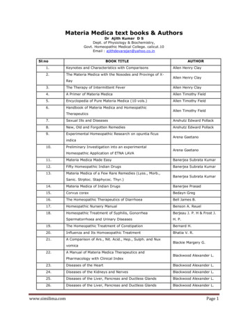
Research ArticlePage 1 of 7AUTHORS:Patrick S. Randolph-Quinney1,2*Scott A. Williams2,3Maryna Steyn1Marc R. Meyer4Jacqueline S. Smilg2,5,6Steven E. Churchill2,7Edward J. Odes1,2Tanya Augustine1Paul Tafforeau8Lee R. Berger2,9Primary osteogenic tumour in Australopithecus sedibaOsteogenic tumour in Australopithecus sediba:Earliest hominin evidence for neoplastic diseaseCenter for the Study of Human Origins,Department of Anthropology, New YorkUniversity, New York, New York, USAWe describe the earliest evidence for neoplastic disease in the hominin lineage. This is reportedfrom the type specimen of the extinct hominin Australopithecus sediba from Malapa, South Africa,dated to 1.98 million years ago. The affected individual was male and developmentally equivalentto a human child of 12 to 13 years of age. A penetrating lytic lesion affected the sixth thoracicvertebra. The lesion was macroscopically evaluated and internally imaged through phase-contrastX-ray synchrotron microtomography. A comprehensive differential diagnosis was undertakenbased on gross- and micro-morphology of the lesion, leading to a probable diagnosis of osteoidosteoma. These neoplasms are solitary, benign, osteoid and bone-forming tumours, formed fromwell-vascularised connective tissue within which there is active production of osteoid and wovenbone. Tumours of any kind are rare in archaeological populations, and are all but unknown inthe hominin record, highlighting the importance of this discovery. The presence of this diseaseat Malapa predates the earliest evidence of malignant neoplasia in the hominin fossil record byperhaps 200 000 years.Department of Anthropology, School ofSocial & Behavioral Sciences, ChaffeyCollege, Rancho Cucamonga, California, USAIntroductionAFFILIATIONS:School of Anatomical Sciences, University ofthe Witwatersrand, Johannesburg, South Africa1Evolutionary Studies Institute, Schoolof Geosciences, University of theWitwatersrand, Johannesburg, South Africa234School of Radiation Sciences, University of theWitwatersrand, Johannesburg, South Africa5Department of Radiology, Charlotte MaxekeAcademic Hospital, Johannesburg, South Africa6Department of Evolutionary Anthropology,Duke University, Durham, North Carolina, USA7European Synchrotron Radiation Facility,Grenoble, France8DST/NRF South African Centre of Excellencein Palaeosciences, University of theWitwatersrand, Johannesburg, South Africa9*Current address: School of Forensicand Applied Sciences, University ofCentral Lancashire, Preston, Lancashire,United KingdomCORRESPONDENCE TO:Patrick kPOSTAL ADDRESS:School of Forensic and Applied Sciences,University of Central Lancashire, Preston,Lancashire, PR1 2HE, UKDATES:Received: 11 Dec. 2015Revised: 16 Mar. 2016Accepted: 17 Mar. 2016KEYWORDS:Malapa; palaeopathology; neoplasia;taphonomy; osteoma; malignantHOW TO CITE:Randolph-Quinney PS, Williams SA, Steyn M,Meyer MR, Smilg JS, Churchill SE, et al.Osteogenic tumour in Australopithecus sediba:Earliest hominin evidence for neoplasticdisease. S Afr J Sci. 2016;112(7/8), Art.#2015-0470, 7 pages. http://dx.doi.org/10.17159/sajs.2016/20150470 2016. The Author(s).Published under a CreativeCommons Attribution Licence.South African Journal of Sciencehttp://www.sajs.co.zaA neoplasm (‘new-growth’ or tumour) is defined as a mass of localised tissue growth, the cellularproliferation of which is no longer subject to the effects of normal growth-regulating mechanisms.1-3A neoplasm may be benign or malignant. Malignant tumours are often referred to colloquially as cancer,although the term ‘malignant neoplasia’ is more clinically appropriate.1 In the developed world, death frommalignancy is second only to cardiovascular disease and is often perceived as a disease of modernity.4Neoplastic disease would have been prevalent in the past (e.g. Odes et al.5), but most likely occurredat much lower levels of incidence than today, given the shorter life expectancy for victims1,6,7 and thediffering environmental context. Both these factors strongly influence the incidence and prognosis of anycancer.3,8 The preserved signatures of neoplasms of any kind are rare in archaeological populations, andare all but unknown in the hominin record. Here we present the earliest fossil evidence for neoplasticdisease in the human lineage, with a detailed description and diagnosis of a tumorous lesion affectingthe spine of a juvenile male Australopithecus sediba, Malapa Hominin 1 (MH1).9,10 This species has beenpostulated as a possible ancestor of the genus Homo.9 The clinical and evolutionary implications of thediagnosed condition are discussed.The Malapa hominin siteThe Malapa site is one of several hominin-bearing Plio-Pleistocene cave deposits located within theCradle of Humankind World Heritage Site to the northwest of Johannesburg, South Africa. The regionincludes sites such as Sterkfontein11, Swartkrans12, Kromdraai13, Gladysvale14 and Rising Star15. Thefossil deposits in these caves were formed in roughly similar fashion as debris cone accumulationsdeposited beneath vertical cave openings, which formed phreatically within the dolomites of the MalmaniSubgroup.15,16 At Malapa, the main hominin-bearing deposits have been dated using uranium-lead datingof flowstones, combined with palaeomagnetic and stratigraphic analyses of flowstones and underlyingsediments, to 1.977 0.002 million years ago (Ma).17 The cave deposits comprise five sedimentaryfacies, termed A to E, from stratigraphically lowest to highest.Facies A and B occur below a central flowstone sheet, and are overlain by an erosion remnant (facies C),which in turn is overlain by the main hominin-bearing breccia, facies D. This has yielded well-preservedmacro- and micro-mammal fossils (such as carnivores, equids and bovids18), including the fossilisedremains of at least six hominins. Two of these, MH1 and MH2, have been reported in the literature asrepresentatives of a new hominin species, Australopithecus sediba9. Taphonomically the site has beeninterpreted as a complex cave system with open deep vertical shafts that operated as death traps for animalson the surface of the landscape. This death-trap scenario might have been the process by which the Malapahominins entered the cave system17,18, as evidenced by peri-mortem damage on the skeletons of MH1and MH2, consistent with a fatal fall19. Furthermore, both skeletons present partial anatomical articulationconsistent with rapid incorporation into the cave sediments early in the decomposition process.18Case study: Vertebra U.W. 88-37A pathological lesion affects the spine of Malapa Hominin 1 (MH1), the type specimen ofAustralopithecus sediba. This individual (Figure 1) was male, and at death he was at a developmentalstage equivalent to that of a human child aged 12 to 13 years9. The pathological specimen (U.W. 88-37)is a complete vertebra originally assigned to T5-T710, now considered to represent the sixth thoracicvertebra10. The dorsal surface of the right-side lamina exhibits a rounded penetrating defect (Figure 2),measuring approximately 6.7 mm supero-inferiorly and 5.9 mm medio-laterally.1Volume 112 Number 7/8July/August 2016
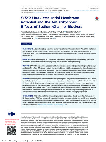
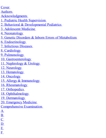
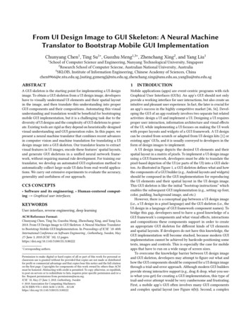
Research ArticlePage 2 of 7Figure 1:Primary osteogenic tumour in Australopithecus sedibaSurviving skeletal elements attributed to Malapa Hominin 1 (at time of writing).Figure 3:Figure 2:Vertebra U.W. 88-37. Photographs of surface morphology ofU.W. 88-3 showing position of lesion on right side of vertebrallamina: (a) right lateral aspect, (b) left lateral aspect, (c) inferioraspect, (d) superior aspect, (e) posterior aspect, (f) anterioraspect. Note that apertures seen on lateral aspects of thevertebral body in images (a), (b) and (f) represent normal vascularforamina infilled with residual breccia matrix. Images produced byPeter Schmidt.The lesion initially widens directly under the oval opening, but thennarrows as it progresses anteriorly. The base of the lesion appearssmooth and sclerotic under microscopic evaluation insofar as thepresence of residual breccia allows. The spinous process deviatesslightly to the right, but appears in keeping with slight asymmetrynoted elsewhere in the surviving thoracic vertebrae. This deviation fallswithin normal variation; we do not consider it significant enough tocause scoliosis or other vertebral misalignment, and it is unlikely thatthis asymmetry was related to the pathology.The defect presents as a lytic lesion that extends ventrally into thelamina for much of its length, the most anterior portion of which remainsinfilled with breccia matrix (Figure 3). On the surface, the lesion haswell-rounded edges with a somewhat sclerotic appearance. There is noevidence of periosteal or reactive bone formation on the cortex of thespecimen. Viewing the right lamina from above, it appears thicker thanthe left lamina and bulges laterally over the lesion, indicating a reactiveremodelling response to the presence of the defect.South African Journal of Sciencehttp://www.sajs.co.zaVertebra U.W. 88-37. Multi-focus (composite image stack)micrograph of surface morphology of U.W. 88-37 showingsub-angular penetrating defect on the right vertebral lamina.The lesion has well-rounded edges with lateral bulging ofthe cortex over the lesion, indicating a reactive remodellingresponse to the presence of the defect. Note that anteriorportion of defect remains infilled with breccia matrix.Micrograph taken with Olympus SZX Multi-focus microscope,magnification 7x. Scale bar 10 mm. Image courtesy ofAlexander Parkinson.2Volume 112 Number 7/8July/August 2016
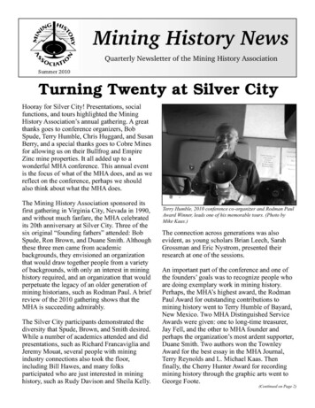
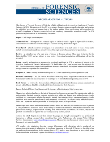
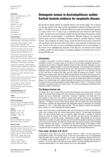
Research ArticlePage 3 of 7Primary osteogenic tumour in Australopithecus sedibaBecause of the presence of breccia within the lesion, the internalmorphology of the specimen was assessed using phase-contrastX-ray synchrotron microtomography (performed at the EuropeanSynchrotron Radiation Facility, ESRF) and a specific acquisitionprotocol applied for high-quality imaging of large fossils (seeSupplementary Appendix materials and methods). From themicrotomographic volume, the maximum long axis of the lesion inthe transverse plane measures 11.8 mm x 4.9 mm along the minoraxis, with a cross-sectional area of 45.6 mm, and in the sagittalplane the lesion measures 14.7 mm x 7.9 mm, with a cross-sectionalarea of 68.6 mm. The internal linear dimensions are consistentlyless than 20 mm in diameter, which has important implications forfinal diagnosis.at the approximate level of the superior articular facet. The internalmorphology shows no involvement of the transverse process orpedicle, and the lesion does not penetrate the vertebral canal. Nomineralised focal point or nidus was discerned. The edges of the firsttwo-thirds of the lesion (moving dorsal to ventral) display scleroticcharacteristics, with circumscribed margins of well-integratedcortical bone, abutted and intersected by trabecular striae (Figure 5and Supplementary Appendix). This pattern is indicative of a slowforming bony process, with remodelling and reorganisation ofposterior aspects of the lesion. The shape of a lesion is indicative ofits growth rate, with lesions that are long and oriented with the longaxis of a bone indicating a nonaggressive benign process. The ventralthird of the lesion, however, displays a geographic pattern of bonedestruction, showing a sharp non-sclerotic margin and evidenceof active osteolytic processes, with sharply-defined transectionof individual trabeculae, and active osteolytic penetration into theanterior portion of the lamina. A volume-rendered negative surfacemodel of the lesion (Figure 6) demonstrates the clear distinctionbetween the dorsal sclerotic zone and the ventral lytic zone within thebody of the active lesion.Key: S – quiescent sclerotic zone, O – active osteolytic zone, B – remaining brecciamatrix infill.Figure 5:Figure 4:Vertebra U.W. 88-37. Sixth thoracic vertebra of juvenileAustralopithecus sediba (Malapa Hominin 1). Partially trans parent image volume with the segmented boundaries ofthe lesion rendered solid pink. Volume data derived fromphase-contrast X-ray synchrotron microtomography. (a) leftlateral view, (b) superior view, (c) right lateral view. Imagesproduced by P.T.Figure 4 shows the microtomographic imaging, with a semi-trans parent volume-rendered image row. The imaging indicates that thelesion is highly penetrative and extends ventrally within the right-sideof the spinous process, penetrating the lamina before terminatingSouth African Journal of Sciencehttp://www.sajs.co.za3Transverse slices through vertebra U.W. 88-37 derived fromphase-contrast X-ray synchrotron microtomography. Relativeposition and anatomical orientation of orthoslices (a), (b)and (c) shown on the volume-rendered model. The posteriorportion of the lesion is sclerotic with circumscribed marginsof well-integrated cortical bone, abutted and intersectedby trabecular striae, with remodelling and reorganisationof the cortex. The anterior portion of the lesion displays ageographic pattern of bone destruction, showing a sharp nonsclerotic margin and evidence of active osteolytic processes,with sharply defined transection of individual trabeculaeand active osteolytic penetration into the anterior portion ofthe lamina. Image produced by P.S.R.Q.Volume 112 Number 7/8July/August 2016
Research ArticlePage 4 of 7Primary osteogenic tumour in Australopithecus sedibasurviving cranial and post-cranial elements of MH1 excludes a diagnosisof specific or non-specific systemic infection, such as brucellosis, nonspecific osteitis, haematogenous osteomyelitis or treponemal osteitis.There is no evidence of deformation or callus formation associated withskeletal trauma such as a healed fracture, and the lesion does not presentmorphology consistent with post-traumatic processes such as corticalhypertrophy or the development of a cloaca. It is therefore most likelythat this condition represents a primary osteogenic or osseous tumourof the spine. These are rare lesions with a much lower incidence thanmetastases, multiple myeloma or lymphoma.1,2,20,21,23,27,32 Based on ageat death, sex, anatomical location of the lesion, and specific patterns ofexpression and skeletal involvement, conditions such as osteosarcoma,chondrosarcoma or Ewing’s sarcoma can be excluded; these neoplasmsare often more aggressive, with destruction of the cortex1,21,23.Included in the differential diagnosis as the most likely cause ofthe observed lesion are osteoid osteoma, osteoblastoma, giantcell tumour and aneurysmal bone cyst. A number of secondarydiagnoses are possible, specifically enostosis (compact bone island),fibrous cortical defect (fibroxanthoma), plasmacytoma, eosinophilicgranuloma, and hydatid cyst infection. The range of possible differentialdiagnoses and primary diagnostic criteria are detailed in Table S1(Supplementary Appendix).Based on the observed pathological, morphological, and life-historycriteria, the two most likely diagnoses are osteoid osteoma andosteoblastoma. Taking the demographic data for these two tumourtypes into account, both options seem possible: both are primary boneforming tumours, osteoblastic in nature; benign; have a predilection formales; and show the highest prevalence in juveniles and adolescents.Osteoid osteoma resembles the observed lesion in terms of size, asthese tumours are usually less than 20 mm in diameter, with wellcircumscribed margins and being round or oval in form23.Figure 6:McCall22 notes that computed tomography is the most valuable methodto investigate this type of lesion. Under CT imaging of osteoid osteomaa small lucency is often recorded, which may have a central highattenuation as a result of mineralisation, and surrounding sclerotic boneis noted with some thickening of the lamina or pedicle. These are featuresseen in MH1 (Figure 4). On plain radiographs, most osteoid osteomasare osteosclerotic, with or without a visible nidus. By contrast, Kan andSchmidt35 suggest that osteoblastomas are predominantly lucent orlytic in roughly 50% of cases, sclerotic in 30% of cases, and mixed inthe remaining 20% of cases. On plain radiographs, osteoblastomas aretypically expansile with a scalloped or lobulated appearance, and theirmargins are well-defined, with a sclerotic rim evident in approximately30% of patients. A sclerotic rim is therefore much more common inosteoid osteomas than in osteoblastomas. The smooth, sclerotic, welldefined posterior margins of the lesion we studied are fully consistentwith a resolving osteoid osteoma. However, the skeletal distributionof osteoid osteoma might argue against this being the most likelydiagnosis, as osteoid osteomas are most commonly found in the lowerextremities; occurrence in the spine is less likely than that exhibitedin osteoblastoma22.Surface rendered image volume of the U.W. 88-37 lesionderived from phase-contrast X-ray synchrotron microto mography. Images show isosurface derived from segmentedboundaries of the lesion (remaining breccia infill removed).The arrow denotes the interface between the smoother dorsalsclerotic zone and the disorganised ventral lytic zone withinthe body of the lesion. (a) right lateral view, (b) medial view.Images produced by P.T.Differential diagnosisDiagnosis was undertaken using palaeopathological and clinicaldiagnostic criteria1,2,20-34. The accumulated evidence for osteolytic andosteosclerotic processes indicates that the disease process was bothchronic and active at the time of death of MH1 (as mentioned, at adevelopmentally equivalent stage to a modern human child of 12 to 13years of age). The lesion was less than 15 mm at the largest diameter,extending deep into the right side of the spinous process and involvingonly the vertebral lamina. The presence of reorganised sclerotic boneindicates a reactive ante-mortem process, and the lesion can thereforenot be attributed to taphonomic, diagenetic or pathology-mimickingeffects or processes1.To quantifiably assess the differential diagnosis, we applied BayesTheorem of conditional probability to the diagnosis of osteoid osteomaand osteoblastoma. Using absolute clinical incidence data of osteoidosteoma36-38 and osteoblastoma25,37-42 to calculate prior and conditionalprobabilities of the disease expression in the vertebral column (asopposed to elsewhere in the skeleton), a conditional probability of0.214 was derived for the likelihood of osteoid osteoma, and 0.068for osteoblastoma. These results indicate a 3.75-fold higher likelihoodthat osteoid osteoma was represented in this case than osteoblastoma(see supplementary online material Table S2 for discussion of Bayesparameters and probability functions used). Given the morphologicaland pathological similarities between the two tumour types, and theage and nature of the specimen under analysis, the results suggestosteoid osteoma firstly and osteoblastoma secondly as the most likelydiagnoses of what was clearly a benign entity of abnormal nature.The morphology of the lesion externally and internally is inconsistent withvertebral osteomyelitis. The absence of a proliferative cortical inflammatoryresponse (such as periosteal and/or endosteal bone hypertrophy) orsecondary lytic lesions across both the U.W. 88-37 vertebra and theSouth African Journal of Sciencehttp://www.sajs.co.za4Volume 112 Number 7/8July/August 2016
Research ArticlePage 5 of 7Primary osteogenic tumour in Australopithecus sedibaDiscussionAs noted above, neoplastic disease in various forms, including osteoidosteoma and osteoblastoma, is an ancient phenomenon. It first appearedduring the Palaeozoic and Mesozoic in extinct fish and members of theDinosauria respectively.3,46,47 However, the fact that reports of cancersor neoplasms remain exceedingly rare in the fossil record of almostany geological epoch1,3,8,46,47,53 may be due to a number of factors,exacerbated by the relative disjunction between osseous tumours andall other forms of neoplasms. Primary bone
Social & Behavioral Sciences, Chaffey College, Rancho Cucamonga, California, USA 5School of Radiation Sciences, University of the Witwatersrand, Johannesburg, South Africa 6Department of Radiology, Charlotte Maxeke Academic Hospital, Johannesburg, South Africa 7Department of Evolutionary A