Transcription
ContemporaryImplant DentistryC HA P T ER 1 4 O FC O N T E M POR AR Y O R A L A N D M A X I L L OFAC IALS U R G E RYB Y : D R A R A S H K HO J AST EH
Dental implant is suitable for: completely edentulous patientspatients missing posterior teethTrauma victims with missing of teeth and bonepatient missing a single tooth
completely edentulous patients
patients missing posterior teeth
Trauma victims with missing of teeth and bone
patient missing a single tooth
osseointegrationa histological definition meaning “ a directconnection between living bone and load bearingendosseous implant at the light microscopic level”
factors required for successful osseointegration:A biocompatible material2. An implant adapted to the prepared bony site3. Atraumatic surgery4. An immobile, undisturbed healing phase1.
Osseointegration
Accepted implant success criteriaThe individual unattached implant is immobilewhen tested clinically. No evidence of periimplant radiolucency is present. The mean vertical bone loss is less than 0.2mmannually after the first year of services. No pain, discomfort or infection The implant design appropriate for prosthesis withan satisfactory to the patient and dentist.
Contraindications to implant placement Acute illnessTerminal illnessPregnancyUncontrolled metabolic diseaseTumoricidal radiation including the implant siteHistory of IV use of Bisphosphonate therapyUnrealistic expectationImproper motivationLack of operator experienceUnable to restore prosthodonticly
Evaluation of implant siteinitial film: panoramic radiograph use of a small radiopaque reference object of knownsize placed at the area of implant placement allowscorrect magnification bonewidth will be evaluated in lateralcephalometric film. cone- Beam computed tomography has becomecommonly available in dental offices.
Bone height, width, and anatomic limitationsmore coritcal bone and denser cancellous bone higher implant success to maximiza the chance for success, there must beadequate bone width to allow 1mm of bone thelingual aspect and 1 mm on the facial aspect ofimplant. specific limitations as a result of anatomic variationsbetween different areas of the jaws must also beconsidered.
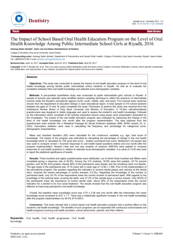
Bone types based on quality of corticalbone and density of cancellous marrow
Anatomic limitation to implant placement
Traditional minimum integration times
Surgical guide templatedelineate embrasure2. locate implant within tooth contour3. aligns implants with long axis of completedrestorations4. identify level of CEJ or tooth emergence from thesoft tissue1.
Surgical guide template
Basic surgicaltechnique
Before implant placementAtraumatic extraction Socket preservation Interim prosthesis design Timing of implant placement
Implant placementpatient preparation : Local anesthesia,preoperative antibiotic prophylaxis, rinse with 15ml 0.12% cholorhexidine gluconate for 30 seconds. Soft tissue incision: usually a simple crestalincision preparation of implant site: with a low speed,high torque hand piece and copious irrigation.Initial position, angulation and depth isestablished with the first twist drill in sequence. Implant placement: implant can be screwed bya very low speed hand piece. Final tightening witha ratchet.
Soft tissue incision
Initial position, angulation, depth is establishedwith first twist drill.
Paralleling pin evaluates position and angulation.
Implant placement
Postoperative carea radiograph should be taken. mild to moderate analgesics use of 0.12% cholorhexidine glunate rinses for 2weeks check up visits every week
Uncovering
Implant uncovering
Complicationsimproper angulation or position perforation of inferior border, the maxillary sinus,the inferior alveolar canal dehiscence of buccocortical or lingocortical plate mandibular fracture soft tissue wound dehiscence
Clinical implant components Implant bodyHealing screwInterim abutmentAbutmentImpression copingImplant analogWaxing sleeveProsthetic retaining screw
Implant body
Healing screw
Interim abutment
Abutment
Impression coping
Implant analogs
Waxing sleeve
Prosthesis- Retaining Screw
Completely Edentulous patientsimplant and tissue supported over denture all implant supported over denture Fixed porcelain metal or resin metal restoration
Implant supported over dentures
Fixed Porcelain metal restoration
Partially Edentulous PatientFree end distal extension Single-tooth implant restoration esthetics antirotation simplicity accessibility variability
Free end distal extension
Implant failure occursat the time of ( or shortly after) stage II ofsurgery: over heating, ill fit implant, infection,excessive pressure, wound healing problems. approximately 18 months after stage II ofsurgery: excessive force, lack of attached tissue andoral hygiene, smoking. more than 18 months after stage II ofsurgery (ailing implant): progressive bone loss
Advanced surgical techniques
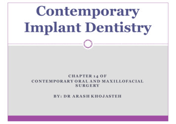
Guided bone regenerationa process that allows bone growth while retardingthe ingrowth of fibrous connective tissue andepithelium. it uses a barrier that is placed over the bone defect. characteristics of an ideal membrane: effective, easeof handling, inexpensive, resorbable, toleratesexposure.
Block bone graftingbone can be harvested from the genial region,mandibular ramus, illiac crest. the defect prepared for grafting by perforatingcortical bone. stabilization of the graft and primary closure isparamount. after 4 to 6 months of healing, the implant surgerycan be accomplished.
Bone graft
Alveolar distractionit is used for anterior maxilla when vertical hard andsoft tissue defects exist. when an osteotomized segment is slowly moved,allowing new bone formation within the gap. its disadvantages: increased cost, compromisedesthetic during distraction.
Alveolar distraction
Transantral grafting (Sinus lift)in the posterior maxilla, crestal resorption isassociated with sinus pneumatization. indirect sinus lift: when only few mm ofaugmentation is needed. direct sinus lift: several implants are placed, morethan 4 to 5 mm augmentation is needed. smoking is a contraindication to sinus lift.
indirect sinus lift
Direct sinus lift
Special situations
Post extraction placement of implant
Anterior maxilla esthetic zoneesthetic concern and compromised bone oftenpresent in the situation of congenitally missing teeth. grafting or corticocancellus blocks must beconsidered.
Atrophic anterior mandibleit is good to place 5 implants, leaving 2 to 3 mmabove the height of the residual bone. the transmandibular implant is effective in theatrophic mandible, with similar remodeling andformation of new bone. if the bone height is less than 6 mm, augmentationwith autogenous graft may be necessary.
Atrophic posterior mandiblewhen less than 8 mm overlying the inferioralveolar nerve is found, implant success iscompromised, and Bone may be grafted. super eruption of posterior maxillary teeth mayresults in adequate interarch space. It this case theinferior alveolar nerve repositioned to allow use ofthe entire height of mandibular body. It carries the risk of permanent anesthesia orpainful dysesthesia.
Inferior alveolar nerve positioning
Atrophic maxillaimplant placed bilaterally in the posterior maxilla,a prosthesis with ideal esthetics, phonetics andhygiene access an be created. a new technique is to place long implants into thebody of the zygoma, along with shorts anteriorimplants.
The Zygomaticus implant
Implants in growing patientsimplant can be placed as soon as patient is oldenough to cooperate with hygiene requirement. (7years) no implant should be placed until eruption ofnatural teeth and alveolar growth are completed. implant placed before this time behave similar to anankylosed tooth.
Implant in irradiated bonean implant supported prosthesis could improveesthetic and function. careful soft tissue handling and perioperativehyperbaric oxygen treatments is necessary.
Early loadingimportant factors for minimizing time: bonequality, implant material, surface and prosthesisconfiguration. the most extreme variation on them of early loadingis immediate loading.
Extra oral implants
CONTEMPORARY ORAL AND MAXILLOFACIAL SURGERY BY: DR ARASHKHOJASTEH Contemporary Implant Dentistry. Dental implant is suitable for: completely edentulous patients patients missing posterior teeth Trauma victims with missing of teeth and bone . Implant placement patient preparation: Local anesthesia, preoperative antibiotic prophylaxis, rinse .