Transcription
Anesthesiology Clin N Am22 (2004) 687 – 711Anesthesia for liver transplant surgeryRandolph H. Steadman, MDDepartment of Anesthesiology, David Geffen School of Medicine,University of California at Los Angeles, P.O. Box 951778, BH-431 Center for the Health Sciences,Los Angeles, CA 90095-1778, USAIronically. . . liver replacement, which was once considered the mostformidable of the whole organ transplantation procedures and the least likelyto be practical, has become the flagship of new principles that are applicable torecipients of all whole organs. . .—Thomas Starzl, 1996Liver replacement represents the sole definitive treatment for end-stage liverdisease. Medical treatment does little to improve survival; when life-threateningcomplications of liver failure such as encephalopathy, gastrointestinal bleeding,or uremia develop, the 5-year survival rate is b 50% [1]. Acute variceal bleedingis a particularly ominous prognostic sign, with a hospital mortality rate similar tothat of myocardial infarction. The 3-year survival is b 30%, despite endoscopicand pharmacologic advances [2].The dismal prognosis in end-stage liver disease led to the search for improvedtherapy, including hepatic replacement through whole-organ transplantation.In 1963, shortly after the effectiveness of azathioprine and prednisone wasestablished for renal transplantation, Starzl performed the first human livertransplant [3]. The recipient, a 3-year-old child with biliary atresia, died in theoperating room from massive hemorrhage caused by venous collaterals anduncontrollable coagulopathy. In 1967, Starzl successfully transplanted a liver intoan 18-month-old infant suffering from hepatocellular carcinoma. With thedevelopment of cyclosporine in 1979, survival after liver transplantationimproved significantly to a 1-year survival rate of over 70%. With the intro-E-mail address: rsteadman@mednet.ucla.edu0889-8537/04/ – see front matter D 2004 Elsevier Inc. All rights reserved.doi:10.1016/j.atc.2004.06.009
688R.H. Steadman / Anesthesiology Clin N Am 22 (2004) 687–711duction of tacrolimus in 1989, the incidence of adverse drug events related toimmunosuppression decreased [4]. Presently, the use of murine monoclonalantibodies directed against CD3 receptors or the interleukin-2 receptor delaystime to the first rejection and decreases the incidence of steroid-resistant rejection[5]. Over the last decade, continued improvements in surgical technique, themanagement of coagulopathy, the prevention of biliary complications, and thetreatment of infections have contributed significantly to decreasing morbidityand mortality.Indications for liver transplantationEnd-stage liver disease is the fourth leading cause of death in the United Statesfor individuals aged 45–54 years; it is surpassed only by cancer, heart disease,and unintentional injury. Among all age groups, liver disease accounted for over27,000 deaths in 2001, making it the 12th leading cause of death [6]. Livertransplantation is the second most common transplant surgery, accounting for21% of all organ transplants. Although the number of transplants has beenrelatively stable at 5000 per year, the number of patients waiting fortransplantation continues to grow, increasing ninefold over the last decade [7].The primary diagnoses for cadaveric liver recipients and their percentages of totalcadaveric liver transplants for a 5-year period (1997–2001) are non-cholestaticdisease (61%), cholestatic liver disease (11%), acute hepatic necrosis (9%),biliary atresia (4%), metabolic disease (3.9%), and malignant neoplasms (3%).Other diagnoses account for the remaining 7% of transplants.The category of non-cholestatic disease includes chronic hepatitis C (21% oftotal transplants between 1987–1998, which is presently the leading indicationfor transplantation in the United States), alcoholic liver disease (17%), alcoholicliver disease and hepatitis C (4.4%), chronic hepatitis B (5.5%), cryptogeniccirrhosis (11%), and autoimmune hepatitis (5%) [8–10]. Absolute contraindications to transplantation include extrahepatic malignancy, cholangiocarcinoma, active untreated sepsis, advanced cardiopulmonary disease, activealcoholism or substance abuse, and anatomic abnormalities precluding transplantation. With recent reports [11,12] of successful transplantation in recipientswith positive HIV serology, this condition is no longer an absolute contraindication. Living donor transplants increased markedly in 1999 and by 2001comprised 11% of total liver transplants. Although adult-to-pediatric living donortransplantation has been performed for over a decade, most of the increase citedabove is the result of the introduction of adult-to-adult living donor transplantation. Recipients of living donor organs, compared with cadaveric graftrecipients, have a higher frequency of cholestatic liver disease (18% versus 10%)and biliary atresia (9% versus 3%) and a lower incidence of non-cholestaticdisease (53% versus 60%) [7]. Management considerations for living donors arediscussed elsewhere in this issue.
R.H. Steadman / Anesthesiology Clin N Am 22 (2004) 687–711689Pathophysiology of liver failurePatients with end-stage liver disease have secondary dysfunction of virtuallyall other organ systems, and anesthetic management must include protection ofother organs damaged by liver failure.Central nervous systemUp to 80% of patients with acute liver failure develop cerebral edema andincreased intracranial pressure [13]. The cerebral symptoms of chronic liverfailure are not believed to be associated with cerebral edema, but increasedintracranial pressure can occur [14,15]. These reports support the belief that theencephalopathy found in chronic liver disease may have a common underlyingpathophysiology with the cerebral edema of acute liver failure, with only the rateand magnitude of change accounting for the clinically observed differences [16].Further supporting this belief, a number of similarities exist in both acute andchronic encephalopathy. The failure of hepatic clearance leads to an accumulationof toxins, such as ammonia and manganese, and to alterations in endogenoustransmitters and messengers, including g-aminobutyric acid (GABA), glutamate,and nitric oxide. The enzymes of the urea cycle are absent in the brain. Theresulting accumulation of glutamine, an osmotic compound, targets the glialastrocytes and results in cerebral edema in acute liver failure. Glutamine alsoaccumulates in chronic liver disease; however, compensatory changes probablyaccount for the absence of cerebral edema [17]. Other reports [18] have shownthrough magnetic resonance spectroscopy that counter-regulatory mechanismsare not always sufficient to prevent glial swelling, the cellular equivalent of lowgrade cerebral edema. Recently, the blood breakdown products hemin andprotoporphyrin IX have been suggested as possible endogenous benzodiazepinescontributing to hepatic encephalopathy because they are potent activators ofGABA receptors [19].Cardiovascular systemUp to 70% of patients with end-stage liver disease develop a hyperdynamicstate characterized by increased cardiac output and arteriolar vasodilatation [20].Vasoactive substances bypassing normal hepatic metabolism are most likelyresponsible. A recent study [21] in cirrhotic animals suggests that cannabinoidsmay contribute significantly to the hemodynamic alterations characteristic of endstage liver disease. The clinical improvement seen after total hepatectomy inpatients with acute liver failure suggests that toxic substances released fromnecrotic liver may be involved. Nitric oxide and guanosine 3V,5V-cyclicmonophosphate (cGMP) have been implicated as mediators [22]. Cardiomyopathy has been associated with alcoholic cirrhosis and hemochromatosis. Rhythmdisturbances may result from electrolyte or acid–base abnormalities. As thecriteria for transplantation have expanded, upper age limits for recipients have
690R.H. Steadman / Anesthesiology Clin N Am 22 (2004) 687–711been liberalized, making preoperative evaluation for ischemic heart disease moreimportant. Atherosclerotic coronary artery disease is at least as common inpatients with cirrhosis as in patients without liver disease [23]. Dobutamine stressecho (DSE) is the preferred preoperative screening study because it assesses theadequacy of myocardial oxygen supply, valvular function, and the presence ofintrapulmonary shunting or portopulmonary hypertension [24]. This test has a92–97% negative predictive value. A negative DSE predicts a good prognosisduring orthotopic liver transplantation (OLT), that is, a low likelihood ofperioperative cardiac events [25,26]. The presence of coronary artery disease isassociated with high mortality and morbidity (50% and 81%, respectively) duringOLT, making DSE screening a routine preoperative test for adult transplantcandidates in most centers [27].Pulmonary systemThe pulmonary complications associated with liver disease include restrictivelung disease, intrapulmonary shunts, ventilation–perfusion (V/Q) abnormalities,and pulmonary hypertension. Restrictive disease results from ascites or pleuraleffusions and frequently responds to fluid removal, at least transiently.Hypoxemia occurring in the absence of ascites or intrinsic lung disease isreferred to as hepatopulmonary syndrome (HPS). Vasodilation of unclear etiologyis associated with the syndrome. A contrast (bubble) echocardiogram may definethe cause of room air hypoxemia. In the case of cardiac shunts, microbubbles areseen almost immediately in the left atrium after venous injection of contrast. Inthe presence of intrapulmonary shunting, microbubbles appear three or morebeats after injection, whereas with V/Q defects the bubbles are absorbed in thelungs [28]. Although HPS resolves after transplantation, the persistence ofhypoxemia with 100% oxygen administration contraindicates transplantation inpatients with this syndrome [29]. In eight patients with HPS who underwenttransplantation at the author’s center, preoperative oxygen dependency resolvedin all patients over a range of 2 to 9 months [30].Approximately 2% of patients with chronic liver disease have portopulmonary hypertension (PPH). PPH is defined as a mean pulmonary artery pressureN 25 mm Hg or pulmonary vascular resistance N 120 dyned sd cm 5 in the presence of a normal pulmonary capillary wedge pressure. Mild (mean pulmonaryartery pressure [PAP], 25–35 mm Hg) or moderate (mean PAP, 35–45 mm Hg)pulmonary hypertension does not contraindicate transplantation, particularlywhen the pulmonary arterial pressures are responsive to a pharmacologic trial ofvasodilators [31]. The outcome of transplantation in patients with severe PPH ispoor [32]. When mean PAP is N 35 mm Hg and PVR is N 250 dyned sd cm 5,OLT is associated with increased perioperative mortality caused by right heartfailure or hepatic failure [33]; however, successful transplantation has beenperformed in patients with severe pulmonary hypertension who have undergonelong-term vasodilator treatment with epoprostenol that resulted in a decrease inpulmonary artery pressure [34,35].
R.H. Steadman / Anesthesiology Clin N Am 22 (2004) 687–711691Renal systemPreoperatively, it is important to identify patients with advanced renal diseasewho need combined liver-kidney transplants and to treat any preexisting acid–base abnormalities and plasma volume defects. If left untreated, less advancedrenal disease might worsen in the perioperative period [36]. The hepatorenalsyndrome (HRS), a functional cause of renal failure, is common in patients endstage liver disease. Diagnosing this syndrome requires the absence of primaryrenal disease, proteinuria, hypovolemia, or hemodynamic causes of renalhypoperfusion. A urinary sodium level b 10 mEq/L or a fractional excretion ofsodium b 1% is typical. The renin-angiotensin-aldosterone system is stimulated,and as the liver disease progresses, an increase in antidiuretic hormone results inan impairment of free water excretion and dilutional hyponatremia. In advancedstages of cirrhosis, intense renal vasoconstriction decreases glomerular filtrationrate (GFR), resulting in HRS [37]. Endothelin may be responsible for afferent arteriolar constriction, whereas increased nitric oxide levels decrease theefferent arteriolar tone, further decreasing GFR [38]. Paradoxically, the currenttreatment of HRS is to administer vasoconstrictors to reverse splanchnicvasodilatation. Terlipressin, a vasopressin analog, has been shown to decreaseserum creatinine, increase mean arterial pressure, and reverse the stimulation ofrenin [39,40]. Nephrotoxic antibiotics and contrast used for diagnostic studiesshould be avoided if possible. Cyclosporine adversely affects renal functionpostoperatively, typically decreasing the GFR by 30% to 50%. Some centerswithhold cyclosporine for the first 48 to 72 hours postoperatively to allowfunctional renal impairment to improve.Gastrointestinal systemEsophageal varices, portal hypertension, and ascites are common. Sclerotherapy or portosystemic shunts may be required. Gastric emptying is delayed, anddrug metabolism is affected (see ‘‘Drug metabolism’’ below). In cirrhotic patientsundergoing nontransplant surgery, the preoperative treatment of ascites (diuretics,paracentesis, and albumin administration) decreases the mortality rate. Thisstrongly suggests that the preoperative condition of liver transplant candidatesshould be optimized before portosystemic shunt procedures or nontransplantsurgery [41].Hematologic and coagulation systemAnemia commonly occurs as a result of chronic disease, malnutrition, orbleeding, is common. Coagulation defects result from multiple causes includingquantitative and qualitative platelet defects, decreased synthesis of clotting factors and their inhibitors, vitamin K deficiency, synthesis of abnormal clotting
692R.H. Steadman / Anesthesiology Clin N Am 22 (2004) 687–711factors, decreased clearance of activated factors, hyperfibrinolysis, and disseminated intravascular coagulation (DIC) [42].Splenic sequestration of platelets decreases the number of circulating plateletsand is the main cause of thrombocytopenia. Low levels of thrombopoietin, aliver-produced cytokine responsible for platelet formation, may also play a role[43,44]. Sepsis (bone marrow suppression) or DIC (consumption) also may leadto low platelet counts. Although a functional defect of platelet aggregation hasbeen described, its clinical significance is unclear [45,46]. All clotting factorsexcept von Willebrand factor are synthesized in the liver. As a result, liver failurepatients have low levels of all factors except fibrinogen, an acute phase reactant,and factor VIII. Decreasing levels of fibrinogen and factor VIII suggest thepresence of primary fibrinolysis or DIC. Prolonged prothrombin time (PT)correlates with the severity of liver disease and is one of the variables commonlyused as a prognostic indication of perioperative risk (Table 1) [47]. Ongoingfibrinolysis may occur, caused by low levels of antiplasmin and inadequateclearance of tissue plasminogen activator. Whether the fibrinolysis is primary orcaused by the activation of clotting (low-grade DIC) is controversial [48]. Usingspecialized assays, including thrombin-antithrombin and plasmin-a2-antiplasmincomplexes [49], a condition termed accelerated intravascular coagulation andfibrinolysis has been identified in 30% of patients with end-stage liver disease;yet, the clinical diagnosis of DIC hinges on a triggering event (eg, surgery, sepsis,or shock) and compatible laboratory findings (worsening PT, partial thromboplastin time, and platelet counts). The use of antifibrinolytics, common duringOLT, is contraindicated in patients with DIC.Drug metabolismEnd-stage liver disease patients tend to be sensitive to drugs, although theymay be resistant to some drugs (eg, pancuronium), a condition caused byincreased binding to globulin. The action of many drugs (eg, opioids, lidocaine,Table 1Pugh’s modification of the Child-Turcotte classificationbPoints rombin time (sec prolonged)Albumin (g/dL)Bilirubin (mg/dL) for cholestatic .52–34–103–4ModerateN6b2.8N3N10Child-Pugh class A, 5–6; class B, 7–9, class C, 10–15.Adapted from Weisner RH, McDiarmid SV, Kamath PS, Edwards EB, Malinchoc M, Kremers WK,et al. MELD and PELD: application of survival models to liver allocation. Liver Transpl 2001;7(7):567–80 (p. 568).
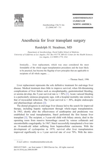
R.H. Steadman / Anesthesiology Clin N Am 22 (2004) 687–711693propranolol) are prolonged because of an increased volume of distribution ordecreased metabolism. Most opioids undergo oxidation in the liver. Morphine,which undergoes glucuronidation, is an exception. Oxidation of opioids isdecreased in end-stage liver disease, whereas glucuronidation is less affected. Themetabolism of fentanyl, however, is largely unaffected by liver disease [50]. Thedisposition of remifentanil, which undergoes ester hydrolysis, is independent ofthe liver.Patient selectionIn January 2004, there were over 17,000 candidates awaiting liver transplantation, with 10% of these prospective recipients dying annually [7]. Thisreality, caused by a growing number of indications for liver transplantation and anonexpanding cadaveric donor pool, highlighted the need for an improved graftallocation system. In 1998, the Department of Health and Human Services issuedthe ‘‘Final Rule’’ [51], defining the principles of organ allocation: the effect ofcandidate waiting times should be minimized in favor of allocation based onmedical urgency, whereas futile transplantations should be avoided to promotethe efficient use of scarce donor organs. Pugh’s modification of the ChildTurcotte (CTP) classification [52] (see Table 1), which has been in use as anassessment of disease severity since minimal listing criteria were first defined in1998, was originally developed to assess operative risk in end-stage liver diseasepatients undergoing portosystemic shunt surgery. Based on the CTP classificationsystem, the United Network of Organ Sharing (UNOS) allocation policy definedthree categories of disease severity. When this score was applied to liver allocation, limitations were apparent, notably the lack of distinction between mildlyand severely abnormal laboratory values (for example, all bilirubin levels over 3were treated similarly), the subjective nature of the assessment for encephalopathy and ascites, and the limited discrimination afforded by only three UNOSdisease severity categories (with the need for waiting time to serve as a tiebreakerwithin each of the three categories) [51]. These shortcomings led to the development of survival models for organ allocation [53].The model for end-stage liver disease (MELD) and pediatric end-stage liverdisease (PELD) models were adopted in February 2002 to allocate organs basedon medical urgency and to decrease the number of waiting list patient deaths[51,54,55]. The MELD score is based on three laboratory results: bilirubin,creatinine, and the international normalized ratio (INR). These tests have beenshown to be predictive of 3-month waiting list mortality (Fig. 1). Initial validationof the MELD score indicates that it is at least as good as the CTP score, usingmore objective, readily available variables [51]. There are known limitations tothe MELD score, such as the effect of body mass on the serum creatinine(a significant contributor to the final score, which may put nutritionally wastedcandidates at a disadvantage), the scoring for candidates with hepatocellular
694R.H. Steadman / Anesthesiology Clin N Am 22 (2004) 687–711Fig. 1. Relationship between MELD score and 3-month mortality in patients with cirrhotic liverdisease. (From Wiesner RH, McDiarmid SV, Kamath PS, Edwards EB, Malinchoc M, Kremers WK,et al. MELD and PELD: application of survival models to liver allocation. Liver Transpl2001;7(7):567–80 (p. 578); with permission.)cancer, and the effect of waiting time. However, scoring exceptions for theseissues and other conditions that are unaccounted for, such as hepatopulmonarysyndrome, should allow these populations to compete fairly for donor grafts. Itremains to be seen whether deaths while awaiting transplantation will decrease orwhether the MELD score will predict outcomes after liver transplantation. In2002 there was an 11% decrease in the number of waiting list deaths, the firstsuch decrease. The institution of the MELD system is one possible explanation,whereas improved pretransplant care may also play a role [7].Anesthetic management for liver transplantationAnesthetic preparation and inductionAnesthesia typically begins with a rapid sequence induction which isnecessitated by the emergent nature of the surgery, preoperative administrationof oral immunosuppressants and bowel decontamination antibiotics, and thepresence of ascites. An arterial catheter is placed either before induction orshortly thereafter. Large-bore intravenous access is obtained. At UCLA, two9 French introducers are placed centrally. Sites designated for venovenousbypass are avoided. A pulmonary artery catheter is commonly used in adultpatients. Transesophageal echocardiography (TEE) is a technique that isincreasingly being used during the procedure. Some liver transplant sites avoidpulmonary artery catheter insertion when TEE is used, although the pulmonaryartery catheter may be necessary when continuous intraoperative monitoring ofpulmonary artery pressures is desired or for postoperative hemodynamic and
R.H. Steadman / Anesthesiology Clin N Am 22 (2004) 687–711695fluid management in the intensive care unit (ICU). A rapid infusion systemcapable of high transfusion flow rates (500–1500 mL/min) is typically used. Suchsystems incorporate a reservoir, pump, filters, heat exchanger, and safety featuresdesigned to avoid and monitor for the presence of blood or air embolism, hypothermia, and line occlusion.The effects of the anesthetic technique on patient outcome are unknown.At the author’s center, a balanced anesthetic is used. This typically consists ofa volatile agent in low to moderate concentrations (0.5–1.0 minimum alveolarconcentration [MAC]) to ensure unconsciousness, while an opioid, usually fentanyl, is chosen to blunt the sympathetic response to stimulation and to provide asmooth transition to the postoperative period. In recipients with fulminant hepaticfailure and cerebral edema, volatile agents are avoided or used cautiously in lowconcentrations with intracranial pressure monitoring (see ‘‘Special situations’’below). In either case, periods of hypotension during the surgery may requiretemporary discontinuation of the volatile agent. Midazolam, with minimalhemodynamic effects, may be useful for its amnesiac effects during these hypotensive periods.Historically, the volatile agent of choice has been isoflurane, which preservessplanchnic blood flow better than other volatile drugs [56]. Recent work [57] inhealthy humans has confirmed the vasodilator effects of isoflurane on the hepatic circulation, compared with the vasoconstrictor effects of halothane. Thisbeneficial effect on hepatic oxygen supply may be advantageous to the newlyreperfused graft. The effects of desflurane on hepatic blood flow have beenevaluated with conflicting results. In animals, desflurane decreased total hepaticblood flow in a dose-dependent fashion at concentrations up to 1.0 MAC [58];however, in a human study [56] that excluded patients with liver disease, liverblood flow was slightly greater with desflurane than isoflurane, although thiseffect was not significant. Another study [59] compared the effects of sevofluraneand desflurane on hepatic blood flow and hepatocellular integrity in elderlypatients. Both agents resulted in decreases in gastric mucosal pH and increases incytosolic liver enzymes. The authors conclude that hepatocyte function is wellpreserved (lidocaine metabolism to monoethylglycinexylidide was unaffected byeither agent), but disturbances of hepatocellular integrity and gastric tonometrysuggest that splanchnic perfusion and oxygen delivery to the liver are decreased.Whether the increased metabolism of sevoflurane (100 times that of desflurane) isdetrimental to the liver is unknown, but it seems unlikely that the metabolites ofsevoflurane cause liver damage [60]. Compound A, a breakdown product ofsevoflurane found to be nephrotoxic in animals, has not been shown to causerenal toxicities in humans, even during low-flow sevoflurane administration [61].Cisatracurium may be the preferred neuromuscular blocking agent in patientsundergoing liver transplantation because of its organ-independent elimination anddiminished histamine release [62]. In patients with end-stage liver disease, thevolume of distribution of cisatracurium is greater than that in healthy controlpatients. Hepatic clearance is also increased in patients with liver disease; thisresults in similar elimination half times and similar duration of action (time to
696R.H. Steadman / Anesthesiology Clin N Am 22 (2004) 687–71125% recovery). Other reports have suggested the use of rocuronium during livertransplantation because the duration of the neuromuscular block appears to be auseful predictor of primary allograft function. All patients whose recovery timewas N 150 minutes experienced primary graft dysfunction [63].Preanhepatic stageThe preanhepatic stage begins with surgical incision and ends with crossclamping of the portal vein, the suprahepatic inferior vena cava, the infrahepaticinferior vena cava, and the hepatic artery. This phase involves dissection andmobilization of the liver and identification of the porta hepatis. With abdominalincision and drainage of ascites, hypovolemia typically occurs. Hypovolemiashould be treated in an anticipatory fashion with colloid-containing fluid tominimize changes in preload. In the presence of preexisting coagulopathy, freshfrozen plasma is indicated soon after incision, although some authors havechallenged the need for fresh frozen plasma during OLT [64]. Thromboelastography or standard laboratory tests (prothrombin time, fibrinogen and plateletcount) are used to guide the correction of coagulopathy [65]. Other authorsdisagree with the premise that coagulation monitoring is associated with bloodproduct transfusion requirements during OLT [66]. Considerable institutionalvariation exists in transfusion practices for OLT. These differences in transfusionrequirements are not accounted for by variations in blood loss during theprocedure [67].Fibrinolysis is unusual during the preanhepatic phase of the surgery, socryoprecipitate administration is typically unnecessary. Hyponatremia should notbe corrected rapidly. A perioperative rise of 21–32 mEq/L in the serum sodiumlevel was associated with central pontine myelinolysis in one report, whereas anincrease of b 16 mEq/L was not [68]. Citrate intoxication, ionized hypocalcemiaresulting from the infusion of citrate-rich blood products in the absence of hepaticfunction, is avoided by the administration of calcium chloride. Ionized hypomagnesemia also results from citrate infusion, but values of ionized magnesiumgradually return to normal after graft reperfusion [69]. The clinical significance ofthis remains speculative, but cardiovascular function may be affected. Aggressivetreatment of hypokalemia is best avoided, particularly in preparation forreperfusion and the associated increase in serum potassium. Supplementalglucose is usually not required except in pediatric patients or those with severedisease, such as fulminant hepatic failure. The maintenance of urine output isdesirable; however, the use of low-dose dopamine for this reason is unproven[70]. Hypothermia should be avoided. The use of heated venovenous bypassduring the anhepatic phase permits core temperature control. Bleeding during thisphase of surgery is related to the degree of preexisting coagulopathy, the presence and severity of portal hypertension, and the duration and complexity of thesurgical procedure [71,72]. The presence and severity of adhesions from previous abdominal surgery may add significantly to the complexity of the surgical dissection.
R.H. Steadman / Anesthesiology Clin N Am 22 (2004) 687–711697Intraoperative management: anhepatic stageThe anhepatic stage begins with the occlusion of vascular inflow to the liverand ends with graft reperfusion. Cross-clamping of the suprahepatic andinfrahepatic vena cava (IVC) decreases venous return by as much as 50%.Venovenous bypass (VVB), which diverts inferior vena cava and portal venousflow to the axillary vein, attenuates the decrease in preload, improves renalperfusion pressure, lessens splanchnic congestion, and delays the development ofmetabolic acidosis [73]. The use of VVB is not without risk. Air embolism,thromboembolism, and inadvertent decannulation may be fatal or result insignificant morbidity [74]. VVB is not uniformly used at all centers [75,76]. Theuse of the ‘‘piggyback’’ technique, with inferior vena caval preservation,decreases the need for VVB [77]. Hepatectomy is followed by hemostasis andvascular anastomoses of the supra- and infrahepatic IVC and the portal vein.Despite the absence of hepatic clotting factor production during the anhepaticstage, blood loss is usually limited by vascular clamping of the inflow vessels tothe liver. However, fibrinolysis may begin during this stage, caused by anabsence of liver-produced plasminogen activator inhibitor, which results in theunopposed action of tissue plasminogen activator. The use of antifibrinolyticsvaries among centers (see below).Intraoperative management: neohepatic stageReperfusion of the new liver through the portal vein begins the neohepaticstage. Reperfusion is associated with abrupt increases in potassium and hydrogenion concentrations, an increa
Anesthesia for liver transplant surgery Randolph H. Steadman, MD Department of Anesthesiology, David Geffen School of Medicine, University of California at Los Angeles, P.O. Box 951778, BH-431 Center for the Health Sciences, Los Angeles, CA 90095-1778, USA Ironically. liver replacement, which was once considered the most