Transcription
On Describing Human White Matter Anatomy: TheWhite Matter Query LanguageCitationWassermann, Demian, Nikos Makris, Yogesh Rathi, Martha Shenton, Ron Kikinis, MarekKubicki, and Carl-Fredrik Westin. 2013. “On Describing Human White Matter Anatomy:The White Matter Query Language.” Lecture Notes in Computer Science: 647–654.doi:10.1007/978-3-642-40811-3 81.Published Versiondoi:10.1007/978-3-642-40811-3 81Permanent 38477Terms of UseThis article was downloaded from Harvard University’s DASH repository, and is made availableunder the terms and conditions applicable to Other Posted Material, as set forth at rrent.terms-of-use#LAAShare Your StoryThe Harvard community has made this article openly available.Please share how this access benefits you. Submit a story .Accessibility
On Describing Human White Matter Anatomy:The White Matter Query LanguageDemian Wassermann1 ? , Nikos Makris2 , Yogesh Rathi1 , Martha Shenton1 , RonKikinis1 , Marek Kubicki1 , and Carl-Fredrik Westin112Brigham and Women’s Hospital and Harvard Medical School, Boston, MA, USAdemian@bwh.harvard.eduMassachusetts General Hospital and Harvard Medical School, Boston, MA, USAAbstract. The main contribution of this work is the careful syntactical definitionof major white matter tracts in the human brain based on a neuroanatomist’s expert knowledge. We present a technique to formally describe white matter tractsand to automatically extract them from diffusion MRI data. The framework isbased on a novel query language with a near-to-English textual syntax. This querylanguage allows us to construct a dictionary of anatomical definitions describingwhite matter tracts. The definitions include adjacent gray and white matter regions, and rules for spatial relations. This enables automated coherent labelingof white matter anatomy across subjects. We use our method to encode anatomical knowledge in human white matter describing 10 association and 8 projection tracts per hemisphere and 7 commissural tracts. The technique is shown tobe comparable in accuracy to manual labeling. We present results applying thisframework to create a white matter atlas from 77 healthy subjects, and we use thisatlas in a proof-of-concept study to detect tract changes specific to schizophrenia.1IntroductionDiffusion magnetic resonance imaging (dMRI) is a technique that allows to probe thestructure of white matter the human brain in vivo. dMRI streamline tractography hasprovided the opportunity for non-invasive investigation of white matter anatomy. Mostcommon methods for isolating fiber bundles based on streamline tractography requirethe manual placement of multiple regions of interest (ROIs). These methods include anapproach that starts from seed points within a predefined region of interest, and thencalculates and preserves only tracts that touch other predefined ROIs [1]. A differentapproach creates seed points throughout the entire brain (whole brain tractography)keeping tracts that pass through conjunctions, disjunctions or exclusions of ROIs eitheroff-line [2] or interactively [3]. An alternative method to manual placement of ROIs isto use a clustering approach [4–6]. Clustering methods are in general fully automatic,unguided, and take advantage of the similarity of fiber paths. However, incorporatingprecise information about human anatomy into a clustering method is difficult.The ability to target specific tracts for analyses compared to whole brain studiesincreases the statistical power and sensitivity of the study, and simplifies the interpretation of results, but requires precise consistent delineation of the tracts across subjects. Although several fascicules, such as the cingulum bundle, are widely recognized?Camera ready version for the Medical Image Computing and Computed Assisted Intervention2013 conference
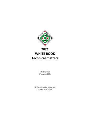
2Demian Wassermann et al.and well defined in field of neuroanatomy, there are others which existence or subdivisions is still a matter of discussion. Examples are the different systems to defineand subdivide the superior longitudinal fasciculus (SLF) proposed by Catani [7] andMakris [8], and the discussion regarding existence of the inferior occipito-frontal fasciculus (IOFF) in humans [9]. Three major challenges make it difficult to extend andreproduce tractography-based dissections and tract-specific analyses: 1) the anatomicaldissection of the white matter into specific fascicles is currently in discussion; 2) comparing fascicle definitions across atlases is a difficult task due to the lack of a system tounify the definitions; 3) semi-automated approaches are usually based on a fixed set offascicles difficult to extend due to amount of technical knowledge needed for the task.The main contribution of this paper is a system to express anatomical descriptionsof white matter tracts in a near-to-English textual language, and we believe this workcan help address the above challenges. We designed this textual language to be humanreadable to make the tract descriptions easy to change and extend without the need ofan engineering background and to be used for automated white matter virtual dissections. The white matter query language (WMQL) proposed in this paper has severalapplications. For example Wakana’s and Catani’s definition of tracts using regions ofinterest (ROIs) [10, 2], can readily be represented using WMQL definitions. This willyield an human readable definition that also can be extended by an anatomist for finerdivision. Another interesting application is to post-process clustering results and automatically label clusters as anatomically known tracts [4–6]. In this paper we presentdefinitions of different tracts from current literature and formulated WMQL descriptions. Our anatomy dictionary currently contains descriptions of 10 association and8 projection tracts per hemisphere, and 7 commissural tracts. An implementation ofWMQL as well as the definitions specified in this publication can be downloaded athttp://demianw.github.com/tract querier2MethodsWe designed the queries in white matter query language (WMQL) to formalize currentdescriptions in anatomy literature. Such descriptions are constructed in terms of different relationships between gyri, sulci, subcortical structures or white matter areas.Theoperations of WMQL, can be divided into 3 groups: 1) anatomical terms stating if atract traverses or ends in a certain brain structure, 2) relative position terms indicatingwhether the tracts are, for instance, medial or frontal to a structure like the amygdala,and 3) logical operations like conjunction, disjunction or exclusion of the previous twotypes of clauses. We illustrate these three types of operations in fig. 1.To apply WMQL queries to dissect dMRI-based tractographies, like in fig. 1, weneed to situate gyri, sulci and other structures used for the queries relative to the tractography. For this, we overlay an atlas of the brain structures on top the tractography,in this work we used the cortical and white matter parcellation based on the Desikanatlas as described by [11] and the neuroanatomic structure segmentation by [12] whichare readily available in FreeSurfer (http://www.freesurfer.org). We provide the detailsof this process in the section MRI Data Acquisition and Processing. It is worth notingthat WMQL does not depend on a particular atlas.
MICCAI 2013, Camera Ready Version3Anatomical Termsa) only(postcentral)b) endpoints in(postcentral)c) postcentralnot in only(postcentral)not in endpoints in(postcentral)Relative PositionTermsd) superior of(amygdala)inferior of(amygdala)e) anterior of(amygdala)posterior of(amygdala)f) lateral of(amygdala)medial of(amygdala)Logical Operationsg) a insula andh) b temporal and(inferior frontal oranterior of(amygdala)middle frontal or orbito frontal)i) uncinate fasciculus a and endpoints in(b)Fig. 1: WMQL Terms (a-f) along with an example construction of a WMQL query (gi). Regions in (g-i): insula (cyan); the orbito-frontal (purple), middle-frontal (brown)and inferior-frontal (dark green) gyri. h) shows the anterior temporal lobe (light green)defined as the section of the temporal lobe anterior to the amygdala (yellow).To implement the white matter query language, we group the tracts in sets representing whether they have an endpoint in each anatomical label (fig. 1b); traverseanatomical label (fig. 1c); or their position relative to each anatomical label (fig. 1d-f).Then, for each anatomical label, like the amygdala, we have six sets:1. endpoints in(amygdala): all tracts with at least an endpoint in the amygdala.2. amygdala: all the tracts traversing the amygdala.3. anterior of(amygdala), posterior of(amygdala), medial of(amygdala),lateral of(amygdala), superior of(amygdala), inferior of(amygdala) : containing thetracts traversing brain areas delimited by their relative position to the amygdalaWe calculate the endpoint sets (1) by computing which label is the tract touching ateach endpoint and adding the tract to the corresponding set. The sets representing labeltraversals (2) are computed by following each tract and adding it to the set corresponding to a particular label, like the amygdala, if tract traverses it. Finally, we compute the6 relative positioning sets (3) by checking the points the tract traverses with respect toa label. For instance, every tract that traverses the area anterior to the amygdala, shownin orange in fig. 1e, will be added to the set anterior of(amygdala). We implement this
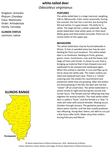
4Demian Wassermann et al.efficiently using a tree of axis-aligned bounding boxes of the structures as a spatial index of the labels and then computing the relative positions of the tracts with respect tothe bounding boxes composing each label [13]. For two sets of tracts a and b and theset of all tracts L, we formalize the WQML logical operations as follows:a or b : {tract : tract a b} only(a) : {tract : tract a tract 6 (L \ a)}a and b : {tract : tract a b} a not in b : {tract : tract a tract 6 b}We have illustrated these operations in fig. 1 a), c); and g)-i). Tracts in WMQL aredefined by using an assignment operation, for instance, we defined the left UF in fig. 1ias assigning a meaning to UF.left:UF.left insula.left and (lateral frontal.left or medial frontal.left or orbito frontal.left)and endpoints in(temporal.left and anterior of(amygdala.left)) not in hemisphere.rightTo simplify the definitions of tracts in both hemispheres with a single assignment, weincluded the suffixes .side and .opposite to WMQL. Creating a bihemispheric definitionbecomes:UF.side insula.side and(lateral frontal.side or medial frontal.side or orbito frontal.side) andendpoints in(temporal.side and anterior of(amygdala.side)) not in hemisphere.oppositeIn this way WMQL allows a single definition for tracts found in both hemispheressimultaneously. The formalization of WMQL as the basic set operations allows us todefine white matter tracts using all the flexibility and expressiveness power of set theory and propositional logic. Finally, to implement the WMQL language, we definedthe WMQL in Backus normal form; we used an LL(1) parser for this grammar whichtransforms WMQL expressions into an abstract syntax tree; and we implemented analgorithm to traverse the tree evaluating the WMQL operations.33.1ResultsDevelopment of WMQLWe are using WMQL to formalize tract descriptions from classic neuroanatomy textbooks and current literature on the anatomy of the white matter [14, 10, 8, 15]. In table 1, we show the queries for 10 association tracts. In the following, for each tractwe describe in WMQL, we provide a description derived from anatomy literature andthe derived WMQL query. The population results of these queries are shown in fig. 2along with the commisural and projection tracts whose definitions we did not includefor space reasons. Using the WMQL, we have constructed a comprehensive atlas basedon high angular MRI (HARDI) tractography [16] . The combination of WMQL withcurrent tractography technologies has enabled us to generalize previous atlases [10, 2]and extend them with white matter tracts not included in these like the middle longitudinal fasciculus (MdLF) or the three different superior longitudinal fasciculi. In total, ouratlas includes 10 long association tracts; 8 projection tracts per hemisphere (7 corticostriatal and the Cortico-spinal tract) and the 7 sections of the corpus callosum accordingto [15].
MICCAI 2013, Camera Ready Version5Table 1: Association Tract Definitions in WMQL: Cingulum bundle (CG); Extreme Capsule(EmC); and the fascicules: Superior Longitudinal (SLF) sections I to III; Arcuate (AF); Inferioroccipito frontal (IOFF); Middle Longitudinal (MdLF); Uncinate (UF)CB.side only((cingular.side or cingular cortex.side) and (middle frontal.side or cuneus.sideor entorhinal.side orsuperior frontal.side or inferior parietal.side or fusiform.side ormedial orbitofrontal.side or lateral orbitofrontal.side or parahippocampal.side orprecuneus.side or lingual.side or centrum semiovale.side))EmC.side (endpoints in(inferior frontal.side or middle frontal.side)andendpoints in(inferior parietal lobule.side) andtemporal.side and insula.side)not inhemisphere.oppositeSLF I.side (superior parietal.side and precuneus.side and superior frontal.side)or(superior parietal.side and precuneus.side and superior frontal.side andlateral occipital.side) not in cingular.side not in temporal.side not in subcortical.side not inhemisphere.oppositeSLF II.side (inferior parietal.side or supramarginal.side or lateral occipital.side) andendpoints in(middle frontal.side)) not in temporal.side not in subcortical.side not inhemisphere.oppositeSLF III.side ((inferior parietal.side or supramarginal.side or lateral occipital.side) andendpoints in(inferior frontal.side)) not in temporal.side not in subcortical.side not inhemisphere.oppositeAF.side (inferior frontal.side or middle frontal.side or precentral.side) and(superior temporal.side or middle temporal.side) not in medial of(supramarginal.side)not insubcortical.side not in hemisphere.oppositeIOFF.side (lateralorbitofrontal.side and occipital.side) and temporal.side not insubcortical.side not in cingular.side not in superior parietal lobule.side not inhemisphere.oppositeILF.side only(temporal.side and occipital.side) and anterior of(hippocampus.side) not inparahippocampal.sideMdLF.side only( (temporal pole.side or superior temporal.side) and (inferior parietal.sideor superior parietal.side or supramarginal.side or precuneus.side or (centrum semiovale.sideand superior parietal.side) or (centrum semiovale.side and inferior parietal.side))UF.side insula.side and (inferior frontal.side or middle frontal.side or orbito frontal.side)and endpoints in(temporal.side and anterior of(amygdala.side))3.2Application of WMQL to Tract Extraction and Statistical AnalysesDiffusion-weighted images (DWI) from 77 healthy subjects (HS) (32.9 12.4 yearsold of age; 64 males; right handed) and 20 male schizophrenic subjects (SZ) pairedwith the HS were acquired. DWI data were acquired on a GE Signa HDxt 3.0T (51directions with b 900 s/mm2, 8 b 0 s/mm2 images, 1.7 mm3 isotropic voxels). A T1MRI acquisition was also performed (25.6cm2 field of view, 1mm3 isotropic voxels).For each DWI image, we obtained a 2-tensor full-brain tractography placing ten seedsper voxel and obtaining an average of one million tracts per subject. We overlaid aparcellation of the cortical and sub-cortical structures and the white matter on DWIimages by processing the T1 images of each subject using FreeSurfer and registeringthe results to the DWI images using ANTS [17]. For each subject, this resulted in thesubcortical structures labeled and the cortex the white matter parcellated [11].
6Demian Wassermann et al.Cingular And Commisural FasciclesCingulum BundleInferior and Lateral Views of the Corpus Callosum Divided in 7 SectionsCorpus CallosumInferior and Lateral Views of the Corpus Callosum Divided in 7 SectionsSuperior LongitudinalFasciclesInferior and Medial Views of Thalamo-CorticalSuperior Longitudinal IIISuperior Longitudinal IISuperior Longitudinal IInferior and Medial Views of Thalamo-CorticalTemporo-Parietal And Temporo-Occipital FasciclesInferior and Medial Views of Striato-Cortical TractsMiddle Longitudinal FascicleInferior LongitudinalArcuate FascicleInferior and Medial Views of Striato-Cortical TractsFronto-Insular-Temporal FasciclessnoitceCallosumS 7 ni dedDividediviD minus7olSectionslaC suproC eht fo sweiV laretaL dna roirefnIInferior and Lateral Views of the CorpusExtreme CapsuleInferior Fronto-Occipital FascicleProjection TractsUncinate FascicleInferior and Medial Views of Thalamo-CorticallacitroC-omalahT fo sweiV laideM dna roirefnIStriato-Cortical TractsInferior and Medial Views of Striato-Cortical TractsCortico-Spinal TractOptic RadiationstcarT lacitroC-otairtS fo sweiV laideM dna roirefnIFig. 2: Iso-surfaces in blue (p-value 0.01, corrected for multiple comparisons) shows 11 association, 8 projection and 7 commissural tracts.Validation To validate our extraction protocol, 2 different experts segmented 5tracts (IOFF, UF, ILF, CST and AF) in each hemisphere of 10 healthy subjects following the protocol in [10]. Overlap between those and the ones extracted was calculatedwith the kappa measure [2]. For all tracts k .7 which is considered good agreement,the worse being the left AF (k .71) and the best the left CST (k .89).White Matter Atlas Generation with WMQL: For each subject we extracted 37white matter tracts using WMQL queries derived from classical and current literatureof the human brain white matter. Some of these are detailed in table 1. Processing eachfull brain tractography took 5 .25 min. to initialize and 7 .02 sec. per query.To generate a tract atlas, we normalized all the FA maps to MNI space, created apopulation FA template using ANTS and then generated group effect maps for eachtract [7]. To obtain the group effect maps, we started by calculating a binary visitationmap for each tract of each subject. This map is a mask in MNI space where a voxel hasa value of one if the tract traverses that voxel and 0 if it doesn’t. We create the groupeffect map for each tract through voxel-wise statistics. The group effect map assessesthe probability that a tract traverses each voxel. For this, we rejected the null hypothesisthat that voxel is not traversed by such tract. We first smoothed the visitation mapswith a 2mm (FWHM) isotropic Gaussian to approximate the distribution of the data toa Gaussian one [18]. Then, we rejected the hypothesis that the voxel does not belongto the tract, i.e. the mean traversal value over all subjects on that voxel is different
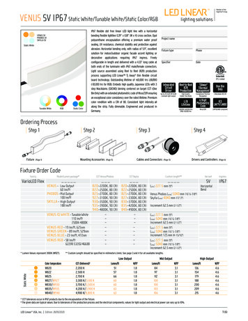
MICCAI 2013, Camera Ready Version7Fig hysubjectssubjects(HS) meanmeanoverdifferentbetweenand schizophrenic(SZ). The p-value was computed using a t-test correcting for age. We observe significant differences on thesubjects(SZ). The p-value was computed using a t-test correcting for age. We observe significantleft and right MdLF, and the left and right SLF II and III tracts.differencesthe leftp-valueand rightand the .001left and right SLF II and III tracts. Notation: theNotation: theon* means .05MdLF,and ** p-value* means p-value .05 and ** p-value .01to 0, by using a voxel wise t-test for a one-sample mean. We calculated the correctedsignificance using permutation testing (10,000 iterations) to avoid a high dependenceon Gaussian assumptions [19]. We set the significance threshold for considering that avoxel belongs to the tract to p-value 0.0001 corrected for multiple comparisons.Group Differences in Schizophrenia As a proof of concept, we analyzed the tractsthat are not included in other atlases, but found in our WM atlas only: MdLF, SLF I, IIand III in 20 SZ subjects and controls extracted from the HS paired by age and gender.We used the group maps to obtain a weighted average of FA resulting in one value persubject and performed a t-test for each tract correcting for age. We show these resultsin fig. 3. Results show agreement with recent discoveries in SZ in the MdLF tract [20]4ConclusionIn this work we have introduced the White Matter Query Language: a tool to representanatomical knowledge of white matter tracts and extract them from full-brain tractographies. This tool is a complement to current semi-automated approaches in tract extraction based in clustering and can be used to implement ROI-based approaches. Theutility of the tool was illustrated by constructing an atlas of white matter tracts from 77healthy subjects and then performing a statistical analysis in schizophrenia. The textualdescriptions in WMQL add transparency between the conceptualization of white matterfiber tracts and their extraction from a dMRI.Acknowledgments: This work has been supported by NIH grants: R01MH074794,R01MH092862, P41RR013218, R01MH097979, P41EB015902 and Swedish ResearchCouncil (VR) grant 2012-3682.
8Demian Wassermann et al.References1. Mori, S., van Zijl, P.C.M.: Fiber tracking: principles and strategies-a technical review. NMRin Biomedicine (2002)2. Wakana, S., Caprihan, A., Panzenboeck, M.M., Fallon, J.H., Perry, M., Gollub, R.L., Hua,K., Zhang, J., Jiang, H., Dubey, P., Blitz, A., van Zijl, P.C., Mori, S.: Reproducibility ofquantitative tractography methods applied to cerebral white matter. NImg (2007)3. Akers, D., Sherbondy, A., Mackenzie, R., Dougherty, R., Wandell, B.: Exploration of thebrain’s white matter pathways with dynamic queries. In: IEEE Viz. (2004)4. Wassermann, D., Bloy, L., Kanterakis, E., Verma, R., Deriche, R.: Unsupervised white matterfiber clustering and tract probability map generation: Applications of a Gaussian processframework for white matter fibers. NImg (2010)5. O’Donnell, L.J., Westin, C.F.: Automatic Tractography Segmentation Using a HighDimensional White Matter Atlas. TMI (2007)6. Wang, X., Grimson, W.E.L., Westin, C.F.: Tractography segmentation using a hierarchicalDirichlet processes mixture model. NImg (2011)7. Thiebaut de Schotten, M., Ffytche, D.H., Bizzi, A., Dell’acqua, F., Allin, M., Walshe, M.,Murray, R., Williams, S.C., Murphy, D.G., Catani, M.: Atlasing location, asymmetry andinter-subject variability of white matter tracts in the human brain with MR diffusion tractography. NImg (2010)8. Makris, N., Kennedy, D.N., McInerney, S., Sorensen, A.G., Wang, R., Caviness, V.S.,Pandya, D.: Segmentation of Subcomponents within the Superior Longitudinal Fasciclein Humans: A Quantitative, In Vivo, DT-MRI Study. Cerebral Cortex (2005)9. Schmahmann, J.D., Pandya, D.: The Complex History of the Fronto-Occipital Fasciculus.Journal of the History of the Neurosciences (2007)10. Catani, M., Thiebaut de Schotten, M.: A diffusion tensor imaging tractography atlas forvirtual in vivo dissections. Cortex (2008)11. Salat, D.H., Greve, D.N., Pacheco, J.L., Quinn, B.T., Helmer, K.G., Buckner, R.L., Fischl, B.:Regional white matter volume differences in nondemented aging and Alzheimer’s disease.NImg (2009)12. Fischl, B., Salat, D.H., Busa, E., Albert, M., Dieterich, M., Haselgrove, C., van der Kouwe,A., Killiany, R., Kennedy, D., Klaveness, S., Montillo, A., Makris, N., Rosen, B., Dale, A.M.:Whole Brain Segmentation: Automated Labeling of Neuroanatomical Structures in the Human Brain. Neuron (2002)13. Bergen, G.v.d.: Efficient Collision Detection of Complex Deformable Models using AABBTrees. Journal of Graphic Tools (1997)14. Parent, A.: Carpenter’s Human Neuroanatomy. Williams & Wilkins (1996)15. Witelson, S.F., Kigar, D.L.: Anatomical development of the corpus callosum in humans: Areview with reference to sex and cognition. In: Brain Lateralization in children: Developmental implications. (1988)16. Malcolm, J.G., Michailovich, O., Bouix, S., Westin, C.F., Shenton, M.E., Rathi, Y.: A filteredapproach to neural tractography using the Watson directional function. TMI (2010)17. Avants, B., Epstein, C., Grossman, M., Gee, J.C.: Symmetric diffeomorphic image registration with cross-correlation: Evaluating automated labeling of elderly and neurodegenerativebrain. MIA (2008)18. Ashburner, J., Friston, K.J.: Voxel-Based Morphometry—The Methods. NImg (2000)19. Nichols, T.E., Holmes, A.P.: Nonparametric permutation tests for functional neuroimaging:a primer with examples. HBM (2002)20. Asami, T., Saito, Y., Whitford, T.J., Makris, N., Niznikiewicz, M., McCarley, R.W., Shenton,M.E., Kubicki, M.: Abnormalities of middle longitudinal fascicle and disorganization inpatients with schizophrenia. Schizophrenia Research (2013)
framework to create a white matter atlas from 77 healthy subjects, and we use this atlas in a proof-of-concept study to detect tract changes specific to schizophrenia. 1 Introduction Diffusion magnetic resonance imaging (dMRI) is a technique that allows to probe the structure of white matter the human brain in vivo. dMRI streamline .