Transcription
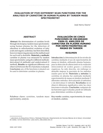
EVALUATION OF FIVE DIFFERENT SCAN FUNCTIONS FOR THEANALYSIS OF CARNITINE IN HUMAN PLASMA BY TANDEM MASSSPECTROMETRYJosé Henry Osorio1ABSTRACTAbstract: the determination of carnitine levelsthrough electrospray tandem mass spectrometryusing human plasma for the detection ofalteration in mitochondrial oxidation of fattyacids and organic acidemias, has been described;however improving precision in the method usedfor such end is desirable. Materials y methods:carnitine in plasma was measured by tandemmass spectrometry using five different methods:derivatized (4 methods) and underivatized (1method). Results: no significant difference wasobserved between the five functions evaluated.Conclusion: any of the functions evaluated canbe used to determine carnitine in plasma.Palabras clave: carnitine, tandem massspectrometry, analysis.EVALUACIÓN DE CINCOFUNCIONES DE ESCANEADOPARA EL ANÁLISIS DECARNITINA EN PLASMA HUMANOPOR ESPECTROMETRIA DEMASAS EN TANDEMRESUMENIntroducción: la determinación de los niveles decarnitina mediante el uso de espectrometría demasas en tándem, utilizando plasma humano,para la detección de alteraciones en la oxidaciónmitocondrial de ácidos grasos y aciduriasorgánicas, ha sido descrita, sin embargo esdeseable mejorar la precisión de los métodosusados para tal fin. Materiales y métodos: lacarnitina en plasma fue analizada medianteespectrometría de masas en tándem mediante 5diferentes métodos: derivatizando (4 métodos)y sin derivatizar (1 método). Resultados: no seobservó diferencia significativa entre las cincofunciones evaluadas. Conclusión: cualquiera delas funciones aquí evaluadas, puede ser utilizadapara determinar carnitina en plasma.Key words: carnitina, espectrometría de masasen tándem, análisis.1Laboratorio de Bioquímica Clínica y Patología Molecular, Departamento de Ciencias Básicas de la Salud, Universidadde Caldas. Correo electrónico: jose.osorio o@ucaldas.edu.coISSN 1657-9550Recibido: febrero 7 de 2013 - Aceptado: abril 1 de 2013Biosalud, Volumen 11 No. 2, julio - diciembre, 2012. págs. 52 - 58
Evaluation of five different scan functions for the analysis of carnitine in human plasma by tandem mass spectrometryINTRODUCTIONThe altered b-oxidation is followed by theaccumulation of representative acylcarnitinesdepending on the type of enzymatic deficiency inthe pathway (1). However, the production of theseacylcarnitines is dependent on the availability offree carnitine in the mitochondria, thus leavingthe possibility of having undetectable alterationsin acylcarnitine accumulation if the cell becomescarnitine depleted (2). It is necessary to knowthe occurrence and distribution of free carnitineand acylcarnitines in blood as an indication ofunusual metabolic conditions. Therefore thediagnosis and management of these metabolicdiseases requires knowledge of both freecarnitine and acylcarnitine concentrations.Carnitine and acylcarnitines contain a quaternaryammonium functional group, making thempreformed positive ions (cations) that are polarand non-volatile (3). Ions produced in thesource are selected by MS1 for transmission tothe collision cell. The fragments produced afterCID are transmitted to MS2 where they are againselected for transmission to the detector. Ionstransmitted by MS1 to the collision cell are calledprecursor ions (commonly referred as “parent”ions), and the fragments produced from CID areproduct ions (known as “daughter” ions) (4).During the derivatization process butyl estersof acylcarnitines are formed. These butyl estersare well suited for analysis by MS/MS since theyalready carry a positive charge and accordinglyno additives are needed in the mobile phase.Both butyl esters derivatives and underivatisedcarnitine and acylcarnitines share a commonproduct ion upon CID, which is singly chargedwith a mass of 85 Da, corresponding to CH2CH CH-COOH. This fragment results from theloss of elements of both (CH3)3N and C4H8 andthe side chain acyl group as RCOOH. Selectiveanalysis of only those molecules that produceand 85 Da product ion is accomplished using Preion scan mode. For the analysis of L-carnitine byMS/MS it is possible to obtain a characteristicfragment of 103 Da derived from the lossof both the trimethylamino group and theelements of C4H8, and both the unlabelled andlabelled forms of carnitine as butyl esters exhibitprominent molecular cations at m/z 218 and 221respectively (5). Some authors use HydrophilicInteraction Liquid Chromatography TandemMass Spectrometry (HILIC), for quantificationof total and free carnitine in human Plasma(6), while others used flow injection tandemmass spectrometry (7). In the present workelectrospray tandem mass spectrometry is usedfor plasma carnitine measurement as it wasperformed before by our group (8) and others(9). At present there is a need for reliable, rapid,and automated procedures for quantitation offree carnitine and individual acylcarnitines fromthe same sample (10). There are several technicalfactors which can influence the concentrationsobtained in tandem mass spectrometry analysis,as the obtained chart shows the carnitine peak,related to an internal standard. During initialexperiments it appeared that the choice ofscan function during analysis had some effecton the intensities of carnitine measured (11).Then increasing precision in any measurementmethod is desired. That is the goal of the presentstudy.MATERIALS AND METHODSRandom blood samples from a normal adultvolunteer were collected by venopuncture andcentrifuged (2300 g x 5 min) to separate theplasma.Preparation of a plasma sample free of endogenouscarnitine: dialysis membrane bags containing 1ml of plasma were placed in isotonic PhosphateBuffered Saline (PBS) (pH 7.4) (250 ml PBS / 1ml sample) for 24 hours at 4oC and gently stirred(PBS composition: 136.9 mM NaCl, 2.68 mMKCl, 1.47 mM KH2PO4, 8.1 mM Na2HPO4). ThePBS solution was changed every 6 hours until24 hours of dialysis.53
José Henry OsorioCard preparation for free and total carnitinemeasurement by tandem mass spectrometry: aninternal standard volume of 20 l (300 mM [d3]Cn in 50% (v/v) methanol:water) was added to180 ml of plasma, and the solution was mixedbefore being left to stand for 15 min. Aliquotsof 20 ml from this plasma were spotted onfilter paper card, and the cards were put in thefume-cupboard for 60 min to dry. These cardswere used for the measurement of free carnitineconcentrations.Plasma sample analysis in microtitre plates: spotswere punched from the card (6.35 mm diameter,corresponding to 12 ml of plasma) into microtitreplates and 500 ml of pure methanol was addedto each sample. The plates were placed on anorbital shaker (setting 750 rpm) for 30 min, andsonicated for 15 min. The plates were returnedto the shaker for a further 2 hours and sonicatedagain for another 30 min. The discs were removedand the resulting eluate was evaporated underair at 450C until dry. To calculate free and totalcarnitine concentration with derivatization, theprocedure was as follows: 50 ml of 1 M ButanolicHCl was added to each sample and incubatedat 60oC for 15 min. Samples were immediatelyreturned to the fume cupboard and evaporatedunder air at 45oC until dry and redissolved in100 ml of 70% (v/v) acetonitrile in water priorto analysis by ESI -MS/MS. In the infusionmode, the sample is simply introduced into acontinuous liquid stream (typically a mixture oforganic and aqueous liquid such as the mixtureused in the present work: 70:30 acetonitrile:H2O)via an injection. Flow rates are usually between0.5 and several microliters per minute. For theanalysis, a scan range of 210-230 (m/z), and forsamples without derivatization, a scan rangeof 150-170 (m/z). At those scan ranges can beChannel1154Parent (Da)218.00221.00Daughter(Da)85.0085.00found the parents of the ions of m/z 85 and m/z103 with derivatization and parents of m/z 85without derivatization respectively. The ratiovalue for carnitine/internal standard intensitieswas obtained and multiplied by 30 mM aftersmoothing the peaks twice (peak width 0.80Da- Savitzky Golay method), centering (minpeak width at half height 2 channels, centroidtop 90%) without background subtraction.Tandem mass spectrometry instrumentation andanalysis: all the analyses were performed using aQuattro II, triple quadrupole mass spectrometer(Micromass, Manchester, UK) equipped withan ion source (ESI) and a micro mass MassLynxdata system. The samples were injected into themass spectrometer using a Jasco AS980 autosampler and a Jasco PU980 HPLC pump (JascoInternational Co., Tokyo, Japan). To evaluatethe linearity of the ion intensity ratio relativeto carnitine concentration, samples of 24 hoursdialysed plasma specimens were enriched withL-carnitine at concentrations of 5, 10, 20, 40, 60,80, and 100 mM, and cards for free carnitineanalysis by MS/MS were prepared. Each samplewas extracted and derivatized in triplicate asdescribed above. In addition one set of standardswas extracted but not derivatized.Derivatized samples were analysed using fourdifferent scan functions as follows:1: Parents of 85 Da, scan range 210-230 (m/z),collision energy 25 eV, cone 30 V, scan time2.0 sec, interscan time 0.1 sec.2: Parents of 103 Da, scan range 210-230 (m/z),collision energy 15 eV, cone 30 V, scan time2.0 sec, interscan time 0.1 sec.3: Multiple Reaction Monitoring of two masspairs:Dwell time (s)0.100.10Biosalud, Volumen 11 No. 2, julio - diciembre, 2012. págs. 52 - 58Collision energy (eV)2525Cone (V)3030ISSN 1657-9550
Evaluation of five different scan functions for the analysis of carnitine in human plasma by tandem mass spectrometryChannel11Parent218.00221.00Daughter Dwell time (s) Collision energy (eV) Cone (V)103.000.102530103.000.102530RESULTSSamples without derivatization were analysedusing the following scan function:4: Multiple Reaction Monitoring of two masspairs:5: Parents of 85, scan range 150-200 (m/z),collision energy 25 eV, cone 30 V, Scan time2.0 sec, interscan time 0.1 sec.To expose any possible differences betweenthe scan functions, the results of measuring20 samples using each scan function werecompared by student t-test.Standard curves for free carnitine (from 0 to100 mM) are shown in Figure 1. The correlationcoefficient values for the 5 scan functions were allclose to 1 and the best correlation was observedusing parent ions of m/z 85 for the derivatizedsamples. The correlation coefficient values for thescan functions with and without derivatizationwere analysed by regression analysis althoughin the presence of a very marginal difference.No significant difference was found at anysignificance level. Regression analysis for the fivecarnitine calibration curves using different scanfunctions is shown in Table 1.120100µM obtained8085DV103DV603MRM DV404MRM DV85 WDV200020406080100120µM expectedFigure 1. Calibration curves for free carnitine in dialysed plasmausing five different scan functions.Samples of 24 hours dialysed plasma specimens were enriched with L-carnitine at concentrations of 5, 10,20, 40, 60, 80, and 100 mM, and cards for free carnitine analysis by MS/MS were prepared. Each samplewas extracted and derivatized in triplicate. Abbreviations: 85 DV, parents of m/z 85 with derivatization(curve 1); 103 DV, parents of m/z 103 with derivatization (curve 2); 3 MRM, Multiple Reaction Monitoringof daughter of 85.00 (curve 3); 4 MRM Multiple Reaction Monitoring of daughter of 103.00 (curve 4); 85WDV, parents of m/z 85 without derivatization (curve 5).55
José Henry OsorioTable 1. Regression analysis for five carnitinecalibration curves using different scan functionsRegression equationsCurve 1y 0.9862x 10.4Curve 2y 1.0029x 8.5Curve 3y 0.9985x 9.9Curve 4y 0.995x 10.2Curve 5y 1.005x 10.51DISCUSIONThe determination of carnitine levels byelectrospray tandem mass spectrometry usingdried blood spot in filter paper, for the detectionof defects in fatty acid oxidation and organicacidaemias, has been described (12-14), thebutylester derivatives and underivatizedcarnitine and acylcarnitines share a commonproduct ion, which is singly charged with amass of 85 Da, and thus allows the use of MS/MS for the analysis of these compounds (15),however the methods used for measuringcarnitine and acylcarnitines can be influencedby several factors when samples are obtainedor even during the laboratory procedure whensamples are analysed (16). It has been shownthat the temperature used during the procedurecan affect the carnitine/acylcarnitine ratio whenmeasured by tandem mass spectrometry (17),or that increasing concentrations of acetonitrileduring measurement increased significantly the56peaks observed (18). Others works analysedthe influence of the anticoagulant used duringsampling (19) or the effect of adding theinternal standard to samples, prior to thepreparation of cards for analysis (20), butthere is no any report in the scientific literaturerelated to the comparison of different scanfunctions for carnitine measurement whenusing tandem mass spectrometry. Typically,MS/MS analysis of carnitine has utilized anacid-catalyzed reaction with butanol to formbutyl esters. This process is not only timeconsuming, but the acidic reaction conditionspromote the hydrolysis of acylcarnitines, andcan result in an overestimation of the carnitineconcentration. Then according to the results ofthe present work, the underivatized process canbe recommended, as it does not require pre- orpost-analytical derivitization. Furthermore,the assay is sensitive, robust, and amenableto high throughput formats as total samplepreparation time is less than 5 min. However itis important to remark that the main advantageof derivatizing samples is that peaks in thespectrum are most clear as the chemical noiseis minor, and the spectrum is cleaner (Figure 2).Using Multiple Reaction Monitoring (MRM)can have advantages as it is a mode by whichdata is recorded in MS/MS. Instead of scanninga mass range both analysers (MS1 and MS2)are programmed to record the current of oneor several ions with selected m/z values,hence increasing the sensitivity. Dwell time isthe length of time that the mass spectrometermonitors a selected ion.Biosalud, Volumen 11 No. 2, julio - diciembre, 2012. págs. 52 - 58ISSN 1657-9550
Relative abundance (%)Evaluation of five different scan functions for the analysis of carnitine in human plasma by tandem mass spectrometrym/z (Da/e)Figure 2. Tandem mass spectra of plasma free carnitine. Peaks for free carnitine and d3-carnitinecorrespond to m/z 161.9 and 165 in spectrum A without derivatization, and to m/z 218.3 and 221.1 inspectrum B with derivatization respectively.BIBLIOGRAPHY1.Osorio JH. Patología molecular de los errores hereditarios de la β-oxidación mitocondrial de los ácidosgrasos: alcances en el diagnóstico y tratamiento. Biosalud 2006; 5:71-83.2.Osorio JH. Carnitine a very important osmolite for mitocondrial fatty acid oxidation. Biosalud 2006;5:85-92.3.Millington DS, Kodo N, Norwood DL, Roe CR. Tandem Mass Spectrometry: A new method foracylcarnitine profiling with potential for neonatal screening for inborn errors of metabolism. J InherMetab Dis 1990; 13:321-324.4.Millington DS, Chace DH. Carnitine and acylcarnitines in metabolic disease diagnosis and management.In: Desiderio DM, ed. Mass spectrometry: clinical and biomedical applications. Vol. I. New York:Plenum Press, New York; 1992. p. 299-316.5.Kodo N, Millington DS, Norwood DL, Roe CR. Quantative assay of free and total carnitine using tandemmass spectrometry. Clin Chim Acta 1989; 186:383-390.6.Sowell J, Fuqua M, Wood T. Quantification of Total and Free Carnitine in Human Plasma by HydrophilicInteraction Liquid Chromatography Tandem Mass Spectrometry. J Chromatogr Sci 2011; 49(6):4638.7.Johnson DW. An acid hydrolysis method for quantification of plasma free and total carnitine by flowinjection tandem mass spectrometry. Clin Biochem 2010; 43(16-17):1362-7.57
José Henry Osorio8.Osorio JH, Pourfarzam M. Plasma free and total carnitine measured in children by tandem massspectrometry. Braz J Med Biol Res 2002; 35:1265-1271.9.Hardy DT, Preece MA, Green A. Determination of plasma free carnitine by electrospray tandem massspectrometry. Ann. Clin. Biochem 2001; 38: 665-670.10. Osorio JH, Loango N, Landazuri P. El tamizaje metabólico en los errores innatos del metabolismo.Biosalud 2008; 7:131-40.11. Osorio JH, Pourfarzam M. Implementación y estandarización de un método para la determinaciónde carnitina en plasma como herramienta diagnóstica de enfermedades hereditarias. Arch Med(Manizales) 2008; 8(2):126-133.12. Houten SM, Wanders RJ. A general introduction to the biochemistry of mitochondrial fatty acidβ-oxidation. J Inherit Metab Dis 2010; 33(5):469-77.13. Osorio JH, Pourfarzam M. Early diagnosis of neurometabolic diseases by tandem mass spectrometry.Acylcarnitine profile from cord blood. Rev Neurol. 2004; 38(1):11-6.14. Thompson JW, Zhang H, Smith P, Hillman S, Moseley MA, Millington DS. Extraction and analysisof carnitine and acylcarnitines by electrospray ionization tandem mass spectrometry directly fromdried blood and plasma spots using a novel autosampler. Rapid Commun Mass Spectrom. 2012;26(21):2548-54.15. Lepage N, Aucoin S. Measurement of plasma/serum acylcarnitines using tandem mass spectrometry.Methods Mol Biol 2010; 603:9-25.16. Osorio JH. Uses of tandem mass spectrometry as a novel tool for research and diagnosis of intermediarymetabolism diseases. Biosalud 2007; 6(1):161-74.17. Osorio JH, Pourfarzam M. Hydrolysis of acylcarnitines during measurement in blood and plasma bytandem mass spectrometry. Acta Bioquim Clin Latinoam 2010; 44(2):15-20.18. Osorio JH. Effect of acetonitrile concentration on acylcarnitines measurement by tandem massspectrometry. Biosalud 2010; 9(1):9-16.19. Osorio JH. Effect of anticoagulant type during the blood sample collection process on the determinationof the acylcarnitine profile in blood and plasma. Biosalud 2010; 9(2):9-13.20. Osorio JH, Pourfarzam M. Effect of adding the internal standard to blood samples, prior to thepreparation of blood spots for acylcarnitine analysis. Biosalud 2010; 9(2):14-20.58Biosalud, Volumen 11 No. 2, julio - diciembre, 2012. págs. 52 - 58ISSN 1657-9550
de Caldas. Correo electrónico: jose.osorio_o@ucaldas.edu.co ISSN 1657-9550 EVALUACIÓN DE CINCO FUNCIONES DE ESCANEADO PARA EL ANÁLISIS DE CARNITINA EN PLASMA HUMANO POR ESPECTROMETRIA DE MASAS EN TANDEM RESUMEN Introducción: la determinación de los niveles de carnitina mediante el uso de espectrometría de
