Transcription
Hindawi Publishing CorporationInternational Journal of EndocrinologyVolume 2013, Article ID 972962, 12 pageshttp://dx.doi.org/10.1155/2013/972962Research ArticleMajor Histocompatibility Class II Pathway Is Not Required forthe Development of Nonalcoholic Fatty Liver Disease in MiceGilles Willemin,1 Catherine Roger,2 Armelle Bauduret,2 and Kaori Minehira1,31Department of Physiology, University of Lausanne, Rue du Bugnon 7, 1005 Lausanne, SwitzerlandCenter for Integrative Genomics, University of Lausanne, 1010 Lausanne, Switzerland3Nestlé Research Center, Vers-chez-les-Blanc, 1000 Lausanne 26, Switzerland2Correspondence should be addressed to Kaori Minehira; kaori.minehira@unil.chReceived 12 February 2013; Accepted 22 March 2013Academic Editor: Jun DingCopyright 2013 Gilles Willemin et al. This is an open access article distributed under the Creative Commons Attribution License,which permits unrestricted use, distribution, and reproduction in any medium, provided the original work is properly cited.Single-nucleotide polymorphisms within major histocompatibility class II (MHC II) genes have been associated with an increasedrisk of drug-induced liver injury. However, it has never been addressed whether the MHC II pathway plays an important role in thedevelopment of nonalcoholic fatty liver disease, the most common form of liver disease. We used a mouse model that has a completeknockdown of genes in the MHC II pathway (MHCIIΔ/Δ ). Firstly we studied the effect of high-fat diet-induced hepatic inflammationin these mice. Secondly we studied the development of carbon-tetra-chloride- (CCl4 -) induced hepatic cirrhosis. After the highfat diet, both groups developed obesity and hepatic steatosis with a similar degree of hepatic inflammation, suggesting no impactof the knockdown of MHC II on high-fat diet-induced inflammation in mice. In the second study, we confirmed that the CCl4injection significantly upregulated the MHC II genes in wild-type mice. The CCl4 treatment significantly induced genes relatedto the fibrosis formation in wild-type mice, whereas this was lower in MHCIIΔ/Δ mice. The liver histology, however, showed nodetectable difference between groups, suggesting that the MHC II pathway is not required for the development of hepatic fibrosisinduced by CCl4 .1. IntroductionMajor histocompatibility class II (MHC II) pathway playsan important role in immune function. The molecules ofMHC II are expressed on the surface of antigen-presentingcells such as macrophages, B cells, and dendritic cells [1].Once the processed antigen loaded onto MHC II moleculesis presented on the surface of the cells, it promotes theCD4 helper T cell recognition. This results in an immuneresponse including the production of inflammatory cytokines[2]. Although the importance of the pathway has beenwidely studied in the immune processing, its specific roleson hepatic inflammation and fibrosis have not been clearlyunderstood.A genome-wide association study identified singlenucleotide polymorphisms (SNPs) in the MHC II pathway inlumiracoxib-treated patients that developed a liver injury [3].The alleles of the genes (HLA-DRB1, 5, -DQB1, and -DQA1)had a strong association with elevated plasma liver enzymesin patients that developed the liver injury after the lumiracoxib treatment. Lumiracoxib, a selective cyclooxygenase-2(COX-2) inhibitor, has been used for osteoarthritis and acutepain treatment [4, 5]. The use of this COX-2 inhibitor iscorrelated with an increased risk of cardiovascular events andan acute hepatotoxicity [6, 7]. However whether the MHCII pathway has been involved in the mechanism of thesediseases has not been clarified.A similar observation has been reported with a treatmentof amoxicillin-clavulanate, an antimicrobial agent [8, 9].Again the SNPs in the region around HLA-DRB1 and HLADQB1 showed a strong association with the drug-inducedliver injury. Interestingly, the alleles in HLA-DQB1 locuswere also strongly associated with primary biliary cirrhosis[10], which is the most common autoimmune liver disease.During the development of biliary cirrhosis, T lymphocytes play an important role [11, 12], and a link between
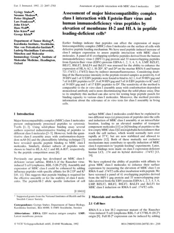
2International Journal of Endocrinology1.61.4209 bp173 bpSTD1.2Fold changeKO PCRBlankWT PCRKO PCRWTWT PCRKO PCRWT PCRKOKO PCRWTWT PCRKO PCRHetWT PCRKO PCRWT PCRWT1.00.80.60.40.2 0H2-Eb1H2-AaH2-Ab1WTMHCIIΔ/Δ(a)(b)Figure 1: MHCII gene genotyping and hepatic expression in wild-type and MHCIIΔ/Δ mice. (a) Two different PCRs were performed for thedetection of wild-type (WT PCR) and knockout (KO PCR: MHCIIΔ/Δ ) fragments; 173 bp for a WT band and 209 bp for a KO band. Thereforeheterozygous (Het) presents both bands. (b) Gene expression of MHC II genes (H2-Eb1, -Aa, -Ab1) in liver samples by real-time RT-PCR. Theexpression was normalized by 𝛽2 microglobulin and compared to the wild-type. Open bars represent wild-type and closed bars MHCIIΔ/Δmice (𝑛 10–14/group). Data are presented as mean SEM. Significantly different from wild-type mice, 𝑃 0.05 (Student t-test).the T lymphocytes hyperactivity and drug-induced liverinjuries has been recently suggested [13]. In addition, severalstudies have demonstrated that the genes in the MHC IIpathway were significantly upregulated in porcine-seruminduced hepatic fibrosis in rats [14, 15]. These facts imply astrong influence of the MHC II pathway on the susceptibilityto develop liver diseases.Nonalcoholic fatty liver disease (NAFLD) is one of themajor liver diseases in industrialized countries. Data aresuggesting a strong increase of NAFLD prevalence in the nextdecades [16, 17]. The disease consists of a diverse spectrumof liver pathologies, starting by hepatic steatosis and thensteatohepatitis, a state of hepatic inflammation [18]. Thenit progresses toward hepatic fibrosis, cirrhosis, and hepaticcarcinoma. Although some molecular pathways that lead tohepatitis in NAFLD can also be activated in the drug-inducedliver injuries, the implication of MHC II pathway has not beenclearly understood in NAFLD model. Given the repeatedreports on the association between alleles on MHC II genesand the susceptibility to a liver inflammation [3, 8–10], thispathway might play an important role in the development ofNAFLD.In the present study, we addressed whether the MHC IIpathway might be required for hepatitis development andfibrosis formation. To this end, we chose to use a mousemodel lacking all conventional genes in MHC II pathway [19].The entire MHC II region (80 kb) was deleted, resulting in theremoval of the genes encoding the MHC II pathway (H2-A𝛽,-A𝛼, -E𝛽, -E𝛽2, and -E𝛼). The mouse genes H2-A𝛽 and -E𝛽have the closest homology to the human HLA-DQB1 gene.These mice are viable and fertile without major anatomicalor physiological abnormalities [19]. We have studied whetherthese mice were protected against a high-fat diet-inducedhepatitis and a chemical-induced hepatic fibrosis.2. Research Design and Method2.1. Animals, Diet, and Chemicals. B6;129S2-𝐻2𝑑𝑙𝐴𝑏1-𝐸𝑎 /J(strain 003584, MHCIIΔ/Δ ) mice in which the 80 kb of MHCII region is deleted were purchased from Jackson Laboratories (Bar Harbor, ME, USA). These mice were bred withC57B6/J mice and the F2 generation was crossbred to obtainMHCIIΔ/Δ and wild-type mice. These mice were housedin ventilated cages at an animal facility of the Universityof Lausanne with 12-hour light and dark cycles and freeaccess to food and water. Five-week-old male mice weresubjected to high-fat diet (D12451, Research diets Inc., NewBrunswick, NJ, USA) for 4 months. The dietary compositionof the high-fat diet was carbohydrates 35%, fat 45%, andprotein 20%. Dietary protein was originated from casein,and fat was mainly from lard. All animal procedures usedin this study were approved by the Swiss cantonal veterinaryservice.Carbon tetrachloride (CCl4 ), chloroform, methanol,EDTA, sirius red, and aprotinin were purchased from SigmaAldrich (Munich, Germany). Kits for measuring plasmaalanine transaminase (ALT), aspartate transaminase (AST),and triglycerides (TG) were purchased from Wako Chemicals(Neuss, Germany). Hematoxylin & eosin was purchased fromMerck (Geneva, Switzerland).
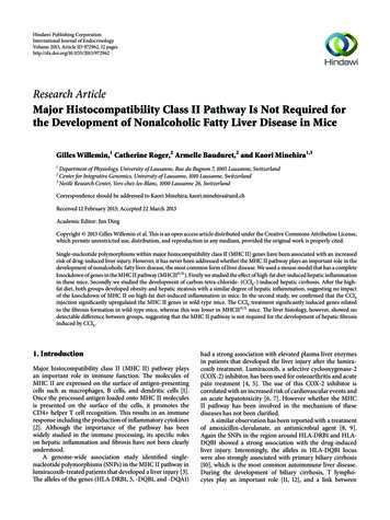
345254020Blood glucose (mM)Body weight (g)International Journal of Endocrinology3530251510520150048Time (weeks)1216030(a)90120(b)3Plasma insulin (𝜇g/L)2.0Liver weight (g)60Time ood glucose (mM)151050MHCIIΔ/ΔWTWTMHCIIΔ/Δ(e)Figure 2: Effect of high-fat diet on metabolic parameters in wild-type and MHCIIΔ/Δ mice. Open circle/bars represent wild-type and closedcircle/bars MHCIIΔ/Δ mice (wild-type: 𝑛 25, MHCIIΔ/Δ : 𝑛 20). Data are presented as mean SEM. (a) Body weight gain during 4-monthdiet. (b) Glucose tolerance test was performed around the 15th week of the intervention. (c) Liver weight at 16th week. (d) Fasting plasmainsulin concentration at 16th week. (e) Fasting blood glucose concentration at 16th week.2.2. High-Fat Diet Experiment. Five-week-old maleMHCIIΔ/Δ mice and wild-type mice were assigned intothe high-fat diet described above. Body weight was measuredevery month. A glucose tolerance test was performed aroundthe 15th week of the intervention. Mice were fasted for 4hours and a solution of 1 g of glucose per kg of mouse wasinjected intraperitoneally. Glycemia was monitored at 0, 15,30, 60, and 120 min using a glucometer (Bayer, Zurich, Switzerland).At the end of the 4-month experiment, mice were fastedfor 4 hours and glycemia was measured by a glucometer.Blood was collected by an intracardiac puncture and placedinto a tube containing EDTA and aprotinin (2 mM and 0.10.2 TIU, respectively) on ice. The plasma was then separated
4International Journal of Endocrinology140240120200160AST (U/L)ALT 1.0Active plasma PAI-1 (𝜇g/L)Liver triglycerides �WTMHCII(c)MHCIIΔ/Δ(d)4.03.5Fold change3.02.52.01.51.00.50IL-6PAI-1F4/80TNF-𝛼 Col-1𝛼1 MMP-14WTMHCIIΔ/Δ(e)Figure 3: Effect of high-fat diet on liver enzymes, lipids, and inflammatory markers in wild-type and MHCIIΔ/Δ mice. Open bars representwild-type and closed bars MHCIIΔ/Δ mice (wild-type: 𝑛 25, MHCIIΔ/Δ : 𝑛 20). Data are presented as mean SEM. (a) Plasma ALT levelat 16th week. (b) Plasma AST level. (c) Liver triglyceride content at 16th week. (d) Fasting active plasma PAI-1 concentration at 16th week. (e)Gene expression related to inflammation and fibrogenesis in the liver at 16th week.by centrifugation and stored at 80 C until analysis. Liver andepididymal adipose tissues were harvested and weighed. Theorgans were flash-frozen into liquid nitrogen and stored at 80 C until analysis.2.3. CCl4 Treatment. To study the development of hepaticfibrosis, 8-week-old wild-type and MHCIIΔ/Δ male mice weretreated by 1.25 𝜇L/g body weight CCl4 (25% in sunflower oil)twice a week during 4 weeks. Another set of mice was treatedby sunflower oil as control vehicle at the same time. Twentyhours following the last treatment, mice were fasted for 4hours and anesthetized for a cardiac puncture. After sacrifice,blood and liver were kept for further analysis. Fresh liverpieces were put into 4% paraformaldehyde solution during
International Journal of Endocrinology5MHCIIΔ/ΔOil red OWTORORH&E(a)F4/80(b)(c)Figure 4: Effect of high-fat diet on liver histology in wild-type and MHCIIΔ/Δ mice. (a) Oil red O staining (red color represents the lipidaccumulation). (b) Hematoxylin and eosin (H&E) staining. (c) F4/80 immunohistochemistry (macrophages were stained in brown: arrows).4 hours. After several washings with PBS, liver samples weredehydrated and parafinated for histological analysis.2.4. Histology. Parafinated liver samples were sliced (4 𝜇m)and stained according to a standard technique with hematoxylin and eosin (H&E) using routine methods. To studythe fibrosis formation, 0.1% sirius red was used to staincollagen I, III, and bile pigment using a protocol describedelsewhere [20]. For the histological samples, photos weretaken with an AXIO Imager M1 with fluo-Axiocam MRm andcolor Axiocam MRc cameras (Carl Zeiss AG, Oberkochen,Germany). Images were treated by Axiovision release 4.8.2.We then quantified the fibrotic area by assessing the ratio ofthe red-stained area (fibrosis) to the total area, the vascularluminal area being subtracted if present, using Photoshop(Adobe Systems, Mountain View, USA) in five images persection of the sample (10 x magnification).Immunohistochemistry by glial fibrillary acidic protein(GFAP) stain was performed by the subsequent incubations of the slices with rabbit anti-GFAP primary antibody(Dako, Glostrup, Denmark) diluted in normal goat serumfor overnight at 4 C, then with goat anti-rabbit coupled withAlexa Fluor 568 (Life Technologies, Carlsbad, USA) for 30minutes in the dark at room temperature. Immunostainedsections were then counterstained with 4 ,6-diamidino-2phenylindole (DAPI), embedded with mowiol and analyzedusing the Zeiss microscope.2.5. Plasma Parameters and Liver Lipids Analysis. Totalplasma ALT and AST were measured by the Roche/Hitachi
6International Journal of Endocrinology10Blood glucose (mM)Liver weight (g)1.51.00.50aab8bc6420WT-oilMHCIIΔ/Δ -oilWT-CCl 4 MHCIIΔ/Δ -CCl �MHCII-oilWT-CCl 4 MHCIIΔ/Δ -CCl 4cbcAST (U/L)ALT (U/L)MHCIIΔ/Δ -oil(b)40000cWT-CCl 4MHCIIΔ/Δ400020000-CCl 4(c)aWT-oilabMHCIIΔ/Δ -oil WT-CCl 4 MHCIIΔ/Δ -CCl 4(d)Figure 5: Effect of 4-week CCl4 treatment on liver and blood glucose in wild-type and MHCIIΔ/Δ mice. Open bars represent oil-treatedwild-type (WT-oil), closed bars CCl4 -treated wild-type (WT-CCl4 ), gray bars oil-treated MHCIIΔ/Δ mice, and semiclosed bars CCl4 -treatedMHCIIΔ/Δ mice (wild-type oil: 𝑛 7, MHCIIΔ/Δ oil: 𝑛 5, wild-type CCl4 : 𝑛 11, MHCIIΔ/Δ CCl4 : 𝑛 9). Data are presented as mean SEM. (a) Liver weight. (b) Fasting blood glucose decreased after the CCl4 treatment in both groups. (c), (d) Strong increase in fastingplasma ALT and AST was observed after the CCl4 treatment. a,b,c 𝑃 0.05, ANOVA and Tukey Kramer HSD test. Different letters indicatethe significant difference between groups.912 instrument with the commercial kits mentioned earlier. Insulin was measured by a Mouse Insulin ELISA kit(Mercodia, Uppsala, Sweden). Plasma active Plasminogenactivator inhibitor-1 (PAI-1) was also measured by an ELISAkit (Molecular Innovations Inc., Novi).From the harvested liver samples, total lipids wereextracted using a modified Folch method [21, 22]. For themeasurement of hepatic TG content, total lipid extract wassubjected to SPE columns (Interchim, Montluçon, France) toseparate TG [23, 24]. TG were then mixed with a chloroformtriton X (1%) solution and dried under N2 gas. TG were thusdissolved into water and the content of TG was measured bythe use of the Wako Chemical kit.2.6. Genotyping and Real-Time Polymerase Chain Reaction(PCR). Ear DNA was extracted by the hotSHOT protocol[25]. Genotyping was performed according to Jackson laboratory’s protocol y, 2 sets of primers were used for the mutatedgene (oIMR1020: 5 -Cgg AAg TgC TTg ACA TTg g-3 ,oIMR1021:5 -gTA TTg ACC gAT TCC TTg Cg-3 ) and wildtype gene (oIMR1273; 5 -AAC CTT CAg gAT CTg TgA TCC3 , oIMR1274; 5 -gTg gCT gTT gCC TTA AgA CC-3 ). AfterPCR cycles, samples were loaded onto 2% agarose gels and themutated band (209 bp) as well as the wild-type band (178 bp)was monitored.Total RNA was extracted from tissues according tothe phenol-chloroform extraction protocol in Tri Reagent(Molecular Research Center, Inc., Cincinnati, USA) [26].cDNA was created by a reverse transcription of 2 𝜇g oftotal RNA using Superscript II Transcriptase from Invitrogen(Life Technologies, Carlsbad, USA) according to the manufacturer’s protocol. Real-time RT-PCR was performed onApplied Biosystems’ 7000 Sequence Detection System (LifeTechnologies, Carlsbad). Twenty-time diluted cDNA samplesand 0.3 𝜇M forward and reverse primers (Microsynth AG,Balgach, Switzerland) in a final 10 𝜇L volume were reactedin the following PCR cycle conditions: 10 minutes at 95 Cfollowed by 40 cycles of 15 seconds at 95 C and 1 minuteat 60 C. Each sample was analyzed in duplicate using theDeltaDeltaCt method [27]. 𝛽2 microglobulin (𝛽2M) was usedas a housekeeping gene to normalize the expression of eachgene.2.7. Statistics. All data are shown in mean standard errorof the mean (SEM). Two groups were compared by Studentt-test. Four groups were compared by one-way analysis ofvariance (ANOVA) and once it reached the significance(𝐹 0.05), a post hoc test (Tukey Kramer HSD test) wasperformed. The statistical analyses were performed by use ofthe JMP software (SAS Institute Inc., Cary).
International Journal of e expressionRelative expression462bb40H2-Ab1ba aa aCol-1𝛼12.52.0Fibrosis (%)Relative expressionMMP-14b ba aTGF-𝛽a ababTIMP-2(b)40302010ab a bc cIL-6aba aCol-3𝛼1(a)500bbPAI-1WT-oilMHCIIΔ/Δ -oilF4/80TNF-𝛼WT-CCl 4MHCIIΔ/Δ -CCl 4(c)1.51.00.50MHCIIΔ/Δ -CCl 4WT-CCl 4WT-oilMHCIIΔ/Δ -oilWT-CCl 4MHCIIΔ/Δ -CCl 4(d)Figure 6: Effect of 4-week CCl4 treatment on hepatic gene expression and fibrosis in wild-type and MHCIIΔ/Δ mice. Open bars represent oiltreated wild-type (WT-oil, 𝑛 7), closed bars CCl4 -treated wild-type (WT-CCl4 , 𝑛 11), gray bars oil-treated MHCIIΔ/Δ mice (MHCIIΔ/Δ oil, 𝑛 5), semiclosed bars CCl4 -treated MHCIIΔ/Δ mice (MHCIIΔ/Δ -CCl4 , 𝑛 9). Data are presented as mean SEM. (a) MHC II geneexpression. (b) Gene expression related to fibrosis formation. (c) Gene expression related to inflammation. (d) No difference in fibrosis(%) between groups treated by CCl4 . The percentage was determined based on the sirius red staining (5 pictures/mice, WT-CCl4 : 𝑛 5,MHCIIΔ/Δ -CCl4 : 𝑛 8). a,b,c 𝑃 0.05, ANOVA and Tukey Kramer HSD test. Different letters indicate the significant difference betweengroups.3. Results3.1. Validation of the Mouse Model. The MHCIIΔ/Δ micewere originally created by the laboratory of Dr. ChristopheBenoist (Institut de Génétique et de Biologie Moléculaireet Cellulaire, Illkirch, France). These mice lack the majorgenes of MHC II pathway and present very low countsof CD4 T lymphocytes in the thymus and spleen [19].The detailed genetic modification and their phenotype havebeen published [19]. These mice have been backcrossed withC57B6/J strain at Jackson laboratory. After purchasing themice from Jackson laboratory, we crossed them with C57B6/Jmice to obtain control wild-type animals. In this model, thePCR for detecting the knockout allele showed a clear band at209 bp. The quantitative PCR analysis showed no or very lowexpression of the genes of the MHC II pathway (Figure 1).groups (Figure 3), suggesting similar liver functions after thehigh-fat diet.To study the inflammatory status of the liver after thelong-term high-fat diet feeding, we have performed H&Estaining and F4/80 for macrophages staining. We observedpositive markers of F4/80 and similar lipid droplet morphology in both groups (Figure 4). The expression of genes relatedto inflammation such as IL-6, TNF-𝛼, PAI-1, and F4/80 wasnot different between groups (Figure 3). The mRNA levelsof fibrosis markers such as collagen type 1𝛼1 (Col-1𝛼1) andmatrix metalloproteinase-14 (MMP-14) were also comparable. We also measured active PAI-1 concentration in theplasma (Figure 3). Again no difference was observed betweenwild-type and MHCIIΔ/Δ mice. All together, blocking theMHC II pathway did not affect the hepatic inflammationinduced by the high-fat diet in mice.3.2. Effect of High-Fat Diet on Inflammatory Status inMHCIIΔ/Δ Mice. Both wild-type and MHCIIΔ/Δ mice similarly gained body weight after 4-month high-fat diet(Figure 2). Liver weight was also similar in both groups.Plasma glucose and insulin concentrations were comparablebetween groups. As expected, both groups had similar glucose tolerance after the long-term high-fat diet. The liver TGcontent was also comparable between groups (Figure 3). Oilred O staining confirmed this result (Figure 4). Plasma ALTand AST levels were equally high after the high-fat diet in two3.3. Effect of CCl4 Injection on the Formation of HepaticFibrosis in MHCIIΔ/Δ Mice. We next studied the developmentof hepatic fibrosis. To this end, we treated wild-type andMHCIIΔ/Δ mice with CCl4 during 4 weeks. The treatmentresulted in a significant decrease of glycemia and a significantincrease of ALT and AST in both groups (Figure 5). Nodifference in liver weight was observed. The CCl4 injectionsalso highly induced the expression of MHC II genes inwild-type mice, while MHCIIΔ/Δ mice had no induction
8International Journal of EndocrinologyMHCIIΔ/ΔCCl4Sirius redOilWTCCl4H&EOil(a)(b)Figure 7: Effect of 4-week CCl4 treatment on liver histology in wild-type and MHCIIΔ/Δ mice. (a) Sirius red staining for collagen. The redstaining represents fibrotic area. (b) Hematoxylin and eosin (H&E) staining.of these genes, as expected (Figure 6). Genes related to thefibrosis formation such as Col-1𝛼1, Col-3𝛼1, and transforminggrowth factor-𝛽 (TGF-𝛽) significantly increased in bothgroups after the CCl4 injections compared to the oil-injectedgroups. The genes such as MMP-14 and tissue inhibitor ofmetalloproteinase-2 (TIMP-2) were also highly induced afterCCL4 treatment in wild-type mice: however, the inductionwas somewhat blunted in MHCIIΔ/Δ mice.The liver histology was analyzed using sirius red stainingfor fibrosis formation. No positive staining was detected inoil-treated groups. The level of positive staining in CCl4 treated livers was quantified using Photoshop. We detecteda comparable level of fibrosis between groups treated by CCl4(Figures 6 and 7). H&E staining also showed inflammatorysigns in the liver treated by CCl4 (Figure 7). Again, noremarkable difference was observed between wild-type andMHCIIΔ/Δ mice. The staining by F4/80 showed a remarkableinfiltration of macrophages in the livers of mice treated byCCl4 , but, again, no difference was observed between wildtype and MHCIIΔ/Δ mice (Figure 8). We further stained stellate cells by GFAP. Interestingly we detected only one positivestaining out of 13 in wild-type mice while we found 7 positivestainings out of 9 samples in MHCIIΔ/Δ mice (Figure 9).4. DiscussionThe present study demonstrated that the MHC II pathway isnot implicated in the development of hepatitis when induced
International Journal of Endocrinology9MHCIIΔ/ΔCCl4CCl4OilWTFigure 8: Effect of 4-week CCl4 treatment on macrophage staining in wild-type and MHCIIΔ/Δ mice. Macrophage staining (F4/80) showeda strong inflammation in both groups treated by CCl4 .by a long-term high-fat diet. We had taken the approach of thehigh-fat diet to mimic our obesogenic lifestyle that is knownto contribute to the nonalcoholic steatohepatitis (NASH). It isalso strongly supported that feeding laboratory animals withhigh-caloric diets such as a high-fat diet can induce NASH[28–30]. As proposed in the pathophysiology of NASH, the“two-hit theory” could also explain the progression of hepaticsteatosis to NASH in our experimental model. The first hit isan infiltration of fat into the hepatocytes (hepatic steatosis)and the second hit is characterized by an infiltration ofimmune cells such as monocytes and lymphocytes becauseof abnormal oxidative stress [31, 32]. Our mice havingundergone a 4-month high-fat diet presented a high amountof fat content in the liver in both groups (first hit). Extensivehigh-fat diet feeding then resulted in a comparable increaseof hepatic inflammation/damage in two groups, judged bymacrophage infiltration (second hit). These data suggest thatthe long-term high-fat diet induced both hepatic steatosisand NASH; however, blocking the MHC II pathway had noinfluence on the development of hepatic steatosis and NASHin these mice.Our long-term high-fat diet also increased the level ofALT and AST in both groups compared to the normalrange of ALT and AST generally observed in chow-fedmice (15–80 U/L and 40–120 U/L, respectively, in our laboratory measurements). This result strongly suggests that themodification in MHC II gene expression affected neither ALTnor AST levels, consistent to their comparable liver histologybetween groups. It has been, however, suggested that somealleles in MHC II pathway were associated with a severity ofNAFLD and ALT, but not AST levels, in a Turkish population[33]. In this study, the investigators could not assess apresence of NASH in the population. To our knowledge, thisstudy is the only report suggesting an association between theHLA allele and NAFLD in general population. Many otherstudies have shown a connection between the HLA allele andplasma ALT or NASH severity in hepatitis C patients or inpatients with a drug-induced liver injury [3, 6, 8, 9, 34, 35].Therefore, MHC II pathway might have a bigger importanceto the development of NASH caused by viral infections ordrugs than by diet-induced NAFLD.Secondly, we focused on the chemically induced fibrosisin mice lacking all conventional MHC II genes. As mentionedbefore, some HLA alleles are associated with an increasedsusceptibility to develop a drug-induced liver injury. TheCCl4 treatment strikingly increased the plasma level of ALT,AST, and the degree of hepatic fibrosis in both genotypes. Thegenes of MHC II pathway were also highly induced by CCl4treatment in wild-type mice, indicating the upregulation ofthe pathway upon the treatment. Despite the lack of theupregulation of the pathway, the MHCIIΔ/Δ mice equallydeveloped a hepatic fibrosis. These data strongly indicate
10International Journal of EndocrinologyCCl 4OilCCl 4MHCIIΔ/ΔWTOilCCl 4MHCIIΔ/ΔCCl 4Figure 9: Effect of 4-week CCl4 treatment on GFAP staining in wild-type and MHCIIΔ/Δ mice. Glial fibrillary acidic protein (GFAP; whitearrows) was stained in red. Increased positive staining was observed in CCl4 -treated MHCIIΔ/Δ mice. Blue: DAPI staining.that the MHC II pathway is not required for the developmentof hepatic fibrosis, at least in the mouse model of CCl4 induced hepatic fibrosis.Although the major genes implicated in the formation offibrosis, namely, Col-1𝛼1, Col-3𝛼1, and TGF-𝛽 were similarlyupregulated in the groups of mice treated by CCl4 , theinduction of some genes such as TIMP-2 and MMP-14 tendedto be lower in the MHCIIΔ/Δ mice compared to the wild-typemice. TIMP-2 expression has been observed during the earlystages of fibrogenesis induced by a porcine serum [36]. Wethought that the deletion of MHC II genes might have affectedthe progression of fibrosis at an earlier time point. In ourstudy, we had tested different periods of injection of CCl4 (2-,4-, 6-, and 8-week treatment) in these mice. In any conditiontested, we did not observe a significant difference in the geneexpression related to the fibrosis formation (data not shown).We also confirmed these results by liver histology. This againsupports the idea that the MHC II pathway is not interferingwith the development of hepatic fibrosis induced by CCl4 .Although histological data suggested that there was nodifference in the severity of the fibrosis between groupstreated by CCl4 , we found a profound increase in the hepaticstellate cells (HSC) staining only in the group of MHCIIΔ/Δmice treated by CCl4 . HSC are nonparenchymal cells inthe liver and are known to play an important role infibrosis and tissue repairing [37]. Upon the quiescent HSCactivation, they are converted into myofibroblasts, which areresponsible for the production of extracellular matrices [38].The importance of the HSC activation in fibrogenesis wasrecently reported by Puche et al. [39]. They have elegantlycreated a transgenic mouse model whose proliferating HSCwere selectively killed. By use of the model, Puche et al.demonstrated that these mice had reduced fibrotic area uponCCl4 treatment compared to their genetically controlledcounterparts. This signifies the important role of HSC in thedevelopment of hepatic fibrosis.In our study, despite the hyperactivation of HSC inMHCIIΔ/Δ mice, the fibrosis formation was strictly comparable between groups. We do not know the exact cause andeffect of this HSC induction in MHCIIΔ/Δ mice treated byCCl4 . We have observed that the expression of the genes ofthe MHC II pathway was very high in HSC fraction whenwe separated different cellular types in the liver (hepatocytes, HSC, Kupffer cells, and endothelial cells) (unpublisheddata). This suggests that the HSC could play an importantrole for the MHC II reaction. We do not know, however,whether the absence of the pathway in MHCIIΔ/Δ mice mighthave affected the HSC proliferation upon CCl4 stimulation.
International Journal of EndocrinologyOn the other hand, the activation of HSC during the development of fibrosis is suggested to be transient [40]. We did notidentify when HSC started to be activated in MHCIIΔ/Δ miceduring the CCl4 treatment. Whether this hyperactivation ofHSC has compensated the lack of the MHC II pathway andcontributed to the fibrosis formation needs to be furtheraddressed.The present study clearly demonstrated that the lack ofMHC II pathway affected neither NASH induced by highfat diet nor fibrosis induced by CCl4 in mice. Despite a largenumber of publications implying the association between thealleles in the MHC II pathway and drug-induced liver injuryand hepatitis C in humans, this study clearly indicated thatthe MHC II pathway is not required in the development ofNASH and fibrosis at least in mice.Conflict of InterestsG. Willemin, C. Roger, A. Bauduret, and K. Minehira have noconflict of interests.Authors’ ContributionK. Minehira contributed to the conception and design of theexperiments. All in vivo experiments and tissue samplingwere carried out in the animal facility of the Bugnon 7/9,University of Lausanne, by G. Willemin and K. Minehira. Histological analysis was carried out by C. R
InternationalJournalofEndocrinology 15 20 25 30 35 40 45 0 4 8 12 16 Body weight (g) Time (weeks) (a) 0 5 10 15 20 25 0 30 60 90 120 Blood glucose (mM) Time (min)