Transcription
(2021) 19:322Ito et al. World J Surg Onchttps://doi.org/10.1186/s12957-021-02434-9Open AccessCASE REPORTEndobronchial brachytherapy as definitivetreatment for endobronchial metastasisafter surgery of non-small cell lung cancerAtsushi Ito1* , Daisuke Yamaguchi1, Shinji Kaneda1, Koji Kawaguchi1, Akira Shimamoto1, Makiko Kubooka2,Yoshihito Nomoto3 and Motoshi Takao1AbstractBackground: Endobronchial metastasis is a very rare type of recurrence after lung cancer surgery. Surgical intervention may be difficult to perform due to the postoperative reduction in the activities of daily living (ADL) and theinvasiveness associated with redo surgery. In such cases, endobronchial brachytherapy (EBBT) plays an important rolenot only as a palliative treatment, but also as a definitive treatment with curative intent.Case presentation: Three men (64, 69, and 74 years old) underwent combination therapy of external beam radiation therapy (EBRT) and EBBT for endobronchial metastasis after lobectomy of stage I–II non-small cell lung cancer(NSCLC): 2 cases of squamous cell carcinoma and 1 of adenocarcinoma. We used a special source-centralizing applicator for EBBT to avoid eccentric distribution of the radiation dose. Follow-up was considered to start from the end ofbrachytherapy. None of our patients experienced severe adverse events, and none needed extensive outpatient treatment. Local control was achieved in all cases by a bronchoscopic evaluation. All patients were alive after 31, 38, and92 months of follow-up, respectively. In the adenocarcinoma patient, two metastases to the lung were discovered 3years after EBBT, and the patient underwent partial wedge resection.Conclusions: EBBT may be a promising treatment with curative intent for endobronchial metastasis after surgery ofNSCLC.Keywords: Endobronchial metastasis, Endobronchial brachytherapy, Definitive treatmentBackgroundDespite the advances in radiation therapy, chemotherapy, and newly developed molecular-targeted therapies,lung cancer remains a major health problem and a leading cause of cancer mortality worldwide. The majority of mortalities in the case of postoperative non-smallcell lung cancer (NSCLC) are due to the developmentof recurrence [1]. Endobronchial metastasis is a veryrare type of recurrence defined in detail by Schoenbaum*Correspondence: i-atsushi@clin.medic.mie-u.ac.jp1Department of Thoracic and Cardiovascular Surgery, Graduate Schoolof Medicine, Mie University, 2‑174 Edobashi, Tsu, Mie 514‑8507, JapanFull list of author information is available at the end of the articleand Viamonte [2]. Patients with endobronchial metastasis often suffer from symptoms of airway obstruction,including coughing, hemoptysis, dyspnea, and wheezing.Therefore, removal of the bronchial obstruction can helpto improve the patient’s clinical status. However, surgical intervention may be difficult due to the postoperative reduction in activities of daily living (ADL) and theinvasiveness associated with redo surgery. In such cases,endobronchial brachytherapy (EBBT) plays an importantrole not only as a palliative treatment, but also as a definitive treatment with curative intent.EBBT is the radiation therapy in which the radioactive material is targeted at the bronchial lumen carrying the target lesion. Historically, EBBT has often been The Author(s) 2021. Open Access This article is licensed under a Creative Commons Attribution 4.0 International License, whichpermits use, sharing, adaptation, distribution and reproduction in any medium or format, as long as you give appropriate credit to theoriginal author(s) and the source, provide a link to the Creative Commons licence, and indicate if changes were made. The images orother third party material in this article are included in the article’s Creative Commons licence, unless indicated otherwise in a credit lineto the material. If material is not included in the article’s Creative Commons licence and your intended use is not permitted by statutoryregulation or exceeds the permitted use, you will need to obtain permission directly from the copyright holder. To view a copy of thislicence, visit http://creativecommons.org/licenses/by/4.0/. The Creative Commons Public Domain Dedication waiver ) applies to the data made available in this article, unless otherwise stated in a credit line to the data.
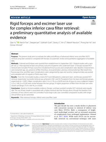
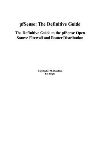
Ito et al. World J Surg Onc(2021) 19:322used for palliative treatment and has been shown to beeffective in improving bronchial stenosis and obstructioncaused by tumors [3]. Recently, several studies have demonstrated that EBBT plays a limited but specific role indefinitive treatment with curative intent in select cases ofearly endobronchial disease, in select advanced inoperable tumors combined with external beam radiotherapy(EBRT), or in the postoperative treatment of small, residual peribronchial disease [3–6].We herein report our experience with EBBT as a definitive treatment for endobronchial metastasis after surgeryof NSCLC to assess the feasibility and efficacy of thisapproach.Case presentationBetween 2013 and 2018, 3 men (64, 69, and 74 years old)underwent a combination therapy of EBRT and EBBTfor endobronchial metastasis after lobectomy of stage I–II NSCLC: 2 cases of squamous cell carcinoma and 1 ofadenocarcinoma. At the timing of the initial surgery, R0resection was achieved in all cases. The diagnosis of cancer recurrence was made by a bronchoscopic biopsy. Inall patients, surgical intervention was considered difficultdue to postoperative reductions in the ADL and the invasiveness associated with redo surgery.The radiation doses of EBRT and EBBT were 40 Gyin 20 fractions and 18 Gy in 3 fractions. BrachytherapyPage 2 of 5was performed once per week using a high dose rateIr-192 after-loading machine (microSelectron , Nucletron, Elekta Co., Sweden). The reference dose pointswere 5–7 mm from the source axis, depending onthe bronchial diameter. Chemotherapy was not performed, and EBRT was performed prior to EBBT. Theaims of EBRT prior to EBBT are to reduce the tumorsize and to obtain a uniform dose distribution to thebronchial wall. In order to keep the interval betweenradiation therapies as short as possible, EBBT wasperformed from the day after EBRT was completed.We used a special source-centralizing applicator (Create Medic Co. Ltd., Yokohama, Japan) developed byNomoto et al. for EBBT to avoid eccentric distributionof the radiation dose (Fig. 1a) [7]. This applicator canhold the source-delivering catheter because the twoself-expandable wings open according to the bronchialdiameter [5]. The applicator is adjusted to the optimalposition such that the tumor is located between the twowings (Fig. 1b) [5]. The distance from the source axis tothe bronchial wall was measured on the planning computed tomography (CT) image [5].Follow-up was considered to start from the end ofEBBT. The local response was evaluated by bronchoscopyperformed 1 to 2 months after the end of EBBT. In addition, CT was performed every 6 months for the first 2years and then yearly for at least 5 years of follow-up.Fig. 1 a A special source-centralizing applicator developed by Nomoto et al. Two self-expandable wings (arrow). b Schema of endobronchialbrachytherapy using the special source-centralizing applicator. The applicator is adjusted to the optimal position such that the tumor is locatedbetween the two wings
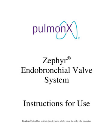
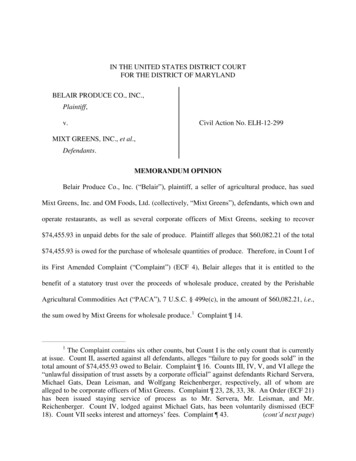
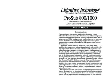
Ito et al. World J Surg Onc(2021) 19:322Case 1A 64-year-old man underwent the right upper lobectomyfor lung cancer of adenocarcinoma (2.0 cm, pT1bN0M0,pStage IA2). The patient had no postoperative complications and was discharged on postoperative day 9. Nineyears after the initial surgery, postoperative follow-up CTshowed a left endobronchial nodule near the peripheraltracheal bifurcation (Fig. 2a). Bronchoscopy confirmeda protruding lesion emanating from the posterior wallPage 3 of 5of the left main bronchus (Fig. 2b). After a diagnosis ofadenocarcinoma by tumor resection using a bronchoscopic snare was made, we performed a combinationof EBRT and EBBT for the small residual disease as adefinitive treatment. The patient had achieved a complete local response without adverse events at the timeof the bronchoscopic evaluation (Fig. 2c). Three yearsafter irradiation, CT revealed a new partly solid noduleat the left lower lobe. The patient then underwent partialwedge resection and is still alive at 90 months since theirradiation.Case 2Fig. 2 a CT findings showed the endobronchial metastasis at the leftmain bronchus (arrow). Bronchoscopic image of b before irradiationand c after irradiationA 69-year-old man underwent the right lower lobectomyfor lung cancer of squamous cell carcinoma (4.0 cm,pT2aN0M0, pStage IB). Two weeks after the surgery, weperformed a right-sided open-window thoracostomy dueto a postoperative bronchopleural fistula. Three monthsafter open-window thoracostomy, he was dischargedafter undergoing thoracoplasty with omental plombage.One year and 6 months after discharge, postoperativefollow-up CT showed a right endobronchial nodule atthe truncus intermedius (Fig. 3a). Fluorodeoxyglucosepositron emission tomography (FDG-PET) revealed anuptake at the lesion (maximum standardized uptake value [SUVmax] 3.8) (Fig. 3b). After a diagnosis of squamous cellcarcinoma was made by a bronchoscopic biopsy (Fig. 3c),we performed a combination of EBRT and EBBT for theendobronchial metastasis as a definitive treatment. Thepatient had achieved a complete local response without adverse events at the time of the bronchoscopicFig. 3 a CT findings showed the endobronchial metastasis at the truncus intermedius (arrow). b PET-CT revealed an uptake in the lesion [ SUVmax3.8]. Bronchoscopic image of c before irradiation and d after irradiation
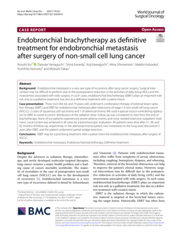
Ito et al. World J Surg Onc(2021) 19:322evaluation (Fig. 3d). The patient has remained diseasefree for 36 months since the irradiation.Case 3A 74-year-old man underwent the left lower lobectomyfor lung cancer of squamous cell carcinoma (5.7 cm,pT2bN0M0, pStage IIA). The patient had no postoperative complications and was discharged at postoperativeday 6. Two years after discharge, postoperative follow-upCT showed a left endobronchial nodule near the peripheral tracheal bifurcation (Fig. 4a). FDG-PET revealed anuptake at the lesion ( SUVmax 7.4) (Fig. 4b). After a diagnosis of squamous cell carcinoma was made by a bronchoscopic biopsy (Fig. 4c), we performed a combinationof EBRT and EBBT for the endobronchial metastasis asa definitive treatment. The patient had achieved a complete local response without adverse events at the timeof the bronchoscopic evaluation (Fig. 4d). The patienthas remained disease-free for 29 months since theirradiation.DiscussionThe frequency of endobronchial metastasis from nonpulmonary malignancies is known to range from about2 to 50%, varying by the definition of metastasis usedand the types of cancer included [8–10]. The overallincidence of tracheal metastasis is 0.44% in surgicallyresected NSCLC, and the incidence of tracheal metastasis is lower in primary lung cancers than in non-pulmonary malignancies [10]. Hemoptysis with coughing is themost common symptom of endobronchial metastasis,Page 4 of 5with an incidence of 41–62% reported [10]. In our cases,all patients with endobronchial metastasis were freefrom symptoms despite the endobronchial nodule shownon postoperative follow-up CT. To determine whetheror not these lesions are sputum, it is useful to examinethe lesion uptake of FDG by PET-CT. In our cases, theuptake of FDG was revealed in endobronchial lesions,but there were no metastatic lesions noted outside thethorax, including in the brain.Schoenbaum and Viamonte report that metastaticspread of malignant tumors to bronchi occurs by directextension from mediastinal lesion, invasion of bronchusby parenchymal mass, or direct metastasis to the wall ofthe bronchi [2]. In our cases, endobronchial metastasisoccurred despite the absence of lymph node metastasis,and the metastatic pathway was suspected of being thebronchial veins in the bronchial wall.Surgery was not indicated in the present cases becauseextensive adhesions were expected on the postoperativeside, and recurrence near the tracheal bifurcation mightrequire extremely invasive and difficult surgery, such ascarinal reconstruction or pneumonectomy. Therefore,we performed a combination of EBRT and EBBT as adefinitive treatment, and none of our patients experienced local recurrence. The most serious adverse eventsof EBBT are fatal hemoptysis and severe bronchitis dueto over-irradiation. These adverse events are caused bythe radioactive source being placed at eccentric locationsin the bronchial lumen, thereby leading to localized hotspots on the bronchial mucous membrane [3]. We therefore used a special source-centralizing applicator to avoidFig. 4 a CT findings showed the endobronchial metastasis at the left main bronchus (arrow). b PET-CT revealed an uptake in the lesion [ SUVmax7.4]. Bronchoscopic image of c before irradiation and d after irradiation
Ito et al. World J Surg Onc(2021) 19:322eccentric distribution of the radiation dose. One monthafter the end of EBBT, bronchoscopy revealed inflammation of the bronchial mucosa; however, none of ourpatients experienced such severe adverse events, andnone needed extensive outpatient treatment. This applicator was also used to protect the bronchial mucosa fromhigh-dose irradiation in the reports of Kawamura et al.[11], Hosni et al. [6], and Nomoto et al. [5].There have been several reports regarding the combination of EBRT and EBBT therapy in either a curative or palliative setting. Nomoto et al. reported that the3-year overall and progression-free survival rates were79% and 77%, respectively, in 15 patients [5]. In addition, Hosni et al. reported that the 2-year overall survivaland local control rates were 67% and 89%, respectively,in 23 patients [6]. While these reports describe relativelyfew patients, these outcomes suggest that EBBT may beexpected to achieve excellent tumor control.ConclusionsEBBT is possibly a promising treatment with curative intent for endobronchial metastasis after surgery ofNSCLC. In particular, the use of the special source-centralizing applicator can prevent over-irradiation of thebronchial mucosa and lead to safe EBBT without severeadverse events.AbbreviationsNSCLC: Non-small cell lung cancer; ADL: Activities of daily living; EBBT: Endobronchial brachytherapy; EBRT: External beam radiotherapy; CT: Computedtomography; FDG-PET: Fluorodeoxyglucose-positron emission tomography;SUVmax: Maximum standardized uptake value.AcknowledgementsNot applicable.Authors’ contributionsAI and MT designed the report, analyzed the data, and wrote the manuscript.The other authors contributed to the acquisition and interpretation of thedata for the work and revised the manuscript. The authors read and approvedthe final manuscript.FundingNoneAvailability of data and materialsNot applicable.DeclarationsEthics approval and consent to participateThis study was approved by the Clinical Research Ethics Review Committee ofMie University Hospital (ethics approval number: H2020-032).Page 5 of 5Consent for publicationWritten, informed consents were obtained from the patients for the publication of this manuscript and any accompanying images.Competing interestsThe authors declare that they have no competing interests.Author detailsDepartment of Thoracic and Cardiovascular Surgery, Graduate Schoolof Medicine, Mie University, 2‑174 Edobashi, Tsu, Mie 514‑8507, Japan.2Department of Radiology, Graduate School of Medicine, Mie University, Tsu,Japan. 3 Department of Radiation Oncology, Graduate School of Medicine, MieUniversity, Tsu, Japan.1Received: 28 May 2021 Accepted: 25 October 2021References1. Taylor MD, Nagji AS, Bhamidipati CM, Theodosakis N, Kozower BD, Lau CL,et al. Tumor recurrence after complete resection for non-small cell lungcancer. Ann Thorac Surg. 2012;93:1813–20 discussion 1820-1811.2. Schoenbaum S, Viamonte M. Subepithelial endobronchial metastases.Radiology. 1971;101:63–9.3. Omori K, Nomoto Y, Kawamura T, Kubooka M, Kawaguchi K, Ii N, et al.Endobronchial brachytherapy combined with surgical procedure forsynchronous multiple primary lung cancer: a case report. Thorac Cancer.2021;12:1252–5.4. Skowronek J. Brachytherapy in the treatment of lung cancer - a valuablesolution. J Contemp Brachyther. 2015;7:297–311.5. Nomoto Y, Ii N, Murashima S, Yamashita Y, Ochiai S, Takada A, et al.Endobronchial brachytherapy with curative intent: the impact of reference points setting according to the bronchial diameter. J Radiat Res.2017;58:849–53.6. Hosni A, Bezjak A, Rink A, Czarnecka K, McPartlin A, Patterson S, et al. Highdose rate brachytherapy as a treatment option in endobronchial tumors.Lung Cancer Int. 2016;2016:3086148.7. Nomoto Y, Shouji K, Toyota S, Sasaoka M, Murashima S, Ooi M, et al. Highdose rate endobronchial brachytherapy using a new applicator. Radiother Oncol. 1997;45:33–7.8. Kiryu T, Hoshi H, Matsui E, Iwata H, Kokubo M, Shimokawa K, et al.Endotracheal/endobronchial metastases: clinicopathologic study withspecial reference to developmental modes. Chest. 2001;119:768–75.9. Youn HC, Kim YH, Lee YK, Kim GY. Multiple endotracheal and endobronchial metastases after pneumonectomy for a primary lung cancer: a casereport. Thorac Cancer. 2013;4:453–6.10. Chong S, Kim TS, Han J. Tracheal metastasis of lung cancer: CT findings insix patients. AJR Am J Roentgenol. 2006;186:220–4.11. Kawamura H, Ebara T, Katoh H, Tamaki T, Ishikawa H, Sakurai H, et al. Longterm results of curative intraluminal high dose rate brachytherapy forendobronchial carcinoma. Radiat Oncol. 2012;7:112.Publisher’s NoteSpringer Nature remains neutral with regard to jurisdictional claims in published maps and institutional affiliations.
hold the source-delivering catheter because the two self-expandable wings open according to the bronchial diameter []. e applicator is adjusted to the optimal 5 position such that the tumor is located between the two wings (Fig. 1b) [5]. e distance from the source axis to the bronchial wall was measured on the planning com-