Transcription
-of-experimental-neurologyJournal of Experimental NeurologyReview ArticleAlzheimer’s Disease: A Brief ReviewSamantha McGirr#, Courtney Venegas#, Arun Swaminathan*University of Nebraska Medical Center, Omaha, NE, USA#Both authors contributed equally to this review*Correspondence should be addressed to Arun Swaminathan; arun.swaminathan@unmc.eduReceived date: June 07, 2020, Accepted date: July 10, 2020Copyright: 2020 McGirr S, et al. This is an open-access article distributed under the terms of the Creative CommonsAttribution License, which permits unrestricted use, distribution, and reproduction in any medium, provided the original authorand source are credited.AbstractIntroduction: Alzheimer’s disease (AD) is the most common cause of dementia globally and imposes a growing burden onhealthcare systems.Historical background: Alois Alzheimer reported the first case of AD in 1907, describing “particular changes in cortical cellclusters” on brain biopsy and attributing the patient’s behavioral changes to these lesions.Pathogenesis: AD is a complex, multifactorial, neurodegenerative disease implicating the interactions of one’s genetic makeup,education, age, and environment. Currently the most accepted theory for the development of AD is the amyloid cascade hypothesis,which attributes clinical signs/symptoms to the overwhelming presence of amyloid beta (Ab) peptides, leading to increased depositioninto amyloid plaques and the eventual result of neuronal damage.Presentation: The most common presentation of those with AD involves an elderly individual with gradual decline in memorycentered cognitive decline.Testing: Though AD remains a clinical diagnosis, imaging such as fluorodeoxyglucose positron emission tomography (PET) andamyloid PET and biomarkers in cerebrospinal fluid (CSF) can be helpful in evaluating some patients.Histology: The histology of AD is composed primarily of extracellular amyloid plaques consisting of misfolded Ab peptides andintracellular neurofibrillary tangles consisting of hyperphosphorylated tau. Eventually, these lead to gross anatomical findings ofatrophy diffusely.Treatment: Current pharmaceutical treatment available for AD include cholinesterase inhibitors as well as memantine. Other aimsinclude increasing one’s cognitive reserve and providing a nutritional approach to prevent or slow the progression of disease.Future Directions: Most therapeutics in development are intended to achieve disease modification by targeting amyloid plaquesor neurofibrillary tangles of tau. There is increasing focus on identifying and prophylactically treating patients with preclinical ADand individuals with risk factors for cognitive decline.IntroductionThe worldwide prevalence of dementia is estimated tobe over 45 million people. Alzheimer’s disease (AD) is themost common cause of dementia, responsible for 60-80%of cases [1]. An estimated 5.8 million Americans ages 65and older have AD, a number which could grow to 13.8million by 2050. As the number of people suffering from ADincreases, so does the economic burden of care. Paymentsfor healthcare and hospice services for Americans 65 andJ Exp Neurol. 2020Volume 1, Issue 3older with dementia are estimated to be 305 billion in2020 [2].The neuropathology of AD consists of extracellular betaamyloid plaque depositions and intracellular neurofibrillarytangles of hyperphosphorylated tau. AD remains a clinicaldiagnosis, although cerebrospinal fluid (CSF) and positronemission tomography (PET) biomarkers can increasediagnostic accuracy [3]. Current treatments, includingcholinesterase inhibitors and memantine, improve quality89
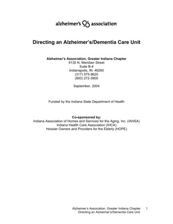
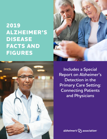
McGirr S, Venegas C, Swaminathan A. Alzheimer’s Disease: A Brief Review. J Exp Neurol. 2020;1(3): 89-98.of life but do not modify or slow the disease course. Currentresearch aims to treat underlying pathology of active ADas well as identify and stage interventions in those withpreclinical, or asymptomatic, AD.Historical BackgroundAlois Alzheimer, a German physician, reported thefirst case of Alzheimer’s disease in 1907 [4]. He first sawAuguste Deter, a 51-year-old woman, in 1901. Auguste’shusband Karl brought her to a mental hospital after shebegan exhibiting unusual behavior, including hidingitems, threatening neighbors, and accusing her husbandof adultery. She also lost the ability to do daily activitiessuch as cooking and housework. Auguste came underAlzheimer’s care at a mental hospital in Frankfurt. Therehe observed and recorded her behavioral patterns: shecould speak but not write her own name, she could nameobjects such as a pencil but not the food she was eating,she was polite sometimes but loud and offensive at othertimes. He diagnosed Auguste with “presenile dementia”[5].Upon her death in 1906, Alzheimer’s biopsy of herbrain revealed diffuse cortical atrophy and “particularchanges in cortical cell clusters” [6]. Alzheimer describedplaques and tangles of nerve fibers which researcherswould identify in the 1980’s as beta amyloid plaques andneurofibrillary tangles of tau [7,8]. That year, Alzheimergave a presentation on Auguste at a German psychiatryconference, asserting these cortical lesions to be the causeof her symptoms. He published a research paper the nextyear, and a psychiatry textbook in 1910 named the disorder‘Alzheimer’s disease.’AD histopathology reveals intraneuronal neurofibrillarylesions made up of tau proteins. Tau proteins are mainlyfound in neurons and are involved in the assembly andstabilization of the neuronal microtubule network. Tauprotein becomes pathological when the phosphorylationregulation becomes unchecked and hyperphosphorylatedtau proteins polymerize into filaments and becomeneurofibrillary tangles. This leads to malfunction of thestructural and regulatory actions of the cytoskeleton andthen leads to abnormal morphology, axonal transport,and synaptic function of neurons, thus leading toneurodegeneration [12].These prior theories paved the way to the widely acceptedhypothesis for the pathogenesis of AD: the amyloid cascadehypothesis. This theory attributes clinical sequelae of thedisease to the overproduction or decreased clearance ofamyloid beta (Ab) peptides, which then leads to increaseddeposition of Ab, furthermore, leading to neuronal damage(Figure 1). The length of Ab varies depending on theposttranslational cleavage pattern of the transmembraneamyloid precursor protein (APP). Ab is generated bycleavage of APP via either b- or g-secretases, resulting inthe infamous insoluble Ab fibrils [13]. Two main typesof Ab polymers play a direct role in the pathology of AD:Ab40 and Ab42. Ab40/Ab42 then oligomerizes, travelsto synaptic clefts, and interferes with synaptic signaling.These eventually further polymerize into insolubleamyloid fibrils that aggregate into amyloid plaques [14].Within the plaques, Ab peptides in b-sheet conformationThe clinical diagnostic criteria for AD were standardizedin the U.S. in 1984 [9]. They were revised in 2011 and2018 to create separate diagnoses for the preclinical, mildcognitive impairment (MCI) and dementia stages of ADand to recognize the role of biomarkers in AD diagnosis[10,11].PathogenesisAD is a complex, multifactorial, neurodegenerativedisease, resulting from complicated interactions of one’sgenetic makeup, education, age, and environment. Manyhypotheses have laid the foundation to gain understandingof the etiology of the disease, with one of the oldest beingthe cholinergic hypothesis. This hypothesis is based uponthe fact that AD patients show reduction in activity ofcholine acetyltransferase and acetylcholinesterase in thecerebral cortex compared with the normal brain [12]. Postmortem brain tissue from patients with AD confirmedthe reduced neurotransmitter pathway activity, revealingthat degeneration of cholinergic neurons and loss ofcholinergic neurotransmission significantly contributesto the cognitive impairment seen in those with AD [12].The Tau hypothesis has also been proposed, consideringJ Exp Neurol. 2020Volume 1, Issue 3Figure 1: The amyloid (Aβ) cascade hypothesis [12].90
McGirr S, Venegas C, Swaminathan A. Alzheimer’s Disease: A Brief Review. J Exp Neurol. 2020;1(3): 89-98.polymerize into structurally distinct forms, includingfibrillar, protofibers and polymorphic oligomers. It is thedeposition of these plaques diffusely throughout the brainthat lead to microglial activation, cytokine release, reactiveastrocytosis, and an overall inflammatory response. Thesestructural changes lead to synaptic and neuronal loss andeventual gross cerebral atrophy [12]. On the other hand,should APP be processed by a-secretase in the healthyadult, soluble b-amyloid is produced, which has beenlinked to play a role in neuronal plasticity/survival, isprotective against excitotoxicity, is important for earlyCNS development, and has been shown to be importantfor promoting synapse formation [15].The genetics of AD should also be considered toplay an influential role in the pathogenesis, alongsideinflammation, apoptosis, and plaque buildup. In fact,the APP gene located on 21q21, mentioned above, wasthe first discovered causative gene of AD [13]. Advancesin genetic research have identified two distinct forms ofAD: Familial Alzheimer’s Disease (FAD) and SporadicAlzheimer Disease (SAD), with the latter making up themajority of cases. Important advances in the 1990s andearly 2000s revealed that FAD is the result of autosomaldominant mutations in APP, PSEN1 and PSEN2 genes,located on chromosome 21, 14, and 1, respectively [12,16].More specifically, PSEN1 and PSEN2 contain the necessaryamino acid residues required for the catalytic active site ofgamma-secretase. Certain mutations of these genes lead toincreased production of Ab peptide and neurodegeneration[12]. Far more commonly, the genetic risk factor for SADwas identified as the type e4 allele on chromosome 19,of the gene for apolipoprotein E (APOE), a low-densitylipoprotein carrier resides. APOE is present in roughly 5060% of patients with AD compared to 20-25% in healthyelderly adults without the history of familial AD. APOEis associated with an approximately three-fold risk ofdeveloping AD if one copy is present, and there is an eightfold risk if two copies are present [16]. These plus othermajor influencing genes for SAD are listed in Table 1 and 2.Genes Involved in Pathogenesis of ADAbbreviationsAmyloid Precursor Protein geneAPPPresenilin genePSApolipoprotein E geneAPOEClusterin genen/aComplement Receptor 1 genen/aPhospholipids Bind to ClathrinProtein genePICALMCholesterol Metabolism geneCH25H,ABCAL, andCH24HSterol O-acyltransferase geneSOAT1Prostaglandin-endoperoxide Synthase2 genePtgs2Angiotensin-Converting Enzyme genen/aSLC26A38 genen/aTable 1: A list of several genes that play a major influentialrole for the development of Sporadic Alzhemer’s Disease(SAD) [17].GeneProposed mechanism of dysfunction contributing to pathogenesis in Alzheimer’s diseaseAPPDefects in synaptic development and neuronal migration [18], A-beta peptide synthesis defects[19]PSEN 1 / 2Altered gamma-secretase activity resulting in elevated amyloid beta-42 levels [20]APOE – epsilon 4Lipid and cholesterol dysmetabolism, synaptic inflammation, impaired clearance of amyloidbeta-42, LDL receptor impairment [21-24]CLUDeposition and metabolism of amyloid beta-42 [25]ABCA7Immune mediated and lipid metabolic response alterations [26]CR1Aggravated senile plaque formation [27]CD33Influences microglia mediated clearance of amyloid beta-42 [28]MS4ADysregulation of intracellular calcium concentration [29]EPHA1Axonal guidance changes [30]SORL1, BIN,CD2AP, PICALMAlterations in lipid metabolism [31]Table 2: A list of relevant genes involved in pathogenesis of Alzheimer’s disease.J Exp Neurol. 2020Volume 1, Issue 391
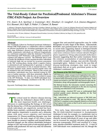
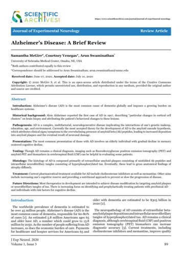
McGirr S, Venegas C, Swaminathan A. Alzheimer’s Disease: A Brief Review. J Exp Neurol. 2020;1(3): 89-98.Figure 2: “Neuronal dysfunction and cell death are responsible for the development of Alzheimer’s disease.Accumulating evidence suggests that abnormally elevated tau hyperphosphorylation, pathogenic Aβ oligomers,and mitochondrial dysfunction cooperate to drive the neuronal dysfunction and cell death that underlie cognitiveimpairment. Although the amyloid hypothesis supports that neurotoxic Aβ primarily induces tau pathology, otherproteolytic fragments of the human APP including sAPPβ, N-APP, and AICD, also appear to contribute to taualterations. In addition, aging, which is tightly associated with mitochondrial dysfunction, can serve as the mostsignificant nongenetic risk factor for Alzheimer’s disease” [32].Clinical PresentationThe most common presentation of AD is that of an elderlyindividual with an insidious progression of cognitivedecline, most centered around memory loss. Declines innon-memory aspects of cognition including word-finding,vision/spatial issues, impaired reasoning or judgment,are also seen in early stages. At this time, patients meetcriteria for mild cognitive impairment [33]. Almost allpatients diagnosed with AD also have neuropsychiatricsymptoms during some stage of their disease, of whichdepression and apathy are the most dominant early on.Verbal and physical aggression are frequently observedthroughout all stages. As the disease progresses, delusions,hallucinations, and aggression are more often seen andadditionally, circadian sleep-wake rhythms are moreexaggerated as compared to those with normal aging [34].As the disease progresses, cognitive difficulties becomeJ Exp Neurol. 2020Volume 1, Issue 3more apparent and widespread, eventually impactingactivities of daily living. The decline in two or more areas,including memory, language, executive and visuospatialfunction, personality, and behavior that lead to the loss ofthe ability to perform instrumental and basic ADL’s pointtowards the diagnosis of AD dementia [33]. For those thatdo survive to the late stages of AD, death is often due toconsequences of the disease itself as well as increasedvulnerability to falls, pressure sores, and infections,ultimately leading to an average of 8 years from diagnosisto death [35]. Tools considered useful for clinical detectionof AD include global cognitive screens, such as the MMSEand MOCA, and more specific tests of memory impairmentlike the five-word test. More formal diagnosis can be doneby specialists, such as neuropsychologists [36].FAD tends to have the typical presentation mentionedabove, although at a much earlier age. Some PSEN192
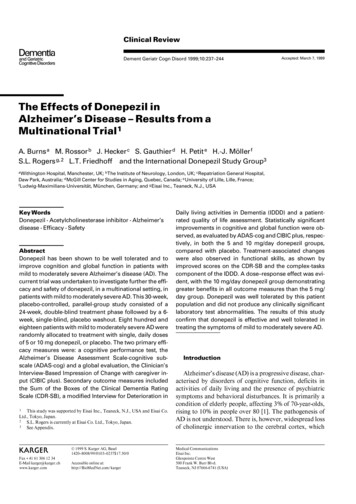
McGirr S, Venegas C, Swaminathan A. Alzheimer’s Disease: A Brief Review. J Exp Neurol. 2020;1(3): 89-98.mutations are associated with other features of disease,including seizures, spastic paresis, and myoclonus [33].There are rarer forms of AD that present atypically,including posterior cortical atrophy, logopenic primaryprogressive aphasia, and frontal variant AD [35]. Posteriorcortical atrophy tends to present with progressive lossof higher visual functions, such as diminished ability tointerpret, locate, or reach for objects via visual guidance.A decreased ability of numeracy, literacy, and praxismay also be present in this variant of the disease [37].Logopenic primary progressive aphasia presents more withlanguage disturbance focus, characterized by slow wordretrieval, word finding difficulties, and impaired sentencerepetition, while motor speech, grammar, and single-wordcomprehension are spared [38]. The frontal variant of ADpresents with stereotyped behaviors, progressive apathy/behavior disinhibition, and executive dysfunction [39].These variant presentations are important to address, butmore than likely, they do result in the more global andtypical picture of memory loss and dementia caused by ADover the course of time.TestingAD remains a clinical diagnosis and is reliant on adetailed history, cognitive assessment and physical exam.Structural imaging is also an important component ofthe assessment for AD. Atrophy of the medial temporallobes on magnetic resonance imaging (MRI) is considereda diagnostic marker for the mild cognitive impairmentstage of AD [40]. Similarly, hypometabolism in theparieto-temporal association area, posterior cingulateand precuneus on fluorodeoxyglucose PET imaging isassociated with AD [41].In 2012, the Food and Drug Administration approved thefirst beta-amyloid tracer, florbetapir, for use in PET scansof suspected Alzheimer’s patients. In one study, florbetapirPET imaging was shown to have a 92% sensitivity and 100%specificity for detecting moderate to frequent plaques inpatients with an autopsy within 2 years of the scan [42].The amyloid PET tracers florbetaben and flutemetamolhave also been approved and show similar sensitivityand specificity [43]. However, amyloid PET scans havehad limited clinical impact due to lack of insurancereimbursement, and they are currently used primarily inresearch trials.Biomarkers in CSF further support a diagnosis of AD.The biomarker profile of AD is increased total tau (T-tau)and phosphorylated tau (P-tau) and decreased amyloidbeta (Ab42). This biomarker pattern can also predictwhich subjects with MCI are likely to progress to AD[44]. Amyloid PET and the CSF marker Ab42 show aconcordance of around 90% across several studies [45].One longitudinal study of healthy elderly and patientsJ Exp Neurol. 2020Volume 1, Issue 3with MCI showed similar diagnostic accuracy betweenCSF and amyloid PET biomarkers and no improvementwhen combining them [46]. Thus, the choice between CSFand amyloid PET biomarkers can be based on availability,cost, patient preference and other factors such as patientsuitability for radiation or lumbar puncture.Currently, there are no blood biomarkers routinely used todiagnose AD. Only a small fraction of brain proteins entersthe bloodstream, and these proteins are diluted by plasmaproteins such as albumin and IgG, making them difficultto measure quantitatively. Additionally, brain proteins inplasma may be degraded or metabolized, so that plasmalevels would not reflect real-time changes in the brain [47].Nonetheless, several plasma proteins are being examinedas potential noninvasive biomarkers of AD. Plasma tau isone such protein. One study with prospective and crosssectional cohorts found higher plasma tau to be associatedwith AD, but with significant overlap in levels of healthycontrols [48]. Another potential blood marker is theaxonal neurofilament light (NFL) protein. A prospectivestudy found high correlation between plasma NFL andCSF NFL, and high diagnostic accuracy of plasma NFL inpatients with AD versus controls [49]. However, plasmaNFL is also increased in other neurological diseasessuch as frontotemporal dementia, limiting its utility fordifferential diagnosis of AD [50]. Finally, despite the utilityof CSF Ab42 as a biomarker, plasma Ab42 has not shownto be predictive of AD development in patients with MCI.Unlike NFL, there is a lack of correlation between levelsof Ab42 in CSF and plasma, possibly due to the release ofAb42 in plasma from peripheral tissues [51].HistologyAmyloid plaques and neurofibrillary tangles are thecardinal features of Alzheimer histopathology. Theamyloid plaques consist of extracellular accumulationsof misfolded Ab sheets consisting of 40 or 42 aminoacids (Ab40 and Ab42), the two by-products of amyloidprecursor protein metabolism [33]. Amyloid plaques arelocated extracellularly, and initially develop in the basal,temporal, and orbitofrontal cortex of the brain, and laterprogress to involve the neocortex, hippocampus, amygdala,diencephalon, and basal ganglia. These aggregates of Abtrigger the formation of neurofibrillary tangles (NFS),which are composed mostly of hyperphosphorylatedtau protein. These NFS are found in the locus coeruleusand transentorhinal and entorhinal areas of the brain.In the last critical stage, these histopathological entitiesare spread to the hippocampus and neocortex [14].Additionally, neuropil threads, dystrophic neurites,associated astrogliosis, microglial activation and evidenceof amyloid angiopathy are also common findings [33].Histology images of Alzheimer’s Disease is illustrated inFigures 3 and 4.93
McGirr S, Venegas C, Swaminathan A. Alzheimer’s Disease: A Brief Review. J Exp Neurol. 2020;1(3): 89-98.Figure 3: A representation of the basic histology of Alzheimer’s Disease, consisting of intracellular neurofibrillarytangles composed of hyperphosphorylated tau and extracellular collections of misfolded Aβ peptide forming amyloidplaques [52].TreatmentCurrently, treatment for Alzheimer’s disease does notimpact the progression or underlying pathology of thedisease. However, with medication and other therapies,steps are taken to increase the quality of life of those withAD. Current therapeutics are based off the CholinergicTheory, which attributes a decrease in cholinergicneurotransmission to a decline in cognitive function [12].At present, there are two classes of pharmacologic therapyavailable for AD: cholinesterase inhibitors (donepezil,rivastigmine, and galantamine) and memantine, which hasactivity as both a non-competitive N-methyl-D-aspartatereceptor antagonist and a dopamine agonist [3]. Thecholinesterase inhibitors are approved for use in patientswith mild, moderate, or severe AD dementia as well asParkinson’s disease dementia. Memantine is approved foruse in patients with moderate to severe AD who may alsohave difficulty with alertness and attention.Other aims of treatment look to modifiable risk factorsin one’s overall health and “cognitive reserve” includingcardiovascular/lifestyle factors, such as a healthy dietand plenty of physical exercise, as well as cognitiveengagement. Cognitive reserve refers to the ability tofend off pathologic insult, meaning the ability to engagealternate synaptic pathways or cognitive strategies to copewith the pathology of AD. By improving one’s physicalwellbeing and mental reserve, this may delay clinicalJ Exp Neurol. 2020Volume 1, Issue 3symptoms of AD [36]. Preventing, halting, or slowingdown the progression of AD with nutrition has been atopic of much research in Alzheimer’s today. Antioxidants,omega-3 fatty acids, B vitamins, folate, medium chaintriglycerides, and combination medical foods, are justa few of the avenues of which are being studied. Forinstance, the variable actions inherent in any givenantioxidant, such as reducing oxidized membrane lipids,limiting damage to nucleic acids, and influencing strepkinase pathways, may alter the pathways of cellular injurythat lead to the positive benefits of having them in the dietof someone with AD [53]. Research has also demonstratedVitamin E to prevent AD-like changes in the brains ofAD genetic models of mice, and other clinical trials haveshown an increase in median survival in patients withAD who were treated with selegiline (15 mg twice daily)and α-tocopherol (1000 IU twice daily) [53]. Several largescale clinical trials have also looked into omega-3 fattyacids, one of which being the MIDAS study which foundthat 900 mg of daily docosahexaenoic acid (DHA) led to a7 year age improvement in cognition over 24 weeks whencompared to placebo. This study and many more point toDHA’s potential direct effects on neurodegeneration inAD as well as its overarching reduction in cerebrovasculardisease [53]. To discuss DHA even further, a randomizedcontrol trial looked at the effect of omega-3 fatty acids hadon B vitamin function. To preface, inadequate B vitaminstatus alone leads to the accumulation of homocysteine, anon-essential amino acid, that when elevated is recognized94
McGirr S, Venegas C, Swaminathan A. Alzheimer’s Disease: A Brief Review. J Exp Neurol. 2020;1(3): 89-98.as a modifiable risk factor for AD and other dementias.This goes along with the results of the VITACOG trial,which showed that B vitamin supplement in elderly adultswith mild cognitive impairment slowed the rate of brainatrophy both globally and regionally [54]. And so it makessense that the group that was randomized to B vitamintreatment (folic acid and vitamins B6 and B12) showedthat B vitamins plus an omega-3 level in the upper rangeof normal interacted to slow cognitive decline, basicallysumizing that elevated omega-3 acids alone significantlyenhanced the cognitive effect of B vitamins [54].Novel treatments for AD today are based off multifaceted strategies and are currently under investigationvia clinical trials. There are three trials currently ongoing,including gantenerumab, crenezumab, and aducanumabas immunotherapy approaches, which aim to effectivelyclear the Ab aggregates [55]. Other future targets fortreatment include targeting mitochondrial oteinaggregations via novel acetylcholinesterase inhibitors orNMDA-Receptor Antagonists, targeting autophagy, andtargeting neuroinflammation, to name a few [55]. Linkshave also been made regarding the gut microbiota and AD,and further research regarding dysbiosis and worseningdisease are being investigated for other novel treatmentapproaches [56].Future DirectionsThe majority of therapeutics in development target thetwo hallmark pathologies of AD: beta-amyloid plaquesand neurofibrillary tangles of tau. As of February 2019, thepipeline of AD drugs includes 132 agents, with 96 (73%)intended to achieve disease modification. Of these, 38have amyloid as a primary or combination target, and 17have tau as a primary or combination target [57].One class of therapeutics against amyloid, monoclonalantibodies, is intended to facilitate amyloid removal fromthe brain. Data have so far failed to show improvement inclinical symptoms despite significant reductions in amyloidload. However, one antibody which had previously failedfutility analyses in Phase III trials later showed positiveresults in a subset of patients. The antibody, aducanumab,is directed against both amyloid fibrils and solubleoligomers, and its manufacturer Biogen plans to apply forFDA approval in 2020 [58]. Another set of therapeuticsinhibit the enzyme that cleaves the beta-amyloid proteinfrom its precursor, amyloid precursor protein (APP).These drugs against the beta-site APP-cleaving enzyme1 (BACE-1) have similarly led to reductions in amyloidlevels but produce worse cognitive decline in AD patientscompared to placebo. The results indicate BACE-1 activitymay be important in preserving normal synaptic function[59].J Exp Neurol. 2020Volume 1, Issue 3Several therapeutics are also in development to targettau, which is downstream of amyloid and thought to be thedirect cause of AD symptoms. Although tau accumulatesintracellularly, several in vivo and in vitro studies haveshown tau can move from one neuron to another in a“prion-like” spread, suggesting a possible therapeutictarget [60]. LMTX, a selective inhibitor of tau proteinaggregation, failed to show cognitive improvement in ADpatients compared to placebo in a Phase III trial, but aseparate Phase II/III trial was started to test a lower doseof the drug [61]. Several anti-tau antibodies are in earlyclinical testing, as are at least two active tau vaccinesdesigned to stimulate antibody production in AD patients[62].Given the underwhelming results of therapeuticsagainst amyloid and tau, drug development has shiftedits focus to the “pre-dementia” space, including patientswith preclinical AD and individuals with risk factors forcognitive decline. A large number of therapeutic trialswith private and public funding are underway in high-riskasymptomatic individuals, including carriers of geneticmutations and those with amyloid-positive PET scans orelevated biomarkers [63]. Data from these trials in the nextseveral years may provide insight into which interventions,if any, can slow or halt the onset of symptomatic AD.Ethical StandardsThis review article was written in a manner consistentwith ethical and scientific guidelines regarding researchand publication.ContributionsBoth Samantha McGirr and Courtney Venegas, fourthyear medical students, were equally involved in theresearch and writing of this paper. Dr. Swaminathan,Neurologist at UNMC who contributed to and edited thepublished review, supervised them both.References1. Crous-Bou M, Minguillón C, Gramunt N, MolinuevoJL. Alzheimer’s disease prevention: from risk factors toearly intervention. Alzheimer’s Research & Therapy. 2017Dec 1;9(1):71.2. Alzheimer’s Association. 2020 Alzheimer’s diseasefacts and figures. Alzheimer’s & Dementia. 2020 Mar 10;16(3):391-460.3. Weller J, Budsonof Alzheimer’s diseaseF1000Research. 2018;7.A. Current understandingdiagnosis and treatment.4. Alzheimer A. Uber eine eigenartige Erkrankung derHirnrinde. Zentralbl. Nervenh. Psych. 1907;18:177-9.95
McGirr S, Venegas C, Swaminathan A. Alzheimer’s Disease: A Brief Review. J Exp Neurol. 2020;1(3): 89-98.5. Yang HD, Kim DH, Lee SB, Young LD. History ofAlzheimer’s Disease. Dementia and NeurocognitiveDisorders. 2016 Dec 1;15(4):115-21.17. Chen YG. Research progress in the pathogenesis ofAlzheimer’s disease. Chinese Medical Journal. 2018 Jul5;131(13):1618.6. Soria Lopez JA, Gonzalez HM, and Leger GC. Chapter13 – Alzheimer’s disease. Handbook of Clinical Neurology2019; 167: 231-255.18. Priller C, Bauer T, Mitteregger G, Krebs B, KretzschmarHA, Herms J. Synapse formation and function ismodulated by the amyloid precursor protein. Journal ofNeuroscience. 2006 Jul 5;26(27):7212-21.7. Glenner GG, Wong CW. Alzheimer’s disease: initialreport of the purification and characterization of anovel cerebrovascular amyloid protein. Biochemicaland Biophysical Research Communications. 1984 May16;120(3):885-90.8. Brion JP, Couck AM, Passareiro E, Flament-DurandJ. Neurofibrillary tangles of Alzheimer’s disease: animmunohistochemical study. Journal of SubmicroscopicCytology. 1985 Jan 1;17(1):89-96.9. McKhann G, Drachman D, Folstein M, Katzman R,Price D, Stadlan EM. Clinical diagnosis of Alzheimer’sdisease: Report of the NINCDS-ADRDA Work Group*under the auspices
McGirr S, Venegas C, Swaminathan A. Alzheimer s Disease: A Brief Review. J Exp Neurol. 2020;1(3): 89-98. eurol Volum ss of life but do not modify or slow the disease course. Current research aims to treat underlying pathology of active AD as well as identify and stage interventions in those with preclinical, or asymptomatic, AD. Historical .