Transcription
Chen et al. BMC Cancer 2014, 2RESEARCH ARTICLEOpen AccessE-cadherin loss alters cytoskeletal organizationand adhesion in non-malignant breast cells but isinsufficient to induce an epithelial-mesenchymaltransitionAugustine Chen1, Henry Beetham1, Michael A Black1, Rashmi Priya2, Bryony J Telford1, Joanne Guest1,George A R Wiggins1, Tanis D Godwin1, Alpha S Yap2 and Parry J Guilford1*AbstractBackground: E-cadherin is an adherens junction protein that forms homophilic intercellular contacts in epithelialcells while also interacting with the intracellular cytoskeletal networks. It has roles including establishment andmaintenance of cell polarity, differentiation, migration and signalling in cell proliferation pathways. Its downregulationis commonly observed in epithelial tumours and is a hallmark of the epithelial to mesenchymal transition (EMT).Methods: To improve our understanding of how E-cadherin loss contributes to tumorigenicity, we investigatedthe impact of its elimination from the non-tumorigenic breast cell line MCF10A. We performed cell-based assaysand whole genome RNAseq to characterize an isogenic MCF10A cell line that is devoid of CDH1 expression dueto an engineered homozygous 4 bp deletion in CDH1 exon 11.Results: The E-cadherin-deficient line, MCF10A CDH1-/- showed subtle morphological changes, weaker cell-substrateadhesion, delayed migration, but retained cell-cell contact, contact growth inhibition and anchorage-dependentgrowth. Within the cytoskeleton, the apical microtubule network in the CDH1-deficient cells lacked the radialpattern of organization present in the MCF10A cells and F-actin formed thicker, more numerous stress fibres inthe basal part of the cell. Whole genome RNAseq identified compensatory changes in the genes involved incell-cell adhesion while genes involved in cell-substrate adhesion, notably ITGA1, COL8A1, COL4A2 and COL12A1,were significantly downregulated. Key EMT markers including CDH2, FN1, VIM and VTN were not upregulatedalthough increased expression of proteolytic matrix metalloprotease and kallikrein genes was observed.Conclusions: Overall, our results demonstrated that E-cadherin loss alone was insufficient to induce an EMT or enhancetransforming potential in the non-tumorigenic MCF10A cells but was associated with broad transcriptional changesassociated with tissue remodelling.Keywords: CDH1, Cytoskeletal modelling, Adhesion, Migration, EMTBackgroundE-cadherin, encoded by the tumor suppressor gene CDH1,is a homophilic cell-to-cell adhesion protein localized to theadherens junctions of all epithelial cells [1]. Its cytoplasmicdomain effectively creates a bridge between the cytoskeletons of adjacent cells by interacting with both cortical actin* Correspondence: parry.guilford@otago.ac.nz1Cancer Genetics Laboratory, Department of Biochemistry, University ofOtago, Dunedin 9054, New ZealandFull list of author information is available at the end of the articlefilaments and the microtubule network [2]. These andother interactions [3] extend E-cadherin’s functionalitybeyond cell-cell adhesion to roles in establishing andmaintaining cell polarity, differentiation, stemness, cell migration and the mediation of signalling through variousproliferation and survival pathways including WNT andEGFR [1-5].Abrogation of CDH1 expression by mutation, deletionor promoter hypermethylation is a feature of many epithelial tumours, including prostate, ovarian, lung and 2014 Chen et al.; licensee BioMed Central Ltd. This is an Open Access article distributed under the terms of the CreativeCommons Attribution License (http://creativecommons.org/licenses/by/4.0), which permits unrestricted use, distribution, andreproduction in any medium, provided the original work is properly credited. The Creative Commons Public DomainDedication waiver ) applies to the data made available in this article,unless otherwise stated.
Chen et al. BMC Cancer 2014, 2hepatocellular carcinomas, and is the hallmark of boththe sporadic and familial forms of diffuse gastric cancer(DGC) and lobular breast cancer (LBC) [1,6]. In bothLBC and DGC, CDH1 inactivation can be an early initiating event [7,8], whereas in other tumour types including prostate, lung, ovarian and colon, its downregulationis usually considered to be a late event that promotes anincrease in invasive capacity [9]. Increased invasivenessfollowing CDH1 downregulation is related, at least inpart, to the central role played by E-cadherin in thede-differentiation process known as the epithelialmesenchymal transition (EMT) [10]. During the EMT,epithelial cells lose polarity and normal cell-cell adhesion, acquiring a mesenchymal phenotype with highermotility and an increase in cell-extracellular matrix(ECM) connections [9,11]. The EMT is associated notonly with increased tumor invasion and metastasis, butalso poor outcome, drug resistance and an increase inthe number of cancer stem-like cells [9,12]. E-cadherindownregulation has been shown to be sufficient to induce an EMT in some [4,9,10,13], but not all [14,15],cancer cell lines/models. However, it remains unclearwhether its loss can induce an EMT in cells which havenot already undergone malignant transformation [16].Clues to the influence E-cadherin loss has on tumorigenesis and the initiation of the EMT come from study ofthe multifocal gastric signet ring cell carcinomas (SRCCs)that occur in Hereditary Diffuse Gastric Cancer (HDGC)families. HDGC is a familial cancer syndrome caused bygermline mutation of the CDH1 gene and is typified byhighly penetrant DGC and an elevated risk of LBC [17].With few exceptions, mutation carriers develop tens tohundreds of gastric foci of SRCC, sometimes with enrichment in the transition zone between the antrum and body[18]. LBC and lobular carcinoma in situ (LCIS) are alsoobserved to be multifocal in female mutation carriers (V.Blair, pers. comm). The multifocal gastric SRCCs are Ecadherin-negative and almost exclusively stage T1a tumours confined to the lamina propria. Lineage markerssuggest that the foci develop from mucous neck cells thathave invaded through the basement membrane of thegastric gland [19]. Invasion is likely to be triggered byinactivation of the wild-type CDH1 allele through mechanisms including promoter hypermethylation [6]. In onemodel [20], E-cadherin loss creates instability in the orientation of the mitotic spindle, leading to a proportion ofthe cell divisions occurring out of the epithelial plane withsubsequent displacement of daughter cells into the laminapropria. The multifocal SRCC foci in the gastric mucosaare known to be relatively indolent, but show unpredictable progression to advanced disease. A small percentageof foci show characteristics of an EMT, and this change isassociated with tumour progression [19]. However, theabsence of an EMT-like phenotype from the majority ofPage 2 of 14SRCC foci suggests that E-cadherin loss alone is insufficient to induce an EMT in this relatively normal geneticbackground.MCF10A is a spontaneously immortalized, nontransformed mammary epithelial cell line derived from human fibrocystic tissue. Although it does carry cytogeneticabnormalities associated with in vitro cultured mammaryepithelial cells, including p16 and p14ARF deletion andMYC amplification [21], MCF10A is considered a “normal”breast epithelial cell due to its near diploid, stable karyotypeand characteristics of normal breast epithelium such aslack of tumorigenicity in nude mice, lack of anchorageindependent growth [22] and ability to form mammospheres in culture [21]. Here we have used cell-basedassays and whole genome RNAseq to characterize anisogenic MCF10A cell line that is devoid of CDH1 expression due to an engineered homozygous 4 bp deletion in CDH1 exon 11. We show that E-cadherin lossdisrupts the organization of the cell’s actin and microtubule cytoskeletons and modifies its adherence andmigration characteristics but is insufficient to inducean EMT.MethodsCell cultureMCF10A cells (product no: CRL 10317), a non tumorigenicmammary epithelial cell line, and the derived isogenicline with CDH1 knock out (MCF10A CDH1-/-) usingCompoZr ZFN technology (product no: CLLS1042)were purchased from Sigma. The MCF10A isogeniclines were cultured in DMEM/F12: (1:1) (Invitrogen)with 5% horse serum (Invitrogen), 10 μg/ml ActrapidPenfil neutral insulin (Novo Nordisk PharmaceuticalsLtd), 20 ng/ml human epidermal growth factor (Peprotech), 100 ng/ml cholera toxin, and 500 ng/ml hydrocortisone (Sigma) [21]. Cells were grown at 37 C with5% CO2, seeded into T75 flasks at densities of 3.0 105and 4.5 105, respectively and passaged at 90% confluency ( 3 days) for a maximum of ten Western blotMCF10A and MCF10A CDH1-/- cells were grown for 72 hto 90% confluency in T25 flasks and lysed using cell culturelysis reagent (Promega) containing cOmplete mini proteaseinhibitor (Roche). BCA assays (Thermo) were performed toequalize total protein loaded. Proteins were separated on10% SDS-PAGE gel for 2 h, followed by blot transfer at100 V for 1 h. Immunoblotting was performed using rabbitanti-E-cadherin antibody (Santa Cruz, SC7870) at 1:200dilution overnight, or rabbit anti-α-actin primary antibody(Sigma) at 1:1,500 dilution overnight followed by antirabbit HRP-linked secondary antibody (Santa Cruz) at1:5,000 dilution for 1 h. Chemiluminescence was performed
Chen et al. BMC Cancer 2014, 2using Pierce ECLplus reagent (Thermo) and imaged usingLAS-3000 (Fujifilm).ImmunofluorescenceMCF10A and MCF10A CDH1-/- cells were seeded onCoverglass slides (Labtek) and grown to confluence for72 h. Cells were fixed with 4% paraformaldehyde thenpermeabilized with 0.2% Triton-X100 in PBS for 5 minat room temperature. Cells were blocked with 10% FBSin PBS for 1 h at room temperature. E-cadherin primaryantibody (Santa Cruz, SC-7870) used at 1:250. Antirabbit secondary antibody conjugated with AlexaFluor488 (Invitrogen) used at 1:750. Immunofluorescenceimages were acquired with an Olympus IX71 microscope, under 40 objective.Proliferation assayMCF10A and MCF10A CDH1-/- cells were seeded atdensities of 2.0 103 and 4.0 103 in three replicates in96 well E-plates and incubated at 37 C in 5% CO2. Thegrowth rate was monitored in real time at 15 min intervals for 96 h using the xCELLigence platform (Roche).Both cell lines were also seeded at the same densitiesinto 96 well flat clear bottom black plates (Corning) andgrown at 37 C in 5% CO2 and imaged every 2 h for 96 husing the IncuCyte 2011A FLR (Essen Bioscience). Confluency was determined using the IncuCyte softwareConfluence v1.5.Cell adhesion assayCell adhesion assays were performed using the IncuCyte2011A FLR. MCF10A and MCF10A CDH1-/- cells wereseeded in six replicates at 2.0 104 cells per well in 96well flat clear bottom plates (Greiner Bio-one) with different surface coatings: no coating for the uncoated,2 μg/ml collagen (Sigma), 2 μg/ml fibronectin (BD Bioscience), 8 μg/ml vitronectin (Invitrogen), 8 μg/ml laminin(Invitrogen) and grown at 37 C, 5% CO2. Images were acquired every 2 h for 8 h using the automated image acquisition software. Cell numbers at each time point were alsodetermined using the Cell Counter plugin l) in ImageJ [23].Scratch wound assayScratch wound assay was performed using the IncuCyte2011A FLR (Essen Bioscience). Briefly, MCF10A andMCF10A CDH1-/- cells were seeded in six replicates atdensities of 2.5 104 and 3.5 104 cells per well, respectively, in 96 well Essen ImageLock Plate (EssenBioscience) with different coating surfaces: no coatingfor the uncoated condition, 2 μg/ml collagen, 2 μg/mlfibronectin, 8 μg/ml vitronectin, 8 μg/ml laminin. Cellswere incubated at 37 C and 5% CO2 and grown to100% confluency. The usage of the Essen imageLockPage 3 of 14plates ensures wounds are automatically located andregistered by the IncuCyte software and analyzed usingwound confluence metrics. Precise and reproduciblewounds were generated using the 96 PTFE pin WoundMaker (Essen Bioscience) on the confluent monolayerand cells returned to the incubator where images of cellswere acquired every 1 h for 35 h under phase contrast microscopy. Wound confluence was graphed over time toquantitatively evaluate the characteristic of wound closingusing the IncuCyte software, Wound Confluence v1.5.Soft agar assayAn overlay of 2.0 104 MCF10A and MCF10A CDH1-/cells and 2.0 103 MCF7 cells in 0.35% agar in mediumwere plated over a base layer of 0.5% agar (Applichem)and grown at 37 C with 5% CO2. Growth medium wasadded the next day and replenished twice a week. After24 days, growth medium was removed and MTT (Sigma)solution was added (final concentration 2 mg/ml), and theplates further incubated at room temperature for 4 h withgentle shaking. The MTT solution was then removed andwashed. Images were taken using Image Scanner 3 and colonies counted. The experiment was performed with at leasttwo technical replicates for each cell line.Immunofluorescence confocal microscopyMCF10A and MCF10A CDH1-/- cells were seeded onglass coverslips coated with fibronectin (Becton Dickinson)and allowed to grow to confluence for 48-72 h. Cells werefixed with ice-cold methanol for 5 min on ice for microtubule staining or fixed with 4% paraformaldehyde incytoskeleton stabilization buffer (10 mM PIPES pH 6.8,100 mM KCl, 300 mM sucrose, 2 mM EGTA, 2 mMMgCl2) on ice for 20 min and then permeabilized with0.25% Triton-X100 in PBS for 5 min at room temperaturefor F-actin staining. Cells were blocked with 5% milk inPBS for 1 h at room temperature. Primary antibodies used:mouse monoclonal antibody (mAb) directed against theectodomain of E-cadherin (HECD-1) (a gift from PeggyWheelock, University of Nebraska, Omaha, NE; withthe permission of M. Takeichi) 1:50; rabbit polyclonalAb (pAB) against E-cadherin (generated in-house) [24]1:1000; rat monoclonal [YOL1/34] antibody against tubulin(Abcam, # ab6161); 1:100 rabbit polyclonal antibody againstZO-1 (Invitrogen, # 61-7300). F-actin was stained withAlexaFluor 488-phalloidin, 1:500 (Invitrogen). Secondaryantibodies were species-specific antibodies conjugated withAlexaFluor-488, -594 or -647 (Invitrogen) for immunofluorescence (1:500). For immunofluorescence, confocal imageswere acquired with a Zeiss 710 Meta laser scanning confocal microscope, with a 60 objective, 1.4 NA oil PlanApochromat immersion lens with 0.6-1.0 μm optical sections. Contrast adjustment and z-projections of raw images
Chen et al. BMC Cancer 2014, 2were done using ImageJ software (National Institutes ofHealth) [23] and Illustrator (Adobe).RNASeqMCF10A and MCF10A CDH1-/- cells were seeded atdensities of 2.0 105 and 3.5 105 cells respectively induplicate in a six well dish and grown until 70% confluency, with a medium change at 24 h. Total RNA wasextracted at 48 h post seeding using quick-RNA MiniprepKit (Zymo) according to the manufacturer’s protocol. RNAyield and purity were assessed using Qubit (Invitrogen) andthe Agilent 2100 Bioanalyser. cDNA library preparationwas performed by New Zealand Genomics Limited usingIllumina TruSeq RNA preparation version 2.0. Each libraryhad inserts of 200 bp and sequence reads were generatedfrom one lane of an Illumina HiSeq 2000 run. Bowtie2and Cufflinks version 2.0.1 software packages were used toalign the read data to human genome build GRC37 andannotated with BiomaRt using Ensembl dataset ”hsapiens gene- ensembl”. Unannotated genes were removed andremaining count data was normalized using EdgeR [25].The per gene read counts were imported into the statisticalsoftware package R (www.r-project.org), and analyzed usingthe functionality included in the edgeR and limmapackages. Briefly, TMM (trimmed mean of M values)normalization was applied to generate normalizedcount data, and the lmFit command was used to fit alinear model to the data for each gene. Normalizeddata were converted to log-cpm (counts per millionreads) prior to analysis using the voom command inlimma. Differential expression results for MCF10ACDH1-/- vs MCF10A were written to CSV files, viewable in Excel (limma moderated t- statistic produced foreach comparison, per gene, with FDR p value adjustmentapplied). Gene Ontology (GO) functional enrichment analysis was carried out using GATHER [26].Page 4 of 14distribution distinct from the wildtype MCF10A which hada more elongated morphology (Figure 1d). At full confluence, both MCF10A and MCF10A CDH1-/- cells retainedcobblestone morphology typical of epithelial cell lines. TheMCF10A CDH1-/- cells however, presented gaps, unlikethe even monolayer distribution observed in wildtypeMCF10A (Figure 1d). Both the MCF10A and the MCF10ACDH1-/- cells maintained normal contact growth inhibition(Figure 1d).To estimate the effect of E-cadherin loss on the growthkinetics of the MCF10A cells, the isogenic pair of cell lineswere seeded at a density of 2.0 103 and 4.0 103 cellsper well in 96 well E-plates and growth followed usingthe xCELLigence real time system (Roche, Basel). TheMCF10A CDH1-/- cells showed a prolonged lag phasewhen compared to the wildtype MCF10A cells (Figure 1e).However, once both cell lines achieved log phase growth,the doubling time of the two lines was almost identical withMCF10A being only slightly shorter (13 h) compared to theMCF10A CDH1-/- cells (14 h) (Figure 1e).We also performed scratch wound assays to measurecell migration in real-time over 35 h post wounding.Wound closure was quantitatively evaluated over timeusing Wound Confluence v1.5 (time point t 0 h, corresponds to 2.5 h post wound generation). MCF10ACDH1-/- cells migrated markedly slower and took longerto close the wound (t 27.4 4.1 h) compared to wildtype cells (t 15.0 1.6 h) (Figure 1f ).Finally, the number of nucleoli present per cell werenoticeably reduced in MCF10A CDH1-/- compared towildtype: 87% of MCF10A CDH1-/- cells have one or twonucleoli per cell compared to 42% of MCF10A cells(Additional file 2: Figure S2). The majority of wildtypeMCF10A cells (57%) had three or more nucleoli comparedto 12% of MCF10A CDH1-/- cells (Additional file 2: FigureS2). The reduction in nucleoli number is suggestive of adecreased demand or capability for ribosome biogenesis.ResultsCharacterization of MCF10A CDH1-/- appearance andgrowth characteristicsLoss of CDH1 does not enable anchorage-independentgrowthA MCF10A CDH1-/- cell line carrying a homozygous 4 bpdeletion in exon 11 of CDH1 has recently been developedusing zinc finger nuclease (ZFN) technology (Sigma-Aldrich,Saint-Louis). The 4 bp deletion (Figure 1a) at mRNAposition 1820-1823 results in a frameshift predicted to giverise to a premature termination codon at position 1868,generating a truncated protein of 582 amino acids thatlacks the extracellular cadherin repeat 4 domain, transmembrane region and cytoplasmic domain. Immunoblotting and immunofluorescence confirmed the absenceof E-cadherin expression from the MCF10A CDH1-/line (Figure 1b-c; Additional file 1: Figure S1). Subconfluent MCF10A CDH1-/- cells exhibited a more roundedmorphology and grew in a clustered and contractedTo determine if E-cadherin loss would cause MCF10A cellsto become tumorigenic, we performed soft agar colony formation assays to monitor anchorage–independent growth.After 24 days in soft agar, no colonies were formed forMCF10A cells, consistent with a previous study [22]. Likewise, MCF10A CDH1-/- cells did not show any colonygrowth (Figure 1g). This result showed neither MCF10Anor MCF10A CDH1-/- cells exhibit the ability to divide andproliferate in the absence of adhesion to the substratum.MCF10A CDH1-/- cells show altered actin and tubulincytoskeletal arrangementTo directly observe the effects of E-cadherin knock-outon the cytoskeleton of MCF10A cells, we examined the
Chen et al. BMC Cancer 2014, 2Figure 1 (See legend on next page.)Page 5 of 14
Chen et al. BMC Cancer 2014, 2Page 6 of 14(See figure on previous page.)Figure 1 Characterization of MCF10A CDH1-/- cells. a) CDH1 sequence from MCF10A CDH1-/- and wildtype MCF10A cell lines depicting theengineered 4 bp deletions as determined by RNAseq. The specified deletion was attributed to ZFN editing on exon 11 of both CDH1 alleles inMCF10A CDH1-/- cells. The ZFN binding site is represented by bases in red uppercase and the ZFN cut site is represented in red lowercase. b)Immunoblot of MCF10A CDH1-/- confirming the loss of E-cadherin expression as a result of the 4 bp deletion with α-actin as loading control. Thecropped images are a composite of the same nitrocellulose immunobloted with antibodies against E-cadherin followed by α-actin (Additional file1: Figure S1). c) Immunofluorescence showing loss of E-cadherin from the cell junctions in MCF10A CDH1-/- but not wildtype MCF10A cells. d)Comparison of growth morphology between MCF10A CDH1-/- and wildtype at subconfluence and full confluence. At subconfluence, MCF10ACDH1-/- showed clustered and contracted distribution while some wiltdtype MCF10A cells exhibited more mesenchymal morphology. At fullconfluence, both isogenic cells retained epithelial cobblestone-like morphology, although MCF10A CDH1-/- displayed gaps not observed inwildtype cells. e) Comparing cell proliferation profile between both MCF10A isogenic cells. A measure of cell proliferation was represented bythe normalized cell index taken from impedence measurements generated by cells grown over 96 h on a 96-well E-Plate on the xCELLigence. f)The time course of cell migration was quantified using IncuCyte wound confluence at 1 h intervals over 35 h. MCF10A CDH1-/- cells were shownto take significantly longer in wound closing compared to wildtype cells. g) Soft agar assay to determine anchorage-independent growth as aresult of E-cadherin loss. Anchorage-independent growth was observed only in the positive control MCF-7 cells, but not in either of the MCF10Aisogenic cells. Representative images from one of two biological replicates were presented.microtubule and actin cytoskeletons using immunofluorescence staining for α-tubulin and F-actin. On the apicalsurface of MCF10A cells, the microtubules displayed aprominent radial pattern of organization with minusends anchored densely in the centre and the plus endsextending towards the cell cortex (Figure 2a). Howeverin MCF10A CDH1-/- cells, the microtubules were lessdense and there was a gross defect in the radial patternof organization, often oriented parallel to the cell cortex(Figure 2a). At the basal surface of the cells, the microtubules formed a meshwork-like structure and no striking differences in organization between MCF10A and MCF10ACDH1-/- cells were observed. Apically in MCF10A cells,actin forms a cross-linking filamentous meshwork whilebasally it organizes itself into stress fibre like structures(Figure 2b). The apical actin meshwork looked similarin MCF10A CDH1-/- cells, but basally the stress fibreswere thicker and more numerous in the E-cadherindeficient cells.Loss of CDH1 impacts on the transcription of diverse cellcell adhesion genesTo elucidate the impact of E-cadherin loss at the transcriptional level, we performed genome-wide RNAseqon the MCF10A and MCF10A CDH1-/- cells. Anaverage of 6.55 107 reads were obtained per library.Using a cut off of /- log21.00 and an adjusted p valueof 0.05, a total of 1,388 genes were observed to be differentially regulated. Relative to the CDH1-expressingcells, 715 genes showed significantly increased expression in the CDH1-/- cells and 673 genes had significantly reduced expression. The GO terms [26] moststrongly associated with the 1,388 genes included morphogenesis (Bayes factor 34.1), organogenesis (Bayesfactor 31.5), development (Bayes factor 30.3), cellularmetabolism (Bayes factor 27.8), cell communication(Bayes factor 24.0), cell adhesion (Bayes factor 21.3)and histogenesis (Bayes factor 18.5) (Additional file 3:Table S1).Loss of CDH1 expression was associated with expressionchanges in other cell-cell adhesion genesFour other cadherins showed significant changes in expression: CDH2 and CDH4 were downregulated in theCDH1-/- cells by 2 fold, whereas CDH3 and CDH16were upregulated by up to 3.8 fold. Three of the fournectin genes which encode proteins involved in adhesion at the adherens junction were also markedly upregulated by up to 2.2 fold, most notably PVRL4.Genes encoding adherens junction-associated proteins(e.g. CTNNB1) showed little or no change in expression in the MCF10A CDH1-/- cells (Additional file 4:Table S2). Ten established tight junction genes showedsignificant upregulation with five demonstrating upregulation of 2 fold namely, CLDN1, CLDN4, CLDN7,OCLN and CGN (Table 1). Two further tight junctiongenes showed significant downregulation (JAM3 1.3fold decrease, p 0.004 and CLDN15 2.0 fold decrease,p 0.015), three others had insignificant changes (TJP11.2 fold decrease, p 0.154; TJP2 1.1 fold increase, p 0.054 and CLDN22 1.1 fold, p 0.74), while 17 showednegligible expression in both cell lines (Additional file4: Table S2). Similarly, eight of the eleven expresseddesmosome genes and six of the eight expressed gapjunction genes also demonstrated increased mRNA expression, most notably DSG4, DSC2, JUP, GJA5, GJB2and GJB4, with fold changes up to 3.9 fold (Table 1).Taken together, this transcriptional data demonstratesthat the loss of CDH1 from the adherens junction is associated with an increase in the expression of genes encoding various tight junction, desmosome and gapjunction proteins. This increased expression of othercell-to-cell adhesion genes may partially compensatefor the loss of E-cadherin and explains the retention of
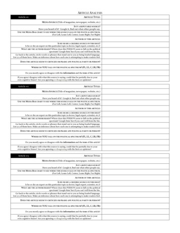
Chen et al. BMC Cancer 2014, 2Figure 2 (See legend on next page.)Page 7 of 14
Chen et al. BMC Cancer 2014, 2Page 8 of 14(See figure on previous page.)Figure 2 E-cadherin loss altered cytoskeletal organization in MCF10A CDH1-/- cells. a) Loss of E-cadherin altered tubulin cytoskeletalarrangement. On the apical surface of MCF10A cells, the microtubules showed radial pattern of organization (indicated by white arrows) withthe minus end densely anchored in the centre and the plus end extending towards the cell cortex. However, in MCF10A CDH1 -/- cells had gross defect in the radial pattern of organization and often oriented parallel to the cortex (indicated by white arrows). At the basal surface, the microtubulesform a meshwork like structure with no striking difference observed between the two cell lines. b) Loss of E-cadherin altered actin cytoskeletalarrangement. On the apical surface of MCF10A cells, actin forms cross-linking filamentous meshwork while basally it organizes itself into stressfibres like structure. Overall apical actin meshwork looks similar in both MCF10A isogenic cells but basally there are more and thicker stressfibres in MCF10A CDH1-/- cells (indicated by white arrows).cell-cell contact and cobblestone epithelial morphologyobserved at full confluency (Figure 1d).Loss of CDH1 from MCF10A cells alters expression of thegenes involved in cell-ECM adhesion and promotes altered adhesion to basement membrane proteinsIn addition to the observed changes in expression ofcell-to-cell adhesion genes, the loss of CDH1 was also associated with significant changes in the expression of cellsubstrate adhesion genes (Table 1). Up to 2.8 fold reductionin the expression of the integrin receptor subunit genesITGA1, ITGA4, ITGA5, ITGAV, ITGB1, ITGB2 was observed. Only ITGA10 and ITGB6 showed significantlyincreased expression (up to 2 fold; Additional file 5:Table S3). Furthermore, many ECM transcripts demonstrated a marked downregulation in the MCF10A CDH1-/cells compared to the wildtype cells. This was evident forCOL4A1, COL4A2, COL4A4, COL6A3, COL8A1, COL12A1,COL18A1, LAMA1, FN1 and VTN. Other members of thelaminin family LAMA5, LAMB1, LAMB2 and LAMC1were also significantly downregulated (Additional file 5:Table S3). Only a small subset of ECM genes were upregulated in MCF10A CDH1-/- cells, namely COL2A1,COL5A3, COL7A1, COL13A1 and LAMA2 (Table 1).Genes encoding focal adhesion components that formlinkages between integrins and the actin cytoskeletonwere also markedly downregulated (Table 1). TLN1 andTLN2, encoding the talin proteins, are key componentsTable 1 Expression profile of cell adhesion genes in MCF10A CDH1-/Cell-cell adhesion genesCell-ECM adhesion genesFocal adhesion genesGeneFCAdj. p valueGeneFCAdj. p valueGeneFCAdj. p 3ITGB1-1.42.91E-04PXN-1.11.65E-02The CDH1 transcript level in MCF10A CDH1-/- cells was markedly reduced by more than 90% compared
beyond cell-cell adhesion to roles in establishing and maintaining cell polarity, differentiation, stemness, cell mi-gration and the mediation of signalling through various proliferation and survival pathways including WNT and EGFR [1-5]. Abrogation of CDH1 expression by mutation, deletion or promoter hypermethylation is a feature of many epi-