Transcription
(2021) 20:65Park et al. Cardiovasc ORIGINAL INVESTIGATIONCardiovascular DiabetologyOpen AccessAngiotensin II‑induced upregulationof SGLT1 and 2 contributes to humanmicroparticle‐stimulated endothelialsenescence and dysfunction: protective effectof gliflozinsSin‑Hee Park1, Eugenia Belcastro1, Hira Hasan1, Kensuke Matsushita2, Benjamin Marchandot2, Malak Abbas2,Florence Toti1, Cyril Auger1, Laurence Jesel1,2, Patrick Ohlmann2, Olivier Morel1,2* and Valérie B. Schini‑Kerth1*AbstractBackground: Sodium-glucose cotransporter 2 (SGLT2) inhibitors reduced cardiovascular risk in type 2 diabetespatients independently of glycemic control. Although angiotensin II (Ang II) and blood-derived microparticles aremajor mediators of cardiovascular disease, their impact on SGLT1 and 2 expression and function in endothelial cells(ECs) and isolated arteries remains unclear.Methods: ECs were isolated from porcine coronary arteries, and arterial segments from rats. The protein expres‑sion level was assessed by Western blot analysis and immunofluorescence staining, mRNA levels by RT-PCR, oxidativestress using dihydroethidium, nitric oxide using DAF-FM diacetate, senescence by senescence-associated beta-galac‑tosidase activity, and platelet aggregation by aggregometer. Microparticles were collected from blood of patientswith coronary artery disease (CAD-MPs).Results: Ang II up-regulated SGLT1 and 2 protein levels in ECs, and caused a sustained extracellular glucose- and Na -dependent pro-oxidant response that was inhibited by the NADPH oxidase inhibitor VAS-2780, the AT1Rantagonist losartan, sotagliflozin (Sota, SGLT1 and SGLT2 inhibitor), and empagliflozin (Empa, SGLT2 inhibitor). AngII increased senescence-associated beta-galactosidase activity and markers, VCAM-1, MCP-1, tissue factor, ACE, andAT1R, and down-regulated eNOS and NO formation, which were inhibited by Sota and Empa. Increased SGLT1 andSGLT2 protein levels were observed in the rat aortic arch, and Ang II- and eNOS inhibitor-treated thoracic aorta seg‑ments, and were associated with enhanced levels of oxidative stress and prevented by VAS-2780, losartan, Sota andEmpa. CAD-MPs promoted increased levels of SGLT1, SGLT2 and VCAM-1, and decreased eNOS and NO formation inECs, which were inhibited by VAS-2780, losartan, Sota and Empa.*Correspondence: olivier.morel@chru-strasbourg.fr; valerie.schinikerth@unistra.fr1Regenerative Nanomedicine, Faculty of Pharmacy, UMR 1260, INSERM(French National Institute of Health and Medical Research), Universityof Strasbourg, 67000 Strasbourg, FranceFull list of author information is available at the end of the article The Author(s) 2021. This article is licensed under a Creative Commons Attribution 4.0 International License, which permits use, sharing,adaptation, distribution and reproduction in any medium or format, as long as you give appropriate credit to the original author(s) andthe source, provide a link to the Creative Commons licence, and indicate if changes were made. The images or other third party materialin this article are included in the article’s Creative Commons licence, unless indicated otherwise in a credit line to the material. If materialis not included in the article’s Creative Commons licence and your intended use is not permitted by statutory regulation or exceeds thepermitted use, you will need to obtain permission directly from the copyright holder. To view a copy of this licence, visit http://creat iveco mmons .org/licen ses/by/4.0/. The Creative Commons Public Domain Dedication waiver (http://creat iveco mmons .org/publi cdoma in/zero/1.0/) applies to the data made available in this article, unless otherwise stated in a credit line to the data.
Park et al. Cardiovasc Diabetol(2021) 20:65Page 2 of 17Conclusions: Ang II up-regulates SGLT1 and 2 protein expression in ECs and arterial segments to promote sustainedoxidative stress, senescence and dysfunction. Such a sequence contributes to CAD-MPs-induced endothelial dysfunc‑tion. Since AT1R/NADPH oxidase/SGLT1 and 2 pathways promote endothelial dysfunction, inhibition of SGLT1 and/or2 appears as an attractive strategy to enhance the protective endothelial function.Keywords: Endothelial senescence and dysfunction, Angiotensin II, Circulating microparticles, SGLT1, SGLT2,EmpagliflozinBackgroundEndothelial dysfunction characterized by impairedendothelial cellular function including blunted nitricoxide (NO) formation, oxidative stress, endothelial senescence, and increased microparticles (MPs) shedding, isan early hallmark of the development of cardiovasculardiseases that is initially affecting highly localized arterialsites which are exposed to disturbed flow and low shearsuch as bifurcations and curvatures [1, 2]. Endothelialdysfunction is an independent predictor of atherothrombotic events in coronary artery disease (CAD) patientsand has lately been proposed as a key determinant ofoutcome in heart failure with preserved ejection fraction(HFpEF) [3–5]. Plasma membrane-derived circulatingMPs have emerged as a surrogate biomarker and effector of endothelial dysfunction and cardiovascular risk [6],including heart failure [7–9] and behave as a biologicaltranscellular signal delivery system promoting vasoconstriction, vascular oxidative stress, fibrosis and remodeling, and also proinflammatory responses [10]. MPs canalso contribute to endothelial senescence and dysfunction especially by modulating the NO/reactive oxygenspecies (ROS) balance in favor of oxidative stress, whichpromotes procoagulant and proinflammatory responses[11, 12]. Indeed, exposure of endothelial cells (ECs) tocirculating MPs from acute coronary syndrome patientsinduced premature endothelial senescence and thrombogenicity through activation of the Ang II/AT1R/NADPHoxidase pathway [13]. Such findings are in good agreement with observations indicating that the angiotensinsystem contributes to the induction of endothelial dysfunction in the inner curvature of the aortic arch [14], inexperimental models of atherosclerosis and aging, hypertension, diabetes and in patients at high cardiovascularrisk [15–17]. ECs express angiotensin-converting enzyme(ACE) that stimulates the conversion of angiotensin I(Ang I) into the biologically active Ang II [18], which, inturn, causes NADPH oxidase-mediated oxidative stressand promotes vasoconstriction, endothelial senescenceand dysfunction, and vascular and cardiac remodeling[19, 20].Recent findings have emphasized the potential role ofsodium-glucose cotransporters (SGLTs), which transport glucose across the plasma membrane via a symportmechanism and the concomitant transfer of sodium inthe development of cardiovascular disease. Several clinical trials have indicated cardiovascular beneficial effectsof SGLT2 inhibitors by lowering mortality from cardiovascular causes and hospitalization for heart failure intype 2 diabetic mellitus patients (T2DM) with established cardiovascular diseases [21–23] and these effectsappeared to be independent of glycemic control [24, 25].Several potential mechanisms have been suggested tocontribute to their beneficial effects including a reduction in blood pressure, arterial stiffness and albuminuria,induction of natriuresis and diuresis, improvement of thelipid profile, myocardial energetics by increasing oxidation of ketone bodies and of visceral adiposity, and weightloss [26, 27]. The fact that high glucose caused the redoxsensitive upregulation of SGLT1 and 2 through the localangiotensin system promoting endothelial senescence[28] suggests also a beneficial effect on the endothelialfunction. Despite of the remarkable cardiovascular benefits, the expression of SGLT1 and SGLT2 on ECs remainspoorly studied as well as their role in the control of theendothelial function.Therefore, the present study examined whether Ang IIand CAD-MPs known to activate the local angiotensinsystem induce SGLT1 and 2 expression in ECs to promote premature senescence and dysfunction. In addition,the possibility that SGLT1 and 2 contribute in a feedforward manner to sustain the pro-oxidant response and thedeleterious effects of both Ang II and CAD-MPs on ECswas evaluated. Moreover to assess the physiological relevance, experiments have investigated the expression levelof SGLT1 and 2 at an arterial site prematurely affected byendothelial dysfunction and exposed to disturbed flowand low shear stress (aortic arch) and an arterial site atlow risk exposed to laminar flow and high shear stress(thoracic aorta), and also in thoracic aorta segmentsexposed to either Ang II or following inhibition of theendothelial formation of NO in adult rats.Materials and methodsMaterialsEmpagliflozin was provided by Boehringer Ingelheim Pharma GmbH & Co KG (Biberach an der Riss,Germany) and sotagliflozin was from CliniSciences
Park et al. Cardiovasc Diabetol(2021) 20:65(Nanterre, France). All other chemicals were from SigmaAldrich (Sigma-Aldrich Chimie SARL, St Quentin Fallavier, France) unless otherwise specified.Animals and ex vivo treatment of rat aortaMale Wistar rats (10 week-old) were obtained from Janvier labs (Le Genest St Isle, France). Only male rats werestudied because of their more stable hormonal profilethan female rats. After 1 week, rats were euthanized byinjection of an overdose of ketamine and xylazine (120and 20 mg/kg, respectively, i.p.). The aortic arch and thoracic aorta were isolated and cleaned of connective tissue. The thoracic aorta was cut into 6 segments (3–4 mmin length). Segments of aortic arch and thoracic aortawere incubated in MCDB 131 medium (Invitrogen) supplemented with fungizone (2.5 µg/ml), penicillin (100 U/ml), streptomycin (100 µg/ml), L-glutamine (2 mM, allfrom Lonza, Levallois-Perret, France) for 15 h. In the caseof the thoracic aorta, segments were incubated in theabsence or presence of either a NADPH oxidase inhibitor(VAS-2870, 1 µM), an AT1R antagonist (losartan, 1 µM),a dual SGLT1 and SGLT2 inhibitor (sotagliflozin, 100nM) or a selective SGLT2 inhibitor (empagliflozin, 100nM) for 30 min before the addition of Ang II (100 nM)or a NO synthase inhibitor ( Nω-nitro-L-arginine, L-NA,300 µM) for 15 h. Thereafter, segments were washed withphosphate-buffered saline solution (PBS) without calcium before being snap frozen or embedded into FSC22Blue Frozen Section Compound (Leica Biosystems,France) and then frozen in liquid nitrogen, and stored at 80 C.Patients and isolation of circulating microparticlesThe Institutional Review Board has approved the studyand all participants gave informed consent. Twenty-sixpatients with CAD (between 50 and 88 years old) wereenrolled at the University Hospital of Strasbourg, France.The extent of CAD was characterized by coronary angiography. Patients with a history of chronic inflammatorydisorders or atrial fibrillation were excluded. Clinicalcharacteristics of patients are given in the Additionalfile 1: Table S1.Blood samples collected by arterial puncture intotubes containing 129 mM sodium citrate were processedwithin 1 h 30 min. Platelet-poor plasma (PPP) samplescontaining circulating MPs were acquired by double centrifugations at room temperature and immediately storedat 80 C until use as previously described [29]. Forex vivo experiments, PPP samples from individual CADpatients were thawed and centrifuged twice at 14,000gfor 1 h at 4 C. The MPs pellets after centrifugation wereconcentrated in calcium and magnesium-free Hank balanced salt solution (HBSS) before the addition to ECs. ToPage 3 of 17determine the effects of modulators, two series of washedMPs were produced from pooled PPP samples of 6 and5 CAD patients. Briefly, 50 ml of PPP samples were subjected to a double centrifugation step (14,000g, 1 h, 4 C)and the final pellet was resuspended in 1.2 ml of HBSS.Two MP isolation procedures were performed, basedon capture by 2 types of biotinylated ligands: annexin-Vor specific antibodies. Ligands were separately insolubilized onto streptavidin-coated microtitration platesbefore incubation with PPP. This capture system allowsthe extensive washing ensuring specific MP binding,lipoproteins are not captured. Each capture procedureshows a specific advantage for the assessment of circulating MPs in clinical subsets. Annexin-V probes PhtdSeraccessibility at the MPs surface, whereas mAbs targetplasma membrane proteins. Because PhtdSer is a ubiquitous feature of MPs, the quantity of MPs captured onannexin-V provides information on the total amount ofcirculating procoagulant MPs, regardless of their cellular origin. The quantity of MPs captured onto specificantibodies identifies the shedding cell type and may giveadditional indication on the cell response to a specificvascular stress, according to the membrane antigenseventually sorted out in MPs. Identical batches of mAb tovarious cell types or phenotypes were used throughout allassays: anti-CD11a for leukocytes, anti-CD31 and CD105for ECs, anti-GPIb for platelets. Background values in thequantification of MPs were obtained with correspondingirrelevant immunoglobulin (Ig) Gs and subtracted. Theconcentration of MPs was measured by prothrombinaseassay using a microplate reader set in kinetics softwareand referred to as nM phosphatidylserine equivalent (nMPhtdSer eq). The phosphatidylserine content of MPs captured onto annexin-V were detected at 405 nm using achromogenic substrate for thrombin. No direct comparison between capture by annexin-V and antibodies canbe done because affinities for the respective ligands andincubation times are different. Variations in measurements are routinely less than 10 % (identical PPP sample assayed on 15 separate occasions), regardless of thepathologic issue and of the capture system (annexin-Vor antibodies) [30]. Finally, analysis of MPs size was performed using qNano Gold system as recommended.Cell culturePorcine hearts were obtained from the local slaughterhouse (SOCOPA, Holtzheim, France) and ECs were isolated from porcine left circumflex coronary arteries asdescribed previously [31]. Briefly, porcine left circumflexcoronary arteries were dissected and cleaned of connective tissues. After washing with PBS without calcium toremove remaining blood, ECs were isolated by type I collagenase (Invitrogen) treatment at 1 mg/ml for 15 min at
Park et al. Cardiovasc Diabetol(2021) 20:6537 C and cultured in a T25 flask containing MCDB 131medium supplemented with 15 % fetal calf serum, fungizone (2.5 µg/ml), penicillin (100 U/ml), streptomycin(100 µg/ml), L-glutamine (2 mM) and grown to 80–90 %confluence for 48–72 h (passage 0). All experiments wereperformed with cultured ECs at passage 1, which weretreated 15 h after passaging. ECs were exposed to serumfree culture medium for 2 h before the addition of Ang IIor CAD-MPs. In some experiments, ECs were pretreatedwith a pharmacological modulator for 30 min before theaddition of Ang II or CAD-MPs. In experiments withCAD-MPs, ECs were incubated with CAD-MPs at 10 nMPhtdSer eq for 48 h.Western blot analysisAfter treatment, ECs washed with cold PBS and frozen segments of the aortic arch and thoracic aorta werehomogenized in extraction buffer (composition in mM:Tris/HCl 20 (pH 7.5), NaCl 150, Na3VO4 1, Na4P2O7 10,NaF 20, okadaic acid 0.01, 1 % Triton X-100 and protease inhibitor cocktail (Complete Mini, Roche)). Totalproteins (15 µg) were separated on 8 or 12 % SDS polyacrylamide gels and transferred electrophoretically ontonitrocellulose membrane (GE Healthcare Life Sciences).After blocking with 5 % bovine serum albumin in Trisbuffered saline containing 0.1 % Tween 20 for 1 h at roomtemperature, membranes were incubated with a primaryantibody against either rabbit polyclonal anti-SGLT1(for porcine; 1:1,000, Abcam, ab14685, for rat; 1:1000;Santa Cruz Biotechnology; sc-98974), rabbit polyclonalanti-SGLT2 (for porcine; 1:1,000, Abcam, ab37296, forrat; 1:1,000; Santa Cruz Biotechnology; sc-98975), rabbit polyclonal anti-angiotensin-converting enzyme (ACE,1:1,000, Abbiotec, 250450), rabbit polyclonal anti-angiotensin type 1 receptor (AT1R, 1:1,000, Abcam, ab124505),mouse monoclonal anti-eNOS (1:5,000, BD TransductionLaboratories, 610297), rabbit monoclonal anti-VCAM-1(1:10,000, Abcam, ab134047), rabbit polyclonal antiMCP-1 (1:1,000, Abcam, ab25124), rabbit polyclonalanti-tissue factor (TF, 1:1,000, Sekisui Diagnostics, 4509),mouse monoclonal anti-KLF4 (1:1,000, Santa Cruz Biotechnology, sc-166238), mouse monoclonal anti-α-Actin(1:1,000, Santa Cruz Biotechnology, sc-32251) or mousemonoclonal anti-β-tubulin (1:20,000, Sigma-Aldrich,T7816) overnight at 4 C. After washing, membraneswere incubated with the secondary antibody (peroxidase-labeled anti-rabbit or anti-mouse immunoglobulinG, 1:10,000, Cell Signaling Technology, #7074, #7076,respectively) for 1 h at room temperature. The immunoreactive bands were developed by enhanced chemiluminescence (ECL, Amersham) using ImageQuant LAS 4000(GE Healthcare).Page 4 of 17ECs were cultured on 8-well Lab-Tek chambers andexposed to either H2O2 (100 µM) or Ang II (100 nM) for24 h. Cells were fixed during 30 min with 4 % (w/v) paraformaldehyde and then incubated with blocking/permeabilizing buffer (PBS containing 1 % BSA (w/v) and 0.5 %Triton X-100 (w/v)) for 30 min at room temperature.After buffer removal, cells were incubated with 1:100dilution of either rabbit anti-SGLT1 or SGLT2 for 1 h at4 C. After washing 3 times with PBS, they were furtherincubated with a 1:250 dilution of a polyclonal goat antirabbit immunoglobulin G coupled to CF 633 (Alexa Fluor633 conjugate, Invitrogen) for 1 h at room temperaturein the dark. After washing 3 times with PBS, cells wereincubated with 1 mg/ml 4’,6-diamidino-2’-phenylindoledihydrochloride (DAPI, Thermo Fisher) during 3 min atroom temperature, in order to counterstain nuclei. Afterdisassembling, slides were mounted with fluorescentmounting medium. Images were acquired using a LeicaTCS SPE confocal microscope.Immunofluorescence stainingECs were cultured on 8-well Lab-Tek chambers and pretreated with either an antioxidant (N-acetyl cysteine, 1mM) for 2 h, VAS-2870 (1 µM), a cyclooxygenase inhibitor (indomethacin, 30 µM), a mixture of mitochondrialrespiratory chain inhibitors (myxothiazol, KCN and rotenone, 0.5, 1, 1 µM, respectively), losartan (1 µM), sotagliflozin (100 nM) or empagliflozin (100 nM) for 30 minbefore the addition of Ang II for 30 min or 24 h. To determine the contribution of glucose and sodium, ECs after a24-h treatment period with Ang II were exposed to different concentrations of glucose (0, 0.344, 1.375, 5.5, 10, 15,20 and 25 mM) for 1 h in either sodium-containing buffer(mM: NaCl 140, KCl 5, C aCl2 2.5, MgSO4 1, KH2PO4 1,and HEPES 10, pH 7.4) or sodium-free buffer with NaClreplaced by N-methyl-D-glucamine-Cl. To evaluate therole of glucose metabolism, a non-metabolizable glucoseanalogue, methyl α-D-glucopyranoside (AMG, 25 mM)was tested and mannitol (25 mM) was used to rule outan osmotic effect. In some experiments, ECs were incubated with a Na /H exchanger (NHE)-1 inhibitor (cariporide, 10 µM), a N a /Ca2 exchanger (NCX) inhibitor(KB-R7943, 10 µM) or a Na /K -ATPase (NKA) inhibitor (ouabain, 10 nM). For in situ experiments, aorta cryosections (25 µm) were incubated with either sotagliflozin(100 nM) or empagliflozin (100 nM) for 30 min at 37 C.Thereafter, cells and aorta sections were exposed to dihydroethidium (5 µM), a redox-sensitive fluorescent dye for30 min at 37 C in the dark. After washing 3 times withPBS, cells and aorta sections were mounted with fluorescent mounting medium. Images were acquired using aLeica TCS SPE confocal microscope.Cellular and in situ level of oxidative stress
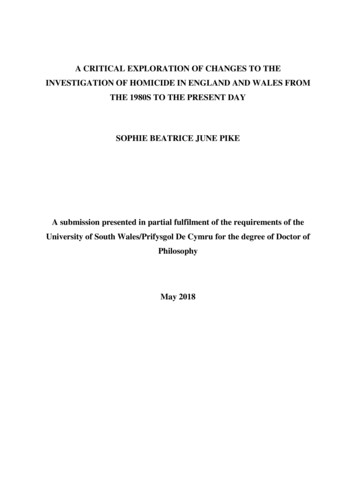
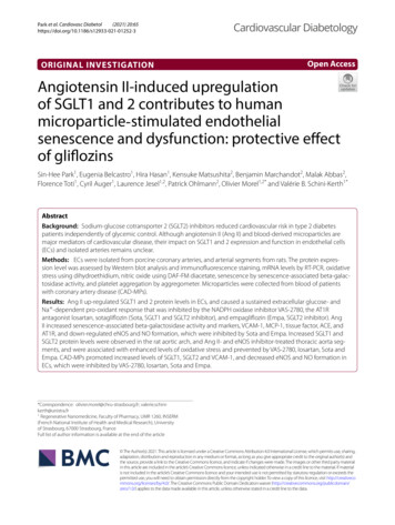
Park et al. Cardiovasc Diabetol(2021) 20:65Transfection of siRNA to ECsECs were transfected with siRNA (40 nM) targetingSGLT2 (Eurogentec; SGLT2 siRNA sense GCC UCA AUC UUU AAC AGC A , antisense UGC UGU UAA AGA UUG AGG C), negative control siRNA sense UCA CCA UGA UCU ACA CUG U, antisense ACA GUG UAG AUC AUG GUG A) for 6 h before the addition of Ang II (100nM) for 24 h. Transfections were conducted using Lipofectamine 3000 (Invitrogen) according to the manufacturer’s instructions.Determination of senescence‐associated β‑galactosidase(SA‑β‑gal) activityFluorescence-based SA-β-gal activity was determinedin ECs using 5-dodecanoylaminofluorescein di-β-Dgalactopyranoside (C12FDG), a membrane-permeablefluorogenic substrate of β-galactosidase, by flow cytometry as described previously [32]. ECs were pretreatedwith either VAS-2870 (1 µM), losartan (1 µM), sotagliflozin (100 nM), empagliflozin (100 nM), cariporide (10µM) or KB-R7943 (10 µM) for 30 min before the addition of Ang II (100 nM) for 24 h. ECs were exposed tochloroquine (300 µM), a lysosomal inhibitory drug, for1 h before the addition of C12FDG (33 µM) for 1 h. Afterwashing twice with PBS, ECs were harvested by trypsinization and centrifuged at 10,000 rpm for 10 min at 4 C followed by resuspension in ice-cold PBS. The relativeSA-β-gal activity was estimated using the MFI of the population determined by BD FACSCelesta flow cytometer.Total RNA was isolated from ECs using NucleoSpin RNA Plus kit (Machery-Nagel). RNA isolated from ECs(500 ng) was used to synthesize cDNA using the MaximaH Minus First Strand cDNA Synthesis Kit, with dsDNase (Thermo Fisher). RT-qPCR was performed withSYBR Green Master Mix (Applied Biosystems) usinga StepOnePlus Real-Time PCR System (Applied Biosystems). Primer sequences are shown in Additional file 1:Table S3. 18s, Hprt and Gusb were used as housekeepinggenes. Relative quantitation was determined by standard 2( ΔΔCT) calculations.mRNA expression by quantitative RT‑PCRCellular level of NOECs were cultured on 8-well Lab-Tek chambers andtreated with either sotagliflozin (100 nM) or empagliflozin (100 nM) for 30 min before the addition of AngII for 24 h or CAD-MPs for 48 h. ECs were exposed toDAF-FM diacetate (4-amino-5-methylamino-2’,7’-difluororescein diacetate, 1 µM), a NO-sensitive fluorescent dye, for 20 min at 37 C in the dark. The formationof NO was induced by the exposure of ECs to bradykinin(100 nM) for 15 min. After washing 3 times with PBS,Page 5 of 17cells were mounted with fluorescent mounting medium.Images were acquired using a Leica TCS SPE confocalmicroscope.Determination of platelet aggregationPlatelets isolated and suspended in Tyrode buffer at310,000 platelets/µl from healthy human blood wereobtained from the Etablissement Français du SangAlsace, Strasbourg. Suspensions of platelets (450 µl)were incubated into a cuvette with stirring at 37 C in anaggregometer (Chronolog 490, Diagnostica Stago SAS,Asnière sur Seine, France). ECs were cultured on Cytodex 3 microcarrier beads and pretreated with eithersotagliflozin (100 nM) or empagliflozin (100 nM) for 30min before the addition of Ang II for 24 h. ECs on Cytodex 3 beads (about 500 cells) were added to suspensionsof platelets for 1 min before the addition of bradykinin(100 nM) to stimulate the endothelial formation of NOfor 1 min. Thereafter, a thromboxane A 2 analog (U46619,0.3 µM) was added to induce platelet aggregation.Statistical analysisValues are expressed as means SEM. Statistical analysiswas assessed by one-way analysis of variance followed byTukey’s multiple comparison post hoc test using GraphPad Prism (Version 7). Group differences were considered statistically significant at P 0.05.ResultsAng II up‐regulates the expression of SGLT1 and 2 in ECsLow signals of SGLT1 and SGLT2 proteins were observedin control ECs (Fig. 1a). These signals increased inresponse to Ang II in a time-dependent manner reaching both about 180 % after a 24-h stimulatory period(Fig. 1a). The response to Ang II at 24 h was concentration-dependent with significant increased levels atconcentrations greater than 10 nM, and reaching about370 and 470 % at 100 nM, respectively (Fig. 1b). HigherSGLT1 and 2 fluorescence signals were also observed inECs in response to Ang II and to H2O2 (Fig. 1c). The stimulatory effect of Ang II on SGLT2 protein levels was notobserved following pre-treatment of ECs with a SGLT2siRNA or with an inactive sequence (Fig. 1d).Role of SGLT1 and 2 in the Ang II‑induced pro‑oxidantresponse in ECsSince Ang II is a potent inducer of the generation of ROSin ECs [33], the possibility that SGLT1 and 2 may contribute to the pro-oxidant response was evaluated usingdihydroethidium. Exposure of ECs to Ang II increasedthe level of ethidium fluorescence after 30 min and thestimulatory effect persisted up to 24 h (Fig. 2 and Additional file 1: Figure S1A and B). Both the short and
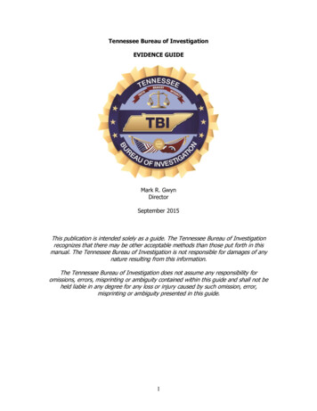
Park et al. Cardiovasc Diabetola(2021) 20:65Page 6 of 17Ang II (100 nM)Control81624hbAng II (24 )cControlH2O2 (100 M) Ang II (100 nSGLT255(kDa)*200#150100500Negative control siRNASGLT2 siRNA- - - - Ang II (100 nM, 24 h)Fig. 1 Ang II up-regulates in a time- and concentration-dependent manner the protein level of SGLT1 and SGLT2 in ECs as assessed by Westernblot (a, b) and immunofluorescence staining (c). SGLT1 and SGLT2 protein staining appears in red and nuclei are stained with DAPI (blue). Exposureof ECs to SGLT2 siRNA (40 nM) for 6 h prevented the stimulatory effect of Ang II on SGLT2 (d). Results are shown as representative immunoblotsand micrography of immunofluorescence staining (upper and left panels) and corresponding cumulative data (lower and right panels). Data areexpressed as mean SEM of n 3. *P 0.05 vs. control and #P 0.05 vs. Ang IIsustained pro-oxidant responses to Ang II were abolished by the antioxidant N-acetyl cysteine, and the AT1Rantagonist losartan (Additional file 1: Figure S1A and B).The characterization of the Ang II-triggered formationof ROS has indicated that the NADPH oxidase inhibitor, VAS-2870, the cyclooxygenase inhibitor, indomethacin and inhibitors of the mitochondrial respiratory chain(combination of myxothiazol, KCN and rotenone) all significantly inhibited both short-term and long-term prooxidant responses, indicating the involvement of severalsources including NADPH oxidase, cyclooxygenases andthe mitochondrial respiratory chain (Additional file 1:Figure S1A and B).Next the possibly that SGLT1 and 2 contribute to thepro-oxidant response to Ang II in ECs was determinedusing a dual SGLT1 and 2 inhibitor, sotagliflozin and aselective SGLT2 inhibitor, empagliflozin. The 24-h butnot the 30-min pro-oxidant response to Ang II was abolished by sotagliflozin and empagliflozin (Fig. 2a, b), indicating that although SGLT1 and 2 do not contribute tothe early pro-oxidant response, they have a crucial role inperpetuating the pro-oxidant response. Sotagliflozin andempagliflozin alone did not affect the low basal formationof ROS after 30 min and 24 h in ECs (Fig. 2a, b).To further characterize the role of SGLT1 and 2 in thepro-oxidant response to Ang II, the role of extracellularglucose and Na was assessed. The pro-oxidant responseof Ang II at 24 h was significantly reduced by decreasing the extracellular concentration of glucose and also byreplacing extracellular Na by N-methyl-D-glucamine,and abolished in the absence of both extracellular glucoseand Na (Fig. 2c), indicating a key role of both extracellular glucose and N a possibly subsequent to their entryvia SGLT1 and 2.In addition, exposure of ECs to increasing concentrations of glucose from 5.5 to 25 mM for 24 h induced apro-oxidant response at concentrations greater than20 mM (Fig. 2d). Moreover, the combined treatment ofECs with Ang II and high glucose resulted in an additivepro-oxidant response (Fig. 2d). A similar potentiating
Park et al. Cardiovasc DiabetolaShort-term (30 min)(2021) 20:65Sotagliflozin(100 nM)Page 7 of 17bEmpagliflozin(100 nM)Long-term (24 h)ControlControlAng II(100 nM, 30 min)Ang II(100 nM, 24 h)cSotagliflozin(100 nM)Empagliflozin(100 nM)deFig. 2 The sustained Ang II-induced formation of ROS in ECs is sensitive to a dual SGLT1 and SGLT2 inhibitor, sotagliflozin, and a selective SGLT2inhibitor, empagliflozin. ECs are incubated with either (a, b) sotagliflozin (SOTA, 100 nM) or empagliflozin (EMPA, 100 nM) for 30 min beforethe addition of Ang II for either 30 min (a) or 24 h (b). For characterization of the role of SGLT1 and 2 in the pro-oxidant response to Ang II,ECs are incubated with Ang II for 24 h before being exposed to (c) the indicated glucose concentrations for 1 h in the presence or absence ofsodium, (d) the indicated glucose concentrations, methyl α-D-glucopyranoside (AMG, a non-metabolizable glucose analogue), or mannitol,and (e) cariporide (a NHE-1 inhibitor, 10 µM), KB-R7943 (a NCX inhibitor, 10 µM), or ouabain (a NKA inhibitor, 10 nM) for 1 h, and the subsequentdetermination of dihydroethidium staining by confocal microscope. Results are shown as representative micrography of dihydroethidium staining(upper panels) and corresponding cumulative data (lower panels). Data are expressed as mean SEM of n 3. *P 0.05 vs. control and #P 0.05 vs.Ang IIeffect of the pro-oxidant response to Ang II was alsoobserved in response to a non-metabolizable glucoseanalogue, AMG, which has been shown to enter cellsvia SGLTs (Fig. 2d) [34, 35]. In contrast, no such effectwas observed with mannitol ruling out an osmotic effect(Fig. 2d). Thus, Ang II and high glucose act togetherto promote an excessive level of oxidative stress that isindependent of glucose metabolism. Moreover, the sustained pro-oxidant response to Ang II was markedlyinhibited by cariporide (Na /H exchanger-1 inhibitor)and KB-R7943 ( Na /Ca2 exchanger inhibitor), but notaffected by ouabain (Na /K -ATPase inhibitor) suggesting the involvement of NHE-1 and NCX, besides SGLT1and 2 (Fig. 2e).
Park et al. Cardiovasc Diabetol(2021) 20:65Page 8 of 17SGLT1 and 2 act in a feedforward manner to sustainthe stimulat
ORIGINAL INVESTIGATION AngiotensinII-inducedupregulation ofSGLT1and2contributestohuman microparticle‐stimulatedendothelial senescenceanddysfunction:protectiveeect ofgliozins Sin‑Hee Park1,Eugenia Belcastro 1,Hira Hasan 1,Kensuke Matsushita 2,Benjamin Marchandot 2,Malak Abbas 2,