Transcription
Digest Journal of Nanomaterials and BiostructuresVol. 16, No. 1, January - March 2021, p. 41 - 49SYNTHESIS OF TIN OXIDE NANOPARTICLES IN ORDERTO STUDY ITS PROPERTIESN. SHAHZADa, N. ALIb*, A. SHAHIDb, S. KHANb, H. ALROBEIc*aNational University of Sciences and Technology (NUST), PakistanbDepartment of Physics, GPG Jahanzeb College Saidu Sharif, Swat, PakistancDepartment of Mechanical Engineering, College of Engineering, Prince Sattam binAbdullaziz University, AlKharj, Saudi ArabiaPure tin oxide nanoparticles ware synthesized via Co-precipitation method. The assynthesized nanoparticles were characterized by Fourier transform infrared spectroscopy(FTIR), thermo-gravimetric analysis (TGA), X-ray diffraction (XRD) and diffusereflectance spectroscopy (DRS). X-ray diffraction shows that tetrahedral shapednanoparticles of crystallite size 47.35nm were prepared successfully. The crystallinity wasestablished due the reduction in distortion ratio and dislocation density. The value of strainshowed that the nanoparticles fabricated were of high stability. The direct and indirectoptical band gap of as-synthesized SnO2 nanoparticles were determined from thereflectance spectra, which were 3eV and 2.65eV respectively.(Received September 10, 2020; Accepted January 8, 2021)Keywords: Tin Oxides, nanoparticles, XRD, DRS, Band gap1. IntroductionNanoscience is the study of matter’s properties at a nanoscale. It is based on the specialsize dependent properties of solid state materials [1]. Nanoscience deals with objects of sizehaving the smallest dimension ranging from a few nanometers to less than 100 nanometers. In thefield of physics and electrical engineering nanoscience is most often related with quantumbehavior, the behavior of electrons and photons in nanoparticles [2]. The word “nanotechnology”was the first time used by Norio Taniguchi and defines nanotechnology as ““Nano-technology”mainly consists of the processing of separation, consolidation, and deformation of materials by oneatom or one molecule”[1]. Nanotechnology is the technology which works on level of nanometer(nm), one billionth of a meter. In the nano region the elements change properties unpredictably. Itis generally related to mechanical engineering on a molecular scale, but it is a confusing term. It issometimes also related to today’s applied nanotechnology because of its numerous uses incosmetics or industrial coating [3]. The term nanomaterial stands for the nano sized structures inwhich at least one of its phases has one or more dimensions, and they include nanometer sizedparticles ranging from 1nm to 100nm [4]. The properties of nanomaterial change due to its size,shape, and morphology. The structure of nanomaterials depends on the method of preparation,nature of solvent, concentration, strength of reducing agent, and temperature [5]. Thenanomaterials are classified as zero dimensional (0-D), one dimensional (1-D), two dimensional(2-D), and three dimensional (3-D).Nanomaterials have wide ranging applications in many areas of studies. Some of them arenarrated below:a.Nanomaterials are used for the elimination of dense metallic ions, and dyes fromwaste water organic, and inorganic pollutants [6].b.Nanomaterials are used in agriculture, and crop production [7].*Corresponding author: h.alrobei@psau.edu.sa
42c.Nanomaterials are used to improve molecular diagnostics such as gene detection,sensitivity, and practicality [8].d.They are used to improve the properties of transistors [9].e.Some nanomaterials such as solid lipid nanoparticles are used for skin hydration,and also have the ability to block UV-Rays [10].f.Nano catalysts are used in the chemical, and pharmaceutical industries forenhancing the efficiency of energy in connection with green technology with the production ofvery less amount of chemical waste production.g.Mesoporous silica shell nanospheres are widely used in hydrogenation catalysis,and magnetic acid catalysis[11]. They are also used for the enhancement of chemical reactions,and separation processes, and for simplification of the processes.h.Metallic nanoparticles, quantum dots, and Nano bubbles have also been widelyused in catalysis [12].i.Consolidated nanocomposites, and nanostructures are used for making very highstrength material with tough structure, tough pliable cements, and interestingly new magnets[13].j.Nanostructured electrodes can enhance the efficiency, and capability of Li-ionbatteries[14].k.Nanotubes can be used as electro chemical actuators because of its supercapacitance [15].Amongst various nanomaterials being researched and used world over, Tin nanoparticlesare also gaining attention of different research studies owing to its wide ranging applications indifferent fields. The research on Tin nanoparticles is extending with passage of time and with therapidly changing advancements in different fields including, construction industry. Tinnanoparticles have a very broad and wide research area. The main reason is that Tin nanoparticlesmay be easily modified by changing their chemical properties.Out of the many advantages of Tin nanoparticles, one key advantage is that theirproperties vary from the majority of the properties of the same composition. Tin nanoparticles maybe coated onto other metals for corrosion prevention. Tin nanomaterials are widely used for manypurposes, such as, enhancing the activities of super capacitors by providing high energy density tothe capacitor [16], enhancing the gas sensing ability due to high sensitivity, response, and lowpriced [17], enhancing the electrochemical properties of other materials such as improving thestorage capacity of a battery [18], enhancement of photocatalytic activities of the materials owingto their good optical transparency, good intrinsic vacancies, and high density [19], acting as chargetransport layer to increase the charge transformation, and for reducing hysteresis [20], cosmeticsmaterials for protecting skin from UV-rays, and other rays effecting the skin [21].Tin oxide (SnO2) nanoparticles have been prepared by many methods by many researchersin recent years. These researchers use different types of precursors, and reagents for their work.Tin oxides prepared by these methods have many applications in different fields.Zhu and co-authors (2000) used sonochemical method for synthesizing SnO2nanoparticles. They used SnCl4 aqueous solution, and azodicarbonamide under ambient air in theirwork, and obtained Tin oxide nanoparticles of the size 3 to 5nm. The particles were spherical inshape, and had band gap of 4.05ev which is greater than the value of band gap (3.7ev) of SnO 2bulk material. The prepared Tin oxide was used as in lithium ion batteries as reversible insertionelectrode for enhancing the performance of battery, and increasing battery life [22].Song and Kang (2000) used homogenous precipitation method for the preparation of SnO2powders. They used tin chloride pentahydrate, and urea in their work. The particles prepared werein powder form, and had large surface area. The particles prepared had size ranging from 3 to 5nm.Because of the large surface area of the SnO2 powders prepared by this method, it was used in gassensors materials, and for enhancing the sensing properties of devices [23].Qui and co-authors (2004) prepared tin nanoparticles using polyol process bysonochemical method. They used tin chloride and ethylene glycol for their work. The particlesprepared were rod shaped having size in range of 50 to 100nm, and length in range of 1 to 3µm.the prepared Tin oxide nanoparticles were used in lithium ion batteries as electrode material andfor enhancing the performance of batteries [24].
43Chiu and Yeh (2007) used hydrothermal method for the preparation of SnO2 nanoparticles.They used tin chloride 2-propanol, and distilled water as solvent for their work. The preparednanoparticles were tetragonal shaped having size of 3nm. The band gap of the particles was 3.6eV,and were used as sensing materials [25].Bagheri-Mohgheghi and co-authors (2008) prepared SnO2 nanoparticles using sol gelmethod. They used hydro-alcoholic solution, ethylene glycol as polymerizing agent, and tinchloride, and citric acid as complexing agent. The particles prepared were tetragonal shaped ofsize ranging from 5 to 25nm with a greater band gap from 4.05 to 4.11eV. The preparednanoparticles were used for the agglomeration of particles [26].Liu and co-authors (2009) used gel combustion method for the preparation of SnO2nanoparticles. They used Tin Chloride, and polyacrylamide gel in their work. SnO2 nanoparticlesprepared were sphere shaped, and had size in range of 8.1 to 27.9nm. The nanoparticles obtainedby this process were used for the detection purposes of gases [27].Krishnakumar and co-authors (2009) used microwave technique for the synthesis of SnO2nanoparticles. They used tin hydroxyl solution with hydrazine hydrate in a precursor solution forobtaining Tin Oxide nanoparticles. The prepared SnO2 nanoparticles were having spherical shape,and size in range of 10 to 11nm [3].Xu and co-authors (2010) also used the homogenous precipitation method for thepreparation of SnO2 nanoparticles. They used tin chloride, urea (carbamide), and aluminum oxiderod for their work. Tetragonal shaped SnO2 nanosheets of size 10nm were prepared. The particlesprepared indicated enhancement in high performance of sensing devices [28].Gondal and co-authors (2010) prepared tin oxide nanoparticles using highly powerful laserbeams. They used tin metal as target material placed in distilled water, and highly powerful laserin their work. The tin oxide nanoparticles prepared were tetragonal shaped of size 3nm, and a bandgap of 4.4eV [29].Parthibavarman and co-authors (2011) prepared SnO2 nanoparticles using microwaveirradiation method. They used tin chloride and NH3.H2O in their work. The prepared SnO2nanoparticles were spherical shaped with size in range of 20 to 25nm with a band gap of 3.7eV.The acquired nanoparticles were used for highly susceptible environmental controlling and inelectronic devices for controlling humidity[30] .Patil and co-authors (2012) also used hydrothermal method for the synthesis of SnO2nanoparticles. They used hydrazine hydrate as a mediator for this process. The obtained SnO 2nanoparticles were of tetragonal shape. The obtained particles were having size 22.4nm, and aband gap of 3.6ev. SnO2 prepared by this method can be used as gas detectors in many gasdetecting devices [31].Honghao and co-authors (2013) prepared SnO2 nanopowder through gas phase detonationreaction. They used stannic chloride as precursor, and mixture of hydrogen, and oxygen gas. TheSnO2 nanopowders prepared were having size of 1 to 10nm, and have spherical morphology.Further details were not discussed as gas phase reaction was not announced by this method.Although, prepared SnO2 nanopowders were having high sensitivity [32].Naje and co-authors (2013) used chemical precipitation method for the preparation ofSnO2 nanoparticles. They used tin chloride and ammonia solution in their work. The preparedSnO2 nanoparticles were in powder form having tetragonal shape, and size was 8 to 10nm with aband gap of 4.3eV higher than that of the band gap of bulk SnO2 [33].Nayef and Hadi (2014) used laser ablation technique for the synthesis of SnO2nanoparticles. They used tin, and methanol for this process. The prepared particles were sphericalshaped, and their size varied according to different experimental conditions. The band gap of theparticles was greater than that of bulk SnO2, and was observed at different laser concentration inrange of 4.16 to 4.26eV [34].Srivastava and Mukhopadhyay (2014) used biological method for the synthesis of SnO2nanoparticles. They used Erwinia herbicola (Gram-negative bacteria), and tin chloride for theirwork. The SnO2 nanoparticles prepared were tetragonal shaped having size in range of 15 to 40nm.The prepared SnO2 nanoparticles showed good photocatalytic behavior, and were used for thephotodegradation of many organic dyes [35].
44Elango and co-authors (2015) used green synthesis for the preparation of SnO2nanoparticles by green synthesis technique. They used plant’s (Persia Americana) seed extract, andstannous chloride as precursor for their work. The SnO2 nanoparticles they obtained weretetragonal shaped with size in range of 4nm, which were used for the degradation of dyes [36].Bhagwat and co-authors (2015) also used sol-gel method for preparing SnO2 nanoparticles.They used tin chloride, ethylene glycol, and liquid ammonia as precursors, and double distilledwater as solvent. The prepared particles were having size in range of 350 to 800nm, and a bandgap of 3.78ev [37].Azade Esmaeili-Bafghi-Karimabad and co-authors (2015) prepared SnO2 nanoparticlesusing simple chemical reaction method. They used tin chloride, and dimethylamine as cappingagent. The particles prepared were tetragonal shaped having size 40nm with a greater band gap of3.82eV. These prepared particles were used for enhancing the thermal stability and reducing theflammability of materials [38].Nagirnyak and co-authors (2016) prepared SnO2 nanoparticles by using chemicaldeposition method. They used tin chloride, and ammonium oxalate as precursors, and preparedtwo samples of SnO2 nanoparticles of size in range of 40 to 200nm, and hexagonal shape. Theband gap was 3.69eV for one sample, and 3.78eV for other. The prepared materials were used assensing materials, and for enhancing the properties of gas sensors [39].Kurniawan and Rahmi (2017) prepared SnO2 nanoparticles using electrochemical method(electrolysis) at a large potential. They used tin bare as electrodes (both anode, and cathode), andhydrochloric acid (HCl) as the electrolytic solution. The particles produced by this method showedtetragonal shape, and size in range of 25 to 150nm [40].Oliveira and co-authors (2018) prepared SnO2 nanoparticles using solvothermal methodusing indium chloride tetrahydrate, and tin chloride pentahydrate as precursors, and ethyleneglycol as solvent. Spherical shaped particles of size in range of 3.4 to 8.2nm with a band gap of 3to 3.4ev were obtained [41].Kim and co-authors (2018) used the liquid phase plasma method for the preparation ofSnO2 nanoparticles. They used tin chloride as a precursor, cetyl trimethyl ammonium bromide, andwater as solvent. The SnO2 nanoparticles were of spherical shape with size in range of 5 to 10nm.These particles were used for enhancing the storing capability, and life time of lithium ionbatteries [42].Diaz and co-authors (2018) prepared SnO2 nanoparticles by using two step solid statesynthesis method. SnCl2.Chitson at different ratio of 1:1, 1:5, and 1:10 with dichloromethane(CH2Cl2) as solvent were used for this process. Rod like nanostructures of SnO 2 of size 2.2 0.2nm using precursors at 1:5. They used the SnO2 prepared by this method in optoelectronicdevices, and solid state sensor devices for enhancing the mechanical properties of these materials,and upgrading their performance [43].2. Materials and methodsAll analytical grade reagents were used. The solutions were prepared in distilled water(E.C. 2.8µS/cm) which was obtained from the distillation apparatus (Model WSB/4, HamiltonLaboratory Glass Ltd.). Tin chloride dihydrate (SnO2.2H2O) (Merck) was used as a precursor.Ethanol (Merck) was used as a solvent.2.1. Synthesis of tin nanoparticlesCo-precipitation method was used for Tin oxide nanoparticles synthesis. Two solutionswere mixed forming a precipitate, which was then washed, annealed to obtain the desired product.Two approaches using ethanol as solvent, and using ethanol and water as solvent, were adoptedusing the same method, to prepare two samples of SnO2 nanoparticles. Detailed procedure is asunder:a.1.12825g of tin chloride, and 50ml ethanol were taken in a beaker, and mixed withthe help of a magnetic stirrer for about 10 to 15minutes to obtain a complete soluble, andtransparent solution.
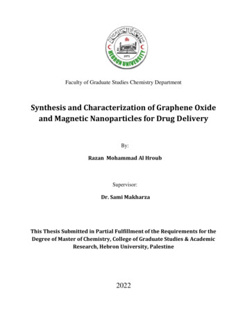
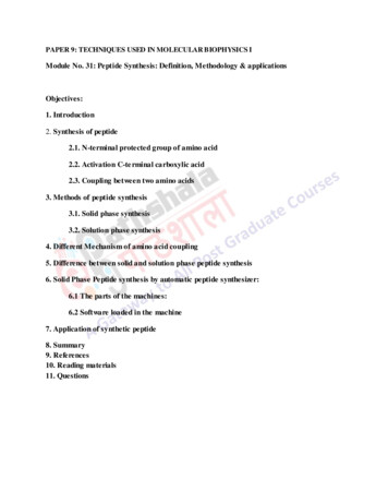
45b.0.4g of sodium hydroxide was dissolved in 100ml of deionized water with thehelp of a magnetic stirrer, and sodium hydroxide solution was obtained (0.1M solution)c.The sodium hydroxide solution was dropwise added to the prepared tin chloridesolution at constant stirring as precipitating agent, and controlling pH of the solution. With theaddition of sodium hydroxide solution, precipitate was formed in the solution, and the pH valuereached 8 with the addition of 109ml of sodium hydroxide solutiond.As the pH of the solution was maintained, further stirring was stopped, and theprecipitate was allowed to settle down by keeping the solution still for 12hrse.After 12hrs the precipitate got concentrated at the bottom, and the ethanol abovethe precipitate was decanted.f.Ethanol was added to the precipitate for washing, and then again kept for 12hrs tosettle down. After 12hrs the ethanol was again removed. This process was repeated three times,and at last a pure precipitate was obtained.g.The precipitation was dried on heating at 90 C for 12 hours.h.After heating, a solid matter was obtained which was grinded into powder andannealed at 800 C for 3 hours.i.After annealing we obtained our desired product which was further investigatedby different characterization techniques.3. Results and discussions3.1. FTIRIn the FTIR spectroscopy graph the band from the 3394 to 3409 cm-1shows the strechingvibration of O-H bond .This band is due to the OH groups and the adsorbed water bound at theSnO2 surface. The band at 1620 to 1630 cm-1 matched to the bending vibration of water molecules,caught in the SnO2 sample. The peak at 521cm-1 matching with the stretching vibrations of theterminal Sn-OH and at the peak 660 to 600cm-1 region shows the stretching modes of the Sn-O-Sn.Due to increase in temperature the peak is also broadened (Fig. 1).Fig. 1. FTIR analysis of SnO2 nanoparticles.3.2. TGA (thermogravemetric analysis)The TGA analysis was carried out and it was observed that at 50 to 120 degrees increasein weight occurred because of oxidation of Sn 2, because of this oxidation the temperature of thefurnace increased and the TGA curve was distorted. The constant weight condition was reachednear 300 C after which the weight goes on decreasing slowly as the temperature increased. At425 to 600 the weight loss further increased because of evaporation of OH- and water moleculesand above 600 the weight loss suddenly increased as shown in the figure.
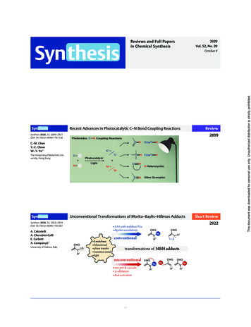
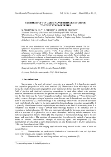
46Fig. 2. TGA OF SnO2.3.3. XRDThe XRD pattern of SnO2 nanoparticles synthesized by co-precipitation method is givenbelow. All of the peaks were matched with diffraction data of the tetragonal structure of tin oxide(JCPDS 01-077-0447 ). Position of the main peak is 2θ 26.68 and the width of the peak is 0.933nm.The average crystallite size (D) of the nanoparticles was estimated using the DebyeScherrer formula as follows:D (i)where, λ, β, θ are the X-ray wavelength of radiation used. The averge crystallite size (D)determined from SnO2 different plane by the Scherrer formula is about 47.35 nm and tetrahedral inshaped.The average dislocaton density of SnO2 particles is determined to be 7.28 1014/m2 by theequation.Dislocation density 1/ D2(ii)The average strain was determined to be 0.206 by the equation.Strain β/4 tan θFig. 3. XRD analysis of SnO2.(iii)
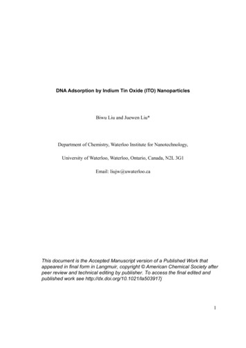
47Table 1. The detailed analysis of SnO2 by 39113.3441 16.9804 19.0016 21.3216 25.9451 27.4194 28.9773 30.9829 32.4046 33.0695 26.314 33.541 37.737 42.516 51.374 54.521 57.668 61.631 64.312 65.827 27.247 34.473 38.436 42.749 52.540 55.104 58.367 62.564 65.244 66.526 60.3495InterplanarSpacing d(A (220)(002)(310)(112)Average particle size 47.35nmStructure TetrahedralJCPDS Card # 01-077-0447Average strain 0.206Dislocation density 1 / D2Average Dislocation density 7.28 1014/m3.4. Optical propertiesThe optical properties were studied by using diffuse reflectance spectroscopy (DRS). Thereflectance spectra of the pure SnO2 nanoparticles are as shown in the figure 4. From the spectra,it is observed that he quantum confinement effect increases with increase of reflectance. Thedecrease in reflectance near the 300nm is due to the optical transition happening in the opticalbandgap. The synthesized SnO2 showed no reflection up to 290nm and washighly reflective abovethe region of 320 nm.The optical bandgap was determined by kubelka-munk relation as shown below, which iscommonly used to study diffuse reflectance spectroscopy.F (R) (iv)where F(R) is the kubelka-munk function which relates to the absorbance, R is the reflectance, k isthe absorbance co-efficient and S is the scattering co-efficient. The optical transition insemiconductors are of two types, the direct and indirect transitions. Therefore direct and indirectbandgap determined by this relation as shown the figure 5 are 3eV and 2.65eV respectively.
48Fig. 4. Reflectance spectra of SnO2.Fig. 5. Bandgap of SnO2 from reflectance spectra.4. ConclusionsSnO2 nanoparticles have been effectively prepared by Co-precipitation method using tinchloride (SnCl) and sodium hydroxide (NaOH) at 800 C. The Fourier transform spectroscopy(FTIR) illustrates that chlorine impurity was entirely removed by washing process. The TGAanalysis showed that sudden mass change occurred at 630 C, showing the breakdown of tin oxide.The structural and optical properties were examined by X-Ray diffraction (XRD) and diffusereflectance spectrum (DRS). X-Ray diffraction (XRD) analysis showed that samples were wellcrystalline and tetrahedral in shape having average crystallite size of 47.35nm. The direct andindirect band gap was found to be 3eV and 2.65eV respectively.AcknowledgmentThis project was supported by the Deanship of Scientific Research at PrinceSattam bin Abdul Aziz University under the research project no. 2020/1/17063. Theauthors are also thankful to NUST Research Directorate for financial support.References[1] P. Mulvaney, P., ACS Nano 9(3), 2215 (2015).[2] G. M. Whitesides, Small 1(2), 172 (2005).[3] T. Krishnakumar et al., Materials Letters 63(11), 896 (2009).[4] J. Tian et al., Journal of Chromatography A 1300, 2 (2013).
49[5] K. S. Siddiqi, A. Husen, R. A. Rao, Journal of nanobiotechnology 16(1), 14 (2018).[6 H. Sadegh et al., Journal of Nanostructure in Chemistry 7(1), 1 (2017).[7] L. R. Khot et al., Crop protection 35, 64 (2012).[8] X. Zhu et al., Biosensors and Bioelectronics 74, 113 (2015).[9] A. D. Franklin, Science 349, 6249 (2015).[10] S. A. Wissing, R. H. Müller, International Journal of Pharmaceutics 254(1), 65 (2003).[11] A. M. Abu-Dief, S. M. Abdel-Fatah, Beni-Suef University Journal of Basic and AppliedSciences 7(1), 55 (2018).[12] N. Sharma et al., Rsc Advances 5(66), 53381 (2015).[13] R. Siegel, B. Kear. Applications: Consolidated Nanostructures. in Nanotechnology ResearchDirections: IWGN Workshop Report. 2000. Springer.[14] N. Mahmood, Y. Hou, Advanced Science 1(1), 1400012 (2014).[15] Y. Gogotsi, Nanomaterials handbook2006: CRC press.[16] V. Velmurugan et al., Materials Research Bulletin 84, 145 (2016).[17] X. Li et al., Nanoscale research letters 13(1), 14 (2018).[18] J. Wang et al., Journal of Alloys and Compounds 748, 1013 (2018).[19] J. Mazloom, F. Ghodsi, H. Golmojdeh, Journal of Alloys and Compounds 639, 393 (2015).[20] Q. Jiang et al., Nature Energy 2(1), 1 (2016).[21] A. Morlando et al., Materials Letters 171, 289 (2016).[22] J. Zhu et al., Chemistry of Materials 12(9), 2557 (2000).[23] K. C. Song, Y. Kang, Materials Letters 42(5), 283 (2000).[24] L. Qiu et al., Ultrasonics sonochemistry 12(4), 243 (2005).[25] H.-C. Chiu, C.-S. Yeh, The Journal of Physical Chemistry C 111(20), 7256 (2007).[26] M. M. Bagheri-Mohagheghi et al., Physica B: Condensed Matter 403(13), 2431 (2008).[27] H. Liu et al., Ceramics International 35(3), 961 (2009).[28] M.-H. Xu et al., Sensors and Actuators B: Chemical 145(2), 875 (2010).[29] M. A. Gondal, Q. A. Drmosh, T. A. Saleh, Applied Surface Science 256(23), 7067 (2010).[30] M. Parthibavarman, V. Hariharan, C. Sekar, Materials Science and Engineering: C 31(5),840 (2011).[31] G. E. Patil et al., International Nano Letters 2(1), 17 (2012).[32] Y. Honghao et al., Rare Metal Materials and Engineering 42(7), 1325 (2013).[33] A. N. Naje, A. S. Norry, A. M. Suhail, International Journal of Innovative Research inScience, Engineering and Technology 2, 7068 (2013).[34] U. M. Nayef, A. J. Hadi, Engineering and Technology Journal 32(6 Part (B) Scientific),1059 (2014).[35] N. Srivastava, M. Mukhopadhyay, Industrial & Engineering Chemistry Research 53(36),13971 (2014).[36] G. Elango et al., Spectrochimica Acta Part A: Molecular and Biomolecular Spectroscopy 145,176 (2015).[37] A. D. Bhagwat et al. Synthesis of Nanostructured Tin Oxide (SnO2) Powders and Thin Filmsby Sol-Gel Method, 2015.[38] A. Esmaeili-Bafghi-Karimabad et al., Journal of Materials Science: Materials in Electronics26(9), 6970 (2015).[39] S. V. Nagirnyak et al., Nanoscale research letters 11(1), 343 (2016).[40] F. Kurniawan, R. Rahmi, Synthesis of SnO2 Nanoparticles by High Potential Electrolysis2017, 6 (2017).[41] F. M. B. D. Oliveira, J. A. Varela, M. O. Orlandi, Materials Research 21(2), 2018.[42] S.-C. Kim et al., Korean Journal of Chemical Engineering 35(3), 750 (2018).[43] C. Diaz et al., Journal of Cluster Science 29(2), 251 (2018).
Naje and co-authors (2013) used chemical precipitation method for the preparation of SnO 2 nanoparticles. They used tin chloride and ammonia solution in their work. The prepared SnO 2 nanoparticles were in powder form having tetragonal shape, and size was 8 to 10nm with a band gap of 4.3eV higher than that of the band gap of bulk SnO 2 [33].