Transcription
St-Amand et al. Lipids in Health and 2020) 19:192RESEARCHOpen AccessTwo weeks of western diet disrupts livermolecular markers of cholesterolmetabolism in ratsRoxane St-Amand1, Émilienne T. Ngo Sock1, Samantha Quinn2, Jean-Marc Lavoie1 and David H. St-Pierre2*AbstractBackground: The present study was designed to test the hypothesis that in the liver, excessive fat accumulationimpairs cholesterol metabolism mainly by altering the low-density lipoprotein-receptor (LDL-R) pathway.Method: Young male Wistar rats were fed standard (SD), high fat (HFD; 60% kcal) or Western (WD; 40% fat 35%sucrose (17.5% fructose)) diets for 2 or 6 weeks.Results: Weight gain ( 40 g) was observed only following 6 weeks of the obesogenic diets (P 0.01). Compared tothe 2-week treatment, obesogenic diets tripled fat pad weight ( 20 vs 7 g) after 6 weeks. Hepatic triglyceride (TG)levels were greater in response to both the WD and HFD compared to the SD (P 0.01) at 2 and 6 weeks and theirconcentrations were greater (P 0.05) in WD than HFD at 2 weeks. Plasma total cholesterol levels were higher (P 0.05) in animals submitted to WD. After 2 and 6 weeks, liver expression of LDL-R, proprotein convertase subtilisin/kexin 9 (PCSKk9) and sterol regulatory element binding protein 2 (SREBP2), involved in LDL-cholesterol uptake, waslower in animals submitted to WD than in others treated with HFD or SD (P 0.01). Similarly, low-densitylipoprotein-receptor-related protein 1 (LRP1) and acyl-CoA cholesterol acyltransferase-2 (ACAT-2) mRNA levels werelower (P 0.01) among WD compared to SD-fed rats. Expression of the gene coding the main regulator ofendogenous cholesterol synthesis, 3-hydroxy-3-methyl-glutaryl-CoA reductase (HMGCoAR) was reduced in responseto WD compared to SD and HFD at 2 (P 0.001) and 6 (P 0.05) weeks. Being enriched in fructose, the WDstrongly promoted the expression of carbohydrate-response element binding protein (ChREBP) and acetyl-CoAcarboxylase (ACC), two key regulators of de novo lipogenesis.Conclusion: These results show that the WD promptly increased TG levels in the liver by potentiating fat storage.This impaired the pathway of hepatic cholesterol uptake via the LDL-R axis, promoting a rapid increase in plasmatotal cholesterol levels. These results indicate that liver fat content is a factor involved in the regulation of plasmacholesterol.Keywords: Western diet, Plasma cholesterol, Lipogenesis, Hepatic steatosis, Liver cholesterol metabolism, Lowdensity lipoprotein-receptor, Proprotein convertase subtilisin/kexin 9* Correspondence: david.stpierre.uqam@gmail.com2Department of Exercise Sciences, Université du Québec à Montréal, 141,Avenue Président-Kennedy, C.P. 8888, succursale Centre-Ville, Montréal,Québec H3C 3P8, CanadaFull list of author information is available at the end of the article The Author(s). 2020 Open Access This article is licensed under a Creative Commons Attribution 4.0 International License,which permits use, sharing, adaptation, distribution and reproduction in any medium or format, as long as you giveappropriate credit to the original author(s) and the source, provide a link to the Creative Commons licence, and indicate ifchanges were made. The images or other third party material in this article are included in the article's Creative Commonslicence, unless indicated otherwise in a credit line to the material. If material is not included in the article's Creative Commonslicence and your intended use is not permitted by statutory regulation or exceeds the permitted use, you will need to obtainpermission directly from the copyright holder. To view a copy of this licence, visit http://creativecommons.org/licenses/by/4.0/.The Creative Commons Public Domain Dedication waiver ) applies to thedata made available in this article, unless otherwise stated in a credit line to the data.
St-Amand et al. Lipids in Health and Disease(2020) 19:192IntroductionNon-alcoholic fatty liver disease (NAFLD), also referredto as hepatic steatosis is characterized by excessive fataccumulation (5 to 10% of liver weight) in absence ofsignificant alcohol consumption [1]. This state is associated with several clinical and pathological manifestations. NAFLD is considered a risk factor for diabetesand cardiovascular diseases (CVD) independently ofother usual risk factors [2, 3]. Hepatic steatosis by itselfis also associated with a proatherogenic lipid profile andproduction of proinflammatory markers [4, 5]. Some authors [6] have even claimed that with regards to body fataccumulation, hepatocytes are likely the first cell typesto display metabolic impairments.The association between liver fat accumulation, CVD,and a proatherogenic lipid profile is well substantiated inthe literature. However, the critical impact of hepaticcholesterol metabolism linking these health conditionsremains largely overlooked. The liver is the master regulator of several biological pathways involved in the control of cholesterol metabolism. These include cholesterolsynthesis, uptake from chylomicron remnants, re-uptakefrom LDL and high-density lipoproteins (HDL) as wellas the release of cholesterol under very-low density lipoproteins (VLDL), and biliary acids production. All ofthese pathways have a major influence on the regulationof plasma cholesterol levels and the subsequentproatherogenic profile associated with fat accumulationin the liver.The association between hepatic steatosis and cholesterol metabolism is most often viewed as how cholesterol metabolism dysregulation contributes to NAFLD inanimal models [7]. However, it was also proposed thatNAFLD impairs cholesterol metabolism in humans [8].Long-term studies do not provide any clear indicationthat fat accumulation in the liver is at the origin of alterations in cholesterol metabolism. On the other hand,several molecular markers of cholesterol metabolism inthe liver were disturbed by a short-term exposure (2weeks) to HFD in young rats [9]. These results supportthe concept that excessive accumulation of fat in theliver may contribute to increased circulating cholesterollevels [9]. A short-term 2-week HFD was used in thatstudy because it has been shown to result in a rapid fataccumulation in the liver. This excessive accumulationof fat in the liver was previously shown to precede boththe occurrence of significant changes in weight gain andthe development of systemic metabolic complicationsclassically observed in response to longer exposure toobesogenic diets [10, 11].The notion that disturbances in lipid metabolism inthe liver play a crucial role in the etiology of plasmahypercholesterolemia is supported by the knowledge thatTG and cholesterol metabolic pathways are closelyPage 2 of 11interrelated in this organ. Cholesterol and fatty acid synthesis both use acetyl-CoA as biochemical starting material, and the reducing equivalent NADPH as a sourceof energy for their synthesis. Therefore, there is potentialcompetition between fatty acid synthesis stored as TGand cholesterol synthesis in the liver. This raises the possibility that de novo lipogenesis resulting in liver fat accumulation may impair hepatic cholesterol metabolism.This hypothesis was recently supported by Ichigo andcolleagues [12] who reported that in comparison to ahigh glucose (65%) diet, exposure to a high fructose(65%) diet for 12 days increased ACAT-2 (enzyme acylCoA acyl transferase 2: involved in cholesterol esterification) while decreasing ATP-binding cassette protein G5(ABCG5) and ABCG8 (dimeric proteins involved in theexportation of cholesterol from the liver to bile ducts)gene expression in the liver. The present study testedthe hypothesis that a short-term exposure (2 weeks) totwo obesogenic diets; a HFD known to rapidly increasefat accumulation in the liver and a WD (40% fat and35% sucrose (17.5% fructose)) known to stimulate denovo lipogenesis disrupts molecular pathways of cholesterol metabolism in the liver. Among the different pathways of cholesterol metabolism, the important receptorLDL-R involved in cholesterol uptake of LDL particles inplasma was investigated along with its posttranscriptional regulator PCSK9 and their transcription factorSREBP2.Material and methodsAnimals care and treatmentsFour-week old male Wistar rats (Charles River, StConstant, Québec, Canada) weighing 190 to 200 g, werehoused individually and had ad libitum access to foodand tap water after they were received at the animal facility. Their environment was controlled in terms of light(12 h light-dark cycling starting at 06:00 AM), humidity,and room temperature (20–23 C). After a 3-day acclimatisation period, rats were randomly submitted eitherto a SD chow diet, a 60% HFD, or a WD (40% fat-35%sucrose) diet for 2 or 6 weeks for a total of 6 experimental groups (n 10/group). Food intake and body weightwere measured every 2 and 3 days, respectively. Theprotocol was approved by the Comité Institutionnel deProtection des Animaux (CIPA; 1218–956-1219) ofUQAM and developed in agreement with the CanadianCouncil on Animal Care’s rules (CCAC-CCPA).At the end of their respective nutritional treatments,rats were euthanized between 09:00 and 11:00 AM aftera 3-h fast. Immediately after complete anaesthesia by inhalation of isoflurane (3% induction and 1.5% maintenance) blood samples were drawn from the heart andcollected in tubes containing 0.5 ml EDTA. The bloodwas centrifuged (1500 RPM for 10 min), and the plasma
St-Amand et al. Lipids in Health and Disease(2020) 19:192was collected to measure TG, cholesterol, and PCSK9.The abdominal cavity was opened, and the median lobeof the liver was freeze-clamped, removed and used forTG, cholesterol and gene expression of several keymarkers of cholesterol metabolism. The mesenteric,retroperitoneal, epididymal, and subcutaneous (sc) fatdeposits were, thereafter, rapidly excised and weighed.The mesenteric fat pad consisted of adipose tissue surrounding the gastrointestinal tract from the gastroesophageal sphincter to the end of the rectum with specialcare taken in distinguishing and removing pancreaticcells. The epididymal fat pad included adipose tissuesurrounding the ureters and bladder. The retroperitonealfat pad represented the distinct deposit behind each kidney, along the lumbar muscles. For sc fat deposit measurement, a rectangular piece of skin was taken on theright side of each animal from the median line of the abdomen to the spine and the right hip to the first rib [13].All tissue and plasma samples were stored at 78 Cuntil analyses.DietsDiets were selected to compare short- and mid-term effects of a standard rodent diet (SD) with two types ofobesogenic diets enriched in: lipids (HFD) or fat and sucrose (WD). Fat and macronutrient contents were notmatched for the selected diets. The energy content (%kcal) of the SD [14] was 13% lipids, 65.6% carbohydratesand 21.4% proteins (Charles River Rodent Diet 5075,Cargill Animal Nutrition, MN, USA). It had a gross energy value of 3970 kcal/kg and a physiological fuel valueof 2885 kcal/kg. Its macronutrient composition (g/100 g)was 55.2% carbohydrates (38% glucose derived fromcorn starch:100% glucose); 13% neutral detergent fiberand 4.1% acid detergent fiber derived from wheat middlings and wheat); 4.5% lipids (poultry fat) and 18% proteins (dehulled soybean meal, fish meal and whey). TheHFD and the WD were obtained from Research Diets(NJ, USA). The HFD consisted of 60% fat, 20% CHOand 20% protein (kcal) while the WD consisted of 40%lipid, 43% CHO (35% sucrose), and 17% protein (kcal).The caloric equivalent of 2.89, 5.21 and 4.67 kcal/g forthe SD, HFD, and WD, respectively was used to reportfood intake in kcal. Details of the HFD and WD are presented in Table 1.Biochemical analysesHepatic TG and cholesterol levels were determinedusing approximately 100 mg of tissue. Overnight 2:1chloroform/methanol solution was used to extract lipids.Following centrifugation, nitrogen flux was used to driedup the lower phase and resuspended with Floch andThesit solution (20%). Quantification of total cholesteroland TG from the liver and plasma was performed usingPage 3 of 11Table 1 Description of the dietsIngredients (g)High Fat dietD12492Western dietD12079BCasein, Lactic, 30 Mesh200195Methionine, DL03Cystine, L30Sucrose, Fine Granulated72.8350Startch, Corn050Lodex 10125100Solka Floc, FCC200 (Fiber)5050Butter, Anhydrous0200Corn oil010Lard2450Soybean Oil, USP250S10026B (Mineral)500S10001A (Mineral)017.5Calcium Phosphate, Dibasic (Mineral)017.5Calcium Carbonate, Light, USP (Mineral)04Choline Bitartrate (Vitamin)22V10001C (Vitamin)11Ethoxyquin (Anti-oxydant)00.04Cholesterol, NF01.5Dye, Blue FD&C #10.050Cholesterol E kits (Wako Diagnostics, Mountain View,CA, USA) and a TG assay (Randox Laboratories Ltd.,Crumlin, UK). Plasma PCSK9 concentrations were measured using Mouse/Rat PCSK9 ELISA kits from CircuLex (Cat# CY-8078).Real-time polymerase chain reactionQuick-frozen tissue samples of liver were powdered witha cold pestle and mortar, and 100 mg was used for theisolation of RNA. Total RNA was extracted usingRNeasy Micro (Qiagen). RNA integrity was validatedusing a Bioanalyzer 2100 (Agilent). Total RNA wastreated with DNase and reverse transcribed using theMaxima First Strand cDNA synthesis kit with ds DNase(Thermo Fisher Scientific). A BioDrop spectrophotometer was used to determine RNA concentrations, and theratio of absorbance at 260 and 280 nm was used to assess purity. RNA integrity was evaluated by visualizationof intact 18S and 28S RNA bands following agarose gelelectrophoresis. SuperScript VILO Master Mix (Invitrogen) was used to synthesize cDNA with 1 μg of RNA per20 μL reaction.Real-time qPCR reactions were performed in triplicateusing 384-well plates in the QuantStudio6 system (LifeTechnologies) with SYBR Select Master Mix kit (AppliedBiosystems). Reaction conditions consisted of 2 μL of a
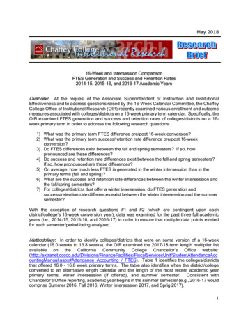
St-Amand et al. Lipids in Health and Disease(2020) 19:1921:10 cDNA dilution and 0.3 μM primers in a final volume of 10 μL. The following cycling protocol wasused: 2 min at 50 C, 2 min at 95 C, followed by 40cycles of 15 s at 95 C, 15 s at 58 C, and 1 min at72 C. The reaction’s specificity was verified by melting curve analysis. Negative controls (RT) and notemplate controls (NTC) were included in the PCRruns. Reference genes were identified by testing 10different genes (picked from literature) and runningTaqMan Array Rat Endogenous Control Plates (Applied Biosystems) on all samples. The 3 reference(housekeeping) genes with the best gene expressionstability measure (Expression Suite, Applied Biosystems) were selected for normalization: Hprt, Actb,and Gapdh. Primers used (Table 2) were designedusing Primer-BLAST (NCBI). Whenever possible,intron-spanning primers were selected to avoid amplification of genomic DNA. A standard curve obtainedby scalar dilution of a cDNA pool was generated toverify PCR efficiency. The triplicate average values ofcycle threshold (Ct) were used for quantification, andtarget genes were determined by 2ΔΔCt calculationmethod. Results are presented as Arbitrary Units (AU;also reported as Fold Change or Relative Expressionin the literature).Statistical analysesAll data are expressed as means standard error of themean (SEM). Statistical comparisons were performedusing a two-way analysis of variance (ANOVA) for nonrepeated measures, using diet and time as main effects.Fisher’s post-hoc test was used in the event of a significant (P 0.05) F ratio.Page 4 of 11ResultsAnthropometric parameters and food intakeBody weight was similar for all dietary groups at the beginning of the experimental period (Table 3). Bodyweight was not statistically modified by the obesogenicdiets after 2 weeks of treatment. However, it was significantly higher (P 0.01) at 6 weeks. Hence, body weightwas greater (P 0.01) in rats fed with the HFD and WDthan those submitted to the SD. Average food intake ing/d was lower (P 0.01) in rats fed the HFD and WD ascompared to rats fed the SD. Food intake in g/d was alsolower (P 0.05) in rats fed the HFD as compared to ratsfed the WD (Table 3). When food intake was calculatedin kcal/d, animals treated with the HFD and WD displayed higher values (P 0.001) than those fed with theSD. The sum of the 3 visceral fat pad weights as well asthe subcutaneous fat deposit were significantly (P 0.05)higher in rats fed the HFD and WD compared to ratsfed the SD after 2 (P 0.05) and 6 weeks (P 0.001;Table 3). HFD and WD resulted in similar body fataccumulations.Liver and plasma triglycerides and cholesterolLiver TG accumulation was significantly higher (P 0.001)in rats fed both the HFD and the WD after 2 as well asafter 6 weeks (Fig. 1a). Liver TG levels were even slightlyhigher (P 0.05) in WD than in HFD-fed animals after the2-week feeding period. Liver TG levels were higher(P 0.01) after 6 weeks than after 2 weeks in the SDfed group. On the other hand, HFD and WD did notaffect plasma TG levels after 2 weeks of feeding butresulted in a slight and significant (P 0.05) decreaseafter 6 weeks of feeding (Fig. 1b).Table 2 Oligonucleotide primers used for quantitative real-time polymerase chain reactionGene name NCBIAliasOligo FWDOligo gatccccatggcaatctg4ActbActb pdhGapdh aattcg80HprtHprt tgaaccagga62
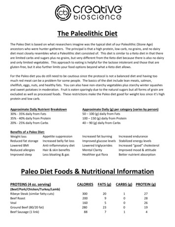
St-Amand et al. Lipids in Health and Disease(2020) 19:192Page 5 of 11Table 3 Anthropometric parameters and food intake2 weeksInitial bodyweight (g)Final bodyweight (g)6 weeksSDHFDWDSDHFD200 3199 4198 3199 3194 3343 4348 6339 6***Food intake (g/day)28.5 0.620.9 0.3Food intake (kcal/day)82 2109 1****Abdominal fat pad weight (g)11.7 0.717.6 1.0Subcutaneous fat pad weight (g)1.7 0.22.6 0.2**509 7*** †††24.3 0.331.2 0.6113 2*** 90 1 †††*18.2 1.033.2 1.72.8 0.4***4.9 0.3†††549 17WD192 3†††**21.9 0.4114 2***††††††48.6 2.57.2 0.6†††***††††††***†††**556 13 †††**26.2 0.6 †††*** 123 3†††*** 52.1 3.0 †††***7.9 0.9†††***Values are mean SEM with 8-10 rats per group: SD standard diet; HFD high fat diet; WD western diet. * Significantly (P 0.05) different from SD; ** P 0.01;*** P 0.001; significantly (P 0.05) different from HFD; P 0.001; ††† significantly (P 0.001) different from the two-week values, respectivelyAlthough some tendencies (P 0.1) can be observed,liver cholesterol concentrations were not changed significantly (P 0.05) by the diets with the exception ofthe group of animals fed the WD for 6 weeks thatshowed significantly (P 0.01) higher levels compared toSD and HFD fed rats (Fig. 1c). On the other hand, plasmatotal cholesterol levels were higher (P 0.05) in WD compared to SD and HFD fed animals after 2 and 6 weeks(Fig. 1d). No significant difference in plasma total cholesterol levels was found between rats fed the SD and theHFD. Overall, plasma total cholesterol levels were higher(P 0.05) with the longer diet duration (Fig. 1d).Fig. 1 Liver and plasma triglyceride (TG) and total cholesterol (TC) concentrations in rats (n 8–10) fed a standard (SD), high fat (HFD), or awestern (WD) diet for two and six weeks. * Significantly (P 0.05) different from SD; *** P 0.001; significantly (P 0.05) different from HFD; P 0.01; P 0.001; significantly (P 0.05) different from the two-week values, respectively; P 0.01
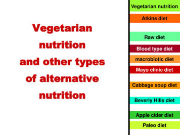
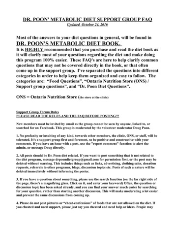
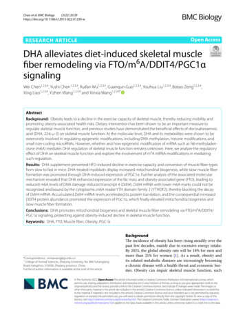
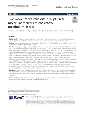
St-Amand et al. Lipids in Health and Disease(2020) 19:192Liver molecular markersAs presented in Fig. 2a, rats submitted to the WDfor 2 weeks depicted lower LDL-R gene expressionlevels as compared to SD (P 0.05) and HFD (P 0.05) animals. LDL-R mRNA levels were also lower(P 0.05) in rats fed the WD compared to those fedthe SD after 6 weeks. The same pattern of responsewas observed for the transcription factor SREBP2(P 0.001) that regulates both LDL-R and PCSK9(Fig. 2b). Similarly, PCSK9 mRNA expression waslower (P 0.01) in rats fed the WD as compared toanimals fed the SD and HFD after 2 and 6 weeks(Fig. 2c). This pattern of response was similar forplasma PCSK9 concentrations after 2 weeks, whileWD-fed animals depicted lower levels as comparedto HFD-fed rats after 6 weeks (Fig. 2d). The transcription of LRP-1, a member of the LDL-R genefamily, was also significantly (P 0.05) lower after 2and 6 weeks in animals submitted to the WD incomparison to animals fed the SD (Fig. 2e). Gene expression of ACAT-2 was also lower (P 0.05) following 2 and 6 weeks of WD compared to SD, wherethe 6-week treatment showed even lower levels thanthe 2-week treatment (Fig. 2f).Transcripts of HMGCoAR, the main enzyme of cholesterol synthesis, were decreased (P 0.001) after 2weeks in rats fed the HFD and the WD while after 6weeks lower levels (P 0.05) were found only in rats fedthe WD (Fig. 3a). Animals fed the SD depicted a lowerlevel of gene expression for HMGCoAR after 6 weeks incomparison to the one observed after 2 weeks. Gene expression of SR-B1, the transporter responsible for theexchange of cholesterol from HDL particles to the liver,was significantly (P 0.001) higher in HFD-fed rats compared to SD- and WD-fed animals in both feeding durations (Fig. 3b). A small but significant (P 0.05) effect ofoverall diet duration was also found with higher valuesobserved after 6 weeks. Gene expressions of ABCG5 andABCG8 were significantly (P 0.01) higher among ratsfed the HFD in comparison to those submitted to 2 and6 weeks of the other diets (Fig. 3c, d). Gene expressionof ACC, an important enzyme regulating the first stepsof de novo lipogenesis, was higher in rats fed the WDcompared to rats fed the two other diets for 2 weeks(Fig. 3e). After 6 weeks of treatment, ACC transcriptionwas also higher (P 0.001) in rats fed the WD than inthose treated with the HFD. ACC mRNA levels werealso lower (P 0.001) in HFD- than in SD-fed rats after2 and 6 weeks. Gene transcription of ChREBP, the transcription factor involved in regulating mediators of denovo lipogenesis, was highly stimulated (P 0.01) amongrats fed the WD compared to those treated with theother dietary regimen for 2 weeks (Fig. 3f). This responsewas not observed after 6 weeks of treatment.Page 6 of 11DiscussionThe main finding of the present study is that animalsfed a WD (high fat high sucrose) for only 2 weeksdepicted lower gene expression of several key markers ofliver cholesterol metabolism including LDL-R, PCSK9,SREBP2, and HMGCoAR compared to animals fed a SDor a HFD. The same response was also observed forPCSK9 measured in plasma. These findings were not observed when rats were fed the HFD. The 2-week WDalso resulted in higher plasma total cholesterol levels.The HFD and even more so the WD promptly (2 weeks)induced excessive TG accumulation in the liver. Theseresults indicate that the WD, which does not containhigher levels of cholesterol, disturbs the mechanisms involved in the regulation of cholesterol metabolism in theliver. The current results indicate that the enrichment insucrose (50% glucose and 50% fructose) and fat of theWD promptly and potently promoted TG accumulationin the liver. Hence, this effect may be put forward as theelement responsible for the alterations observed in theregulation of cholesterol metabolism. From a clinicalperspective, these results suggest that high plasma cholesterol levels may be linked to lipid metabolism in theliver.The first observation of the present study is thatshort-term (2 weeks) exposure to either a HFD (60% fat)or a WD (40% fat 35% sucrose (17.5% fructose)) induced important increases in the liver TG levels and thiseffect remained stable after 6 weeks of treatment. Anearly accumulation of lipids in the liver in response tohigh-fat feeding has been previously observed [11, 15,16]. This response has been interpreted as if the liverwas acting as a buffer to protect the other organs from apotentially deleterious surge of lipids [11, 17]. Accordingly, plasma TG levels were not changed after 2 weeksof HFD or WD. An additional finding of the presentstudy is that liver TG accumulation after 2 weeks waseven higher in the WD than in the HFD. Fructose contained in the WD is a strong inducer of hepatic lipogenicenzymes [18]. Therefore, the present increases in geneexpression of ACC and ChREBP measured in the liver ofanimals submitted to 2 weeks of WD support the assumption that de novo lipogenesis is associated with theexcessive accumulation of fat in this organ.In addition to resulting in a rapid increase in liver TGaccumulation, the short 2-week period of feeding appliedin the present study has the advantage of preceding significant increases in body weight gain and the systemicdevelopment of metabolic complications associated withlonger exposure to obesogenic diets. In support of this,the body weight observed among animals fed the HFDand WD was similar to the body weight observed amongrats fed the SD diet after 2 weeks. Body weight was approximately 50 g higher among the animals fed the
St-Amand et al. Lipids in Health and Disease(2020) 19:192Page 7 of 11Fig. 2 Hepatic gene expression of low-density lipoprotein receptor (LDL-R), proprotein convertase subtilisin/kexin type 9 (PCSK9), low-densitylipoprotein-receptor-related protein 1 (LRP-1), sterol regulatory element binding protein 2 (SREBP2), acyl-CoA cholesterol acyltransferase-2(ACAT-2), and plasma PCSK9 concentrations in rats (n 9–10) fed a standard (SD), high fat (HFD), or a western (WD) diet for 2 and 6weeks. * Significantly (P 0.05) different from SD; *** P 0.001; significantly (P 0.05) different from HFD; P 0.01; P 0.001; significantly (P 0.05) different from the two-week values, respectivelyobesogenic diets after 6 weeks. Moreover, the sum of abdominal and subcutaneous fat accumulation was approximately 3 times higher after 6 weeks of theobesogenic diets than it was after only 2 weeks (7 vs 20g), all while liver TG levels were similar after 2 and 6weeks. The present results support the validity of selecting such an experimental design where animals are submitted to 2 weeks of obesogenic diets. This is reinforcedby the fact that despite no change in body weight wasdetected, hepatic lipid metabolism was strongly alteredby the HFD and WD. For this reason, the present discussion is centered on results derived from the 2-weekfeeding condition.Using this experimental design, the present study aimedto gather evidence for the existence of an association between a rapid increase in liver TG among rats submitted
St-Amand et al. Lipids in Health and Disease(2020) 19:192Page 8 of 11Fig. 3 Hepatic gene expression of 3-hydroxy-3-methyl-glutaryl-CoA reductase (HMGCoAR), scavenger receptor class B member 1 (SR-B1), ATPbinding cassette protein G5 (ABCG5), ATP-binding cassette protein G8 (ABCG8), acetyl-CoA carboxylase (ACC), and carbohydrate-responseelement binding protein (ChREBP) in rats (n 7–10) fed a standard (SD), high fat (HFD), or a western (WD) diet for 2 and 6 weeks. * Significantly(P 0.05) different from SD; ** P 0.01; *** P 0.001; significantly (P 0.05) different from HFD; P 0.001; significantly (P 0.05) differentfrom the two-week values, respectively; P 0.01; P 0.001to short-term (2 weeks) obesogenic diets and disturbancesin gene expression of key regulators of cholesterol metabolism in the liver. The main finding of the present studywas that feeding animals with a WD for both 2 and 6weeks resulted in a decrease in gene expression of threeimportant and related markers of liver cholesterolmetabolism: LDL-R, PCSK9, and the transcription factorSREBP2. The combined action of these 3 molecules regulates transport of highly atherogenic LDL particles. Withapproximately 70% of its expression carried out in theliver, it is clear that LDL-R is crucial for the clearance ofLDL-cholesterol particles from circulation [19]. Therefore,
St-Amand et al. Lipids in Health and Disease(2020) 19:192the increase in plasma total cholesterol observed in ratsfed a WD for 2 or 6 weeks could be the result of the decrease in gene expression LDL-R in the liver. While thepresent data strongly support the physiological relevanceof this mechanism; the importance of other pathways involved in the regulation of circulating cholesterol levelswill also require further attention in future studies.The results also showed that the levels of mRNA andplasma PCSK9 (a protease that down-regulates LDL-R protein expression [20–22]) are reduced at 2 and 6 weeks afterWD feeding. This could have normally contributed to promoting the clearance of plasma cholesterol by increasingLDL-R protein levels. However, because of the decrease inupstream gene expression of SREBP2, a transcription factorthat regulates the expression of both LDL-R and PCSK9[23, 24], the reduction in PCSK9 levels did not positivelyaffect the expression of LDL-R. Consequently, plasma totalcholesterol remained high in rats fed the WD at 2 and 6weeks. Similarly, gene expression of LRP1, responsible forthe removal of circulating lipoprotein remnants enriched incholesterol [25], was reduced in rats fed the WD, thus potentially contributing to the increased plasma cholesterollevels. In additio
RESEARCH Open Access Two weeks of western diet disrupts liver molecular markers of cholesterol metabolism in rats Roxane St-Amand1, Émilienne T. Ngo Sock1, Samantha Quinn2, Jean-Marc Lavoie1 and David H. St-Pierre2* Abstract Background: The present study was designed to test the hypothesis that in the liver, excessive fat accumulation