Transcription
BREASTAcellular Dermal Matrix for the Treatmentand Prevention of Implant-AssociatedBreast DeformitiesScott L. Spear, M.D.Mitchel Seruya, M.D.Mark W. Clemens, M.D.Steven Teitelbaum, M.D.Maurice Y. Nahabedian,M.D.Washington, D.C.; and Los Angeles,Calif.Background: Acellular dermal matrix has been increasingly accepted in prosthetic breast reconstruction. Observed benefits include improved control andsupport of implant position, better implant coverage, and the suggestion of adecreased capsular contracture rate. Based on this positive experience, it is notsurprising that acellular dermal matrix would be applied to other challengingimplant-related problems. This study investigates the use of acellular dermalmatrix for correction or prevention of implant-associated breast deformities.Methods: Patients who underwent primary aesthetic breast surgery or secondary aesthetic or reconstructive breast surgery using acellular dermal matrix andimplants between November of 2003 and October of 2009 were reviewed retrospectively. Patient demographics, indications for acellular dermal matrix, andacellular dermal matrix type and inset pattern were identified. Preoperative andpostoperative photographs, success or failure of the procedure, complications,and need for related or unrelated revision surgery were recorded.Results: Fifty-two patients had acellular dermal matrix placed alongside 77breast prostheses, with a mean follow-up of 8.6 months (range, 0.4 to 30.4months). Indications included prevention of implant bottoming-out (n 6),treatment of malposition (n 32), rippling (n 20), capsular contracture (n 16), and skin flap deficiency (n 16). Seventy-four breasts (96.1 percent) weremanaged successfully with acellular dermal matrix. Three failures consisted ofone breast with bottoming-out following treatment of capsular contracture, onebreast with major infection requiring device explantation, and one breast withrecurrent rippling. There was a 9.1 percent total complication rate, consistingof three mild infections, one major infection necessitating explantation, onehematoma, and one seroma.Conclusion: Based on this experience in 77 breasts, acellular dermal matrix hasshown promise in treating and preventing capsular contracture, rippling, implantmalposition, and soft-tissue thinning. (Plast. Reconstr. Surg. 127: 1047, 2011.)Since 2006, an increasing number of studieshave highlighted the beneficial use of acellulardermal matrix in primary prosthetic breastreconstruction.1–12 Reported benefits include bettercontrol of implant position, better implant supportFrom the Department of Plastic Surgery, Georgetown University Hospital, and the David Geffen School of Medicine atthe University of California, Los Angeles.Received for publication April 18, 2010; accepted August13, 2010.Presented in part at the 26th Annual Meeting of the Northeastern Society of Plastic Surgeons, in Charleston, SouthCarolina, September 23 through 27, 2009, and the 2010Plastic Surgery Senior Residents Conference, in Anaheim,California, January 20 through 23, 2010.Copyright 2011 by the American Society of Plastic SurgeonsDOI: 10.1097/PRS.0b013e31820436afand coverage, and the suggestion of a decreasedfrequency of capsular contracture.1– 6,8 –11,13–15 Gamboa-Bobadilla placed human acellular dermal matrix alongside 13 breast prostheses with poor pectoralis major muscle coverage and found that 92percent of patients had a successful breast recon-Disclosure: Dr. Spear is a paid consultant forLifeCell and Allergan. Dr. Teitelbaum is a consultant for LifeCell, Allergan, and Kythera Biopharmaceuticals. Dr. Nahabedian is on the speaker’sbureau for LifeCell. Drs. Clemens and Seruya haveno financial interest in any of the products, devices,or drugs mentioned in this article.www.PRSJournal.com1047
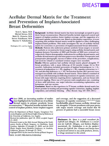
Plastic and Reconstructive Surgery March 2011struction and 73 percent had an “excellent” aesthetic outcome.2 In a series of 76 immediate, singlestage prosthetic breast reconstructions using humanacellular dermal matrix, Salzberg reported a 0 percent contracture rate and a 0 percent “serious” complication rate over a 52-month follow-up period.3Zienowicz and Karacaoglu observed a 0 percent rateof contracture, rippling, symmastia, or bottomingout in a study of 30 immediate implant-breast reconstructions with human acellular dermal matrixover an 18-month mean follow-up period.6 Breuingand Colwell placed human acellular dermal matrixalongside 44 breast prostheses and identified a 0percent contracture rate, a 2.3 percent extrusionrate, and a 4.5 percent infection rate over a 6-monthto 3-year surveillance period.5In addition to investigating the use of acellular dermal matrix in primary prosthetic breastreconstruction, a limited number of studieshave studied the application of acellular dermalmatrix in the treatment of breast implant–associated deformities.5,16 –19 Prosthetic breast surgerycan be complicated by either capsular attenuation,manifesting as implant malposition or rippling, orcapsular contracture, presenting as implant firmness or distortion. To correct breast implant rippling in 34 patients, Duncan used human acellulardermal matrix and observed an improvement inpalpable rippling, an average patient satisfactionof 85 percent, a 2.9 percent rate of capsular contracture, and a 2.9 percent rate of infection.16 Ina series of 10 patients with breast implant–relatedproblems, including rippling, bottoming-out, symmastia, and contracture, Baxter found that 80 percent of revisions were stable with the use of humanacellular dermal matrix.17 Breuing and Colwellreported a 0 percent recurrence of contracture in23 breasts treated with capsulectomy and humanacellular dermal matrix for implant-relatedcontracture.5 Most recently, Maxwell and Gabrieldemonstrated decreased rates of capsular contracture and improved implant cushioning and stabilization using acellular dermal matrix in revisionary aesthetic breast surgery of 78 patients.19Given the safety and efficacy of acellulardermal matrix in primary prosthetic breastreconstruction1– 6,9 –11 and the encouraging preliminary data on the use of acellular dermal matrix inselect cases of secondary prosthetic breastsurgery,5,16 –19 we hypothesize that acellular dermalmatrix may be a useful adjunct in the treatmentand prevention of some implant-associated deformities. The purpose of the present study is toreview experience with acellular dermal matrix forcorrection or prevention of implant-associated1048breast deformities and describe the key technicalaspects. This is especially significant because manyof these problems have proven extraordinarily difficult to consistently correct in the past.20 –28PATIENTS AND METHODSAn institutional review board–approved, retrospective review was carried out on patients operated on between November of 2003 and October of 2009 at Georgetown University Hospital(S.L.S. and M.Y.N.) and in the practice of StevenTeitelbaum, M.D. Patients who underwent primary aesthetic breast surgery or secondary aesthetic/reconstructive breast surgery using acellular dermal matrix and implants were identified.Techniques to address breast implant–related deformities often included a combination of implantexchange, device position change, capsule modification, and placement of acellular dermal matrix. When warranted, devices were exchangedand/or relocated to a subglandular, prepectoral, subpectoral, or neosubpectoral pocket.When necessary, capsule modification was performed by means of capsulotomy, capsulectomy,and/or capsulorrhaphy. Depending on thebreast implant–associated complication, acellular dermal matrix was placed alongside breastimplants using a “reconstructive,” “gutter,”“symmastia,” or “overlay” technique.29To treat difficult/recurrent capsular contracture, rippling, or implant bottoming-out, or toprevent loss of inframammary fold definition andbottoming-out, the reconstructive technique isused (Fig. 1). This involves interpositional (between the pectoralis major muscle and chest wall)placement of acellular dermal matrix, as described previously by the authors.8 In the case ofAlloDerm (LifeCell Corp., Branchburg, N.J.),acellular dermal matrix is oriented with the deepdermal surface facing the skin flaps and centeredover the mid arc of the inframammary/lateralmammary fold, providing maximal coverage andsupport where most necessary. Strattice (LifeCell), in contrast, does not require such orientation. Next, the inferior border of the acellulardermal matrix is sutured to the chest wall tissue,to best control the inframammary and lateralmammary folds. The appropriate device is thenplaced into the pocket and seated accordingly.Care is taken to achieve a “hand-in-glove” fit between the breast device, acellular dermal matrixgraft, and breast skin flaps. The superior border ofthe acellular dermal matrix is then sutured to thepectoralis major muscle, minimizing gap formation and thus the risk of direct exposure of the
Volume 127, Number 3 Acellular Dermis and Breast ImplantsFig. 1. The “reconstructive” technique. Inset of an acellular dermal matrix interpositional graft, lying between the pectoralis major muscle and chest wall. As an inferolateral sling, the acellulardermal matrix aids in supporting the implant and defining thebreast lower pole. (Reprinted with permission from LifeCellCorp.)prosthesis to a mastectomy incision. To reduce therisk of fluid accumulation between the acellulardermal matrix and the breast implant or betweenthe acellular dermal matrix and the breast skinflaps, drains are placed. Similar to a skin graft, theacellular dermal matrix should appear relativelysmooth without major folds or wrinkles andshould be in good contact with the breast skinflaps to ensure proper take and integration.Drains are left in place until the output is less than30 ml over a 24-hour period.To address inferior malposition, the guttertechnique is used to reinforce the fold repair(Fig. 2). Preoperatively, the site of the newly desired inframammary fold is noted and marked onthe patient. The operation begins with a skin incision and removal of the prior device. A capsulorrhaphy is then carried out in one or more rows,tacking the anterior and posterior leaflets of thecapsule together and to the chest wall, therebyrecreating the desired inframammary fold. An implant sizer is placed to verify that the inframammary fold is correctly set. To reinforce the foldrepair, an appropriate piece of acellular dermalmatrix is then selected and inset over the capsulorrhaphy suture line with 1 to 2 cm of overlap.The chosen implant is then placed in the reestablished pocket. A small drain is optional and completes the operation.Fig. 2. The “gutter” technique, with placement of acellular dermal matrix over the capsulorrhaphy suture line to reinforce thefold repair in the setting of lateral or inferior malposition. (Reprinted with permission from LifeCell Corp.)For correction of recurrent medial malposition,the symmastia technique is used (Fig. 3). Preoperatively, the site of the newly desired medial breastborder is marked on the patient. Initial operativesteps include the creation of a neosubpectoralpocket and/or performance of a multilayer breastcapsulorrhaphy to establish the desired medialbreast border, as described previously.20,30 To create a neosubpectoral pocket, the pectoralis majormuscle is dissected away from the anterior leafletof the capsule. The implant is kept in the capsuleas long as possible to facilitate dissection and isremoved through an open capsulotomy when it isno longer helpful. Of importance, the newly es-Fig. 3. The “symmastia” technique, with creation of a neosubpectoral pocket and inset of acellular dermal matrix along thepectoralis major muscle/capsule interface in the context ofmedial malposition. (Reprinted with permission from LifeCellCorp.)1049
Plastic and Reconstructive Surgery March 2011tablished pocket is purposely underdissected medially. Once the neosubpectoral pocket is formed,the original pocket is closed off by tacking theanterior and posterior leaflets of the capsule together and to the chest wall. To prevent migrationof the device into the old implant space, an appropriate piece of acellular dermal matrix is thenplaced medially over the suture line and/or pectoralis/capsule interface generally with 1 to 2 cmof overlap. If implant lower pole support is alsonecessary, the acellular dermal matrix inset can beextended with an inferior cuff to help cradle thedevice. A sizer or implant is used, sometimes inconjunction with temporary or permanent marionette (percutaneous, horizontal mattress) sutures, to ensure that the acellular dermal matrix isin appropriate position and set on appropriatetension. The desired breast implant is then placed,followed by insertion of a single small drain asnecessary.To address thin breast skin flaps, the overlay(between implant and capsule) technique is generally used (Fig. 4). Preoperatively, areas of softtissue deficiency are marked out on the patient.Operative steps begin with an open capsulotomyand removal of the old device. Areas of capsularthinning are noted and verified with the preoperative markings. Acellular dermal matrix is thenplaced inside the device pocket and draped overthe thin capsular regions. With the aid of a sizeror the final implant, the acellular dermal matrix isset on appropriate stretch and free of wrinkles orfolds on inset. Capsule and skin closure is carriedout in routine fashion.Fig. 4. The “overlay” technique. Acellular dermal matrix is parachuted into the device pocket with the use of marionette suturesand draped over thin capsular regions to address areas of softtissue deficiency.1050Generally, patients were discharged on the dayof surgery or, in rare cases, on postoperative day1. Oral antibiotics were continued until drain removal, typically once the output was less than 30ml/day. Marionette (percutaneous, horizontalmattress) sutures, if used, were usually discontinued 7 to 10 days postoperatively under sterile technique. This consisted of swabbing the externalskin sutures with alcohol, cutting one end of thesuture flush at the level of the skin, and pulling theother end of the suture out of the skin. Theseseries of maneuvers may help prevent introduction of skin flora into the breast pocket and therisk of infection.Office charts and hospital records were reviewed retrospectively for patient demographics,indications for acellular dermal matrix, and typeof acellular dermal matrix and inset pattern. Associated demographics included patient age, bodymass index, former or active tobacco use, historyof chemotherapy, and exposure to radiotherapy.Indications for acellular dermal matrix use weresometimes multiple and included the preventionof implant bottoming-out and the correction ofcapsular contracture, rippling, implant malposition, and soft-tissue thinning. Depending on costand availability, human-derived acellular dermalmatrix, specifically AlloDerm, or porcine-derivedacellular dermal matrix (Strattice) was used.Preoperative and postoperative photographs,failure of the procedure, complications, and needfor related and unrelated revision surgery wererecorded. Complications assessed included infection, implant loss or exposure, hematoma, andseroma. Infections were categorized as mild, defined as warmth, swelling, cellulitis, or nonpurulent drainage that was responsive to initial antibiotic therapy; or severe, characterized as persistentor substantial warmth/erythema/swelling despiteantibiotic therapy. Related revision procedureswere defined as those operations performed toaddress an issue directly related to the performance of acellular dermal matrix placement. Unrelated revision operations were defined as thoseprocedures performed to address an issue not associated with the performance of acellular dermalmatrix placement.For statistical analysis, the Fisher’s exact testwas used for evaluation of percentages or frequencies. A value of p 0.05 was considered statisticallysignificant.RESULTSOver a 6-year period, 52 patients (28 aestheticand 24 reconstructive) had acellular dermal ma-
Volume 127, Number 3 Acellular Dermis and Breast Implantstrix placed alongside 77 breast prostheses (43 aesthetic and 34 reconstructive). Patient demographics are summarized in Table 1. The mean age ofthe patients was 44.4 years, with a range of 21 to66 years, and the average body mass index was21.5. Among aesthetic patients, 82.1 percent werenonsmokers, 7.1 percent were former smokers,and 10.7 percent were active smokers. In reconstructive patients, 91.7 percent were nonsmokers,0 percent were former smokers, and 8.3 percentwere active smokers. Among reconstructive patients, 41.7 percent had a history of radiotherapyand 45.8 percent had a history of chemotherapy.None of the aesthetic patients had a history ofradiotherapy or chemotherapy.Indications for acellular dermal matrix aresummarized in Table 2 and included preventionof implant bottoming-out (n 6) and treatmentof malposition (n 32), rippling (n 20), cap-Table 1. Patient DemographicsValue (%)No. of patientsAestheticReconstructiveAge, yrAverageRangeMean BMI, kg/m2No. with preoperative chemotherapyAestheticReconstructiveNo. with preoperative radiotherapyAestheticReconstructiveSmoking historyNo. of nonsmokersAestheticReconstructiveNo. of former smokersAestheticReconstructiveNo. of active smokersAestheticReconstructive28 (53.8)24 (46.2)44.421–6621.50 (0.0)11 (45.8)0 (0.0)10 (41.7)23 (82.1)22 (91.7)2 (7.1)0 (0.0)3 (10.7)2 (8.3)BMI, body mass index.Table 2. Indications for Acellular Dermal Matrix inthe Treatment and Prevention of Implant-RelatedBreast Deformitiessular contracture (n 16), and skin flap deficiency (n 16). Of note, 26.9 percent of patientshad more than one indication for acellular dermalmatrix use. Among the 77 breast prostheses, 39had AlloDerm placement and 38 had Strattice.Complication rates were 7.0, 11.8, and 9.1 percent for aesthetic, reconstructive, and total breastoperations, respectively (Table 3). Complicationsconsisted of three mild infections, one major infection requiring explantation, one hematoma, andone seroma. Total complications were higher in irradiated versus nonirradiated breasts, although thiswas not found to be statistically significant (20.0 percent and 7.4 percent, respectively; p 0.223). Comparison of overall complication rates between Strattice and AlloDerm was not found to be statisticallysignificant (10.3 percent and 7.7 percent, respectively; p 0.71).Five breasts underwent unrelated revision surgery. After successful correction of bilateral implant bottoming-out with interpositional acellulardermal matrix grafts, one patient underwent bilateral revision augmentation-mastopexy becauseof a desire for smaller implants. After successfultreatment of left reconstructed breast (pedicledtransverse rectus abdominis musculocutaneousflap) symmastia with acellular dermal matrix, onepatient underwent transverse rectus abdominismusculocutaneous flap revision with raising of theinframammary fold. After successful correction ofleft implant inferior malposition with interpositional acellular dermal matrix graft, one patient underwent left implant size change and revisionmastopexy to improve symmetry opposite herreconstructed right breast. Finally, after successful correction of bilateral symmastia, one patient underwent right implant size change andleft breast lipoinfiltration to address a superomedial depression.With a mean follow-up of 8.6 months (range,0.4 to 30.4 months), 74 breasts (96.1 percent) weresuccessfully managed with acellular dermal maTable 3. Complications following Acellular DermalMatrix Use in the Treatment and Prevention ofImplant-Related Breast DeformitiesNo.PatientsOperative breastsIndications*MalpositionRipplingCapsular contractureSkin flap deficiencyPrevention of bottoming-out*Indications are per breast and were sometimes multiple.527790322016166Aesthetic Breasts(n ⴝ ructiveBreasts (n ⴝ 34)No.% of BreastsNo.% of 51
Plastic and Reconstructive Surgery March 2011trix. Three failures consisted of one breast withbottoming-out following treatment of capsular contracture, one breast with major infection followingsymmastia repair requiring device explantation anddelayed replacement, and one breast with recurrentrippling necessitating repeat acellular dermal matrixplacement (Table 4). Comparison of failure ratesbetween Strattice and AlloDerm was not found to bestatistically significant (5.3 percent and 2.6 percent,respectively; p 0.62).The first failure consisted of a 43-year-oldwoman with a history of multiple breast augmentations performed at an outside hospital to addressleft-sided infection and capsular contracture, whopresented with recurrent left capsular contracture. The patient underwent left revision augmentation with insertion of a 450-cc, moderate-profile,silicone implant in a neosubpectoral pocket andplacement of acellular dermal matrix (10 16cm) as an interpositional graft between the pectoralis major muscle and the chest wall (reconstructive technique). The acellular dermal matrixwas found intraoperatively to be too small to adequately serve as an inferolateral sling and still beattached at both the pectoralis major muscle andthe inframammary fold. Over the subsequentmonths, the patient presented with bottoming-outof her implant.The second failure consisted of a 31-year-oldwoman with a history of multiple breast augmentations performed by an outside surgeon to correct symmastia and who presented with recurrentsymmastia. The patient underwent bilateral revision breast augmentation with insertion ofsmooth, round silicone implants in a neosubpectoral pocket and placement of acellular dermalmatrix using the above-described symmastia technique. Three weeks postoperatively, the patientpresented with increasing pain, redness, and swelling of the right breast. Fluid aspiration was consistent with a methicillin-resistant Staphylococcusaureus–associated abscess. The patient underwentdevice explantation and curettage of the implantcapsule, with preservation of the acellular dermalmatrix, given its good integration with the surrounding tissue. Six months later, the patient successfully underwent device replacement (smooth,round silicone) and experienced no further episodes of infection.The third failure consisted of a 32-year-oldwoman with a history of bilateral immediate breastreconstruction with tissue expanders and acellulardermal matrix who presented with bilateral upperpole rippling following implant exchange. Thepatient underwent bilateral revision breast reconstruction with placement of acellular dermal matrix as overlay grafts (between capsule and skinflaps) and reinsertion of original devices (smooth,round silicone). One month postoperatively, thepatient presented with erythema and wounddrainage along the left reconstructed breast. Thisinfection was managed by incision and drainage,débridement of 10 percent of the acellular dermalmatrix that was nonadherent, and placement of anew device. Four months later, the patient presented with recurrent superolateral pole ripplingof the left reconstructed breast. This was successfully managed with revision breast reconstruction,placement of acellular dermal matrix graft undertension from the lateral mammary fold to the capsule along the superolateral margin of the breast,and reinsertion of the original device.CASE REPORTSCorrection of Capsular ContractureA 44-year-old woman with a history of bilateral breast augmentation and left mastopexy performed at an outside hospitalpresented with recurrent right capsular contracture after fourprevious failed revisions (Fig. 5). To address this deformity, thepatient underwent bilateral revision augmentation with rightpartial capsulectomy, partial subpectoral (“dual-plane”) placement of smooth, round silicone implants [right, Allergan 15265; left, Allergan 10-210 (Allergan, Inc., Irvine, Calif.)], andinsetting of Strattice (8 16 cm, each) interpositional graftsusing the reconstructive technique. Comparison of preoperative and 10-month postoperative photographs showed absenceof breast distortion and preservation of a natural breast contour.Correction of Medial MalpositionTable 4. Failure Rates following Acellular DermalMatrix Use in the Treatment and Prevention ofImplant-Related Breast DeformitiesIndicationMalpositionRipplingCapsular contractureSkin flap deficiencyPrevention of bottoming-outTotal1052No. of FailedBreast Procedures (%)1/32 (3.1)1/20 (5.0)1/16 (6.3)0/16 (0.0)0/6 (0.0)3/77 (3.9)A 39-year-old woman with a history of multiple bilateralbreast augmentation-mastopexy procedures presented withsymmastia (Fig. 6). To treat the deformity, neosubpectoralpockets were developed; implants were exchanged for smaller,smooth, round silicone implants (Allergan 20-350); and Strattice (8 8 cm, each) was placed using the symmastia techniqueover the pectoralis/capsule interface. Postoperative photographs demonstrated successful treatment of symmastia, whichwas maintained at 8-month follow-up.Correction of RipplingA 21-year-old woman with a history of multiple breastaugmentation procedures performed at an outside hospital
Volume 127, Number 3 Acellular Dermis and Breast ImplantsFig. 5. (Left) Preoperative view of a 44-year-old patient displaying right capsular contracture followingbilateral breast augmentation and left mastopexy at an outside hospital. The operative plan includedbilateral revision augmentation with right partial capsulectomy; partial subpectoral (“dual-plane”)placement of smooth, round silicone implants (right, Allergan 15-265; left, Allergan 10-210); and insetting of Strattice (8 16 cm each) interpositional grafts using the reconstructive technique. (Right)The patient 10 months postoperatively, showing correction of capsular contracture and absence ofbreast distortion.Fig. 6. (Left) Preoperative view of a 39-year-old female smoker with a history of multiple bilateral breast augmentation-mastopexy procedures presenting with symmastia. The operative plan included creation of neosubpectoral pockets; implant exchange for smaller, smooth, round silicone implants (Allergan 20-350); and Strattice(8 8 cm each) placement using the symmastia technique over the pectoralis/capsule interface. (Right) Thepatient at 8 months postoperatively, showing successful treatment of symmastia and stable implant location.to address implant bottoming-out presented with bilateralbreast implant rippling (Fig. 7). Operative steps to correctthese deformities included medial capsulorrhaphies; replacement of the original smooth, round silicone implants(Allergan 10-300) in a new partial subpectoral pocket; andinsetting of AlloDerm (4 16 cm each) both as an inter-positional graft between the inferolateral edge of the pectoralis major muscle and the inframammary fold, and as anoverlay graft between the capsule and the implant. Eighteenmonth postoperative photographs showed no evidence ofrippling or implant bottoming-out, with stable correction ofimplant deformities.1053
Plastic and Reconstructive Surgery March 2011Fig. 7. Preoperative anteroposterior (above, left), oblique (above, right), and magnified (second frombelow) views of a 21-year-old patient displaying bilateral breast implant rippling after multiple subpectoral breast augmentation procedures performed at an outside hospital. Postoperative anteroposterior (second row, left), oblique (second row, right), and magnified (below) views obtained 18 monthsafter bilateral revision augmentation with placement of original smooth, round silicone implants (Allergan10-300) under a partial subpectoral pocket and insetting of AlloDerm as interpositional grafts between the inferolateral edge of the pectoralis major muscle and the inframammary fold and as overlay grafts between thecapsule and implant. Note the correction of implant rippling and appropriate implant position.1054
Volume 127, Number 3 Acellular Dermis and Breast ImplantsPrevention of Bottoming-OutA 36-year-old woman with a history of active tobacco use and100-lb weight loss following bariatric surgery presented for abilateral breast augmentation-mastopexy procedure (Fig. 8).The patient underwent bilateral circumvertical mastopexies;partial subpectoral placement of smooth, round saline implants(Allergan 68MP, 330 cc filled); and placement of AlloDerminterpositional grafts between the pectoralis major muscle andthe inframammary fold using the reconstructive technique.Comparison of preoperative and 9-month postoperative photographs demonstrates stable and appropriate device position,without signs of soft-tissue stretch or implant bottoming-out.DISCUSSIONThe treatment and prevention of implant-associated breast deformities, arising from capsularattenuation and contracture, remain significantchallenges to the plastic surgeon. Capsular attenuation can manifest as implant malposition, bottoming-out, or rippling, whereas capsular contraction can be associated with implant firmnessand/or distortion. In addition, implant placementcan be complicated by surrounding soft-tissuethinning, putting the prosthesis at risk for threatened and/or actual exposure. Unfortunately, pasttechniques to address these various issues have notbeen highly predictable or durable.Capsular contracture remains one of themost frequent complications and is the mostcommon reason for reoperation following aesthetic and reconstructive prosthetic breastsurgery.31,32 Recently, the Inamed 6-year andMentor 3-year silicone breast implant core studiesreported capsular contracture rates of 8.1 to 14.8percent, 18.9 to 20.5 percent, 8.3 to 15.9 percent,and 16.3 percent for primary augmentation, revisionaugmentation, primary reconstruction, and revision reconstruction, respectively.31,32 To addressthis
Disclosure: Dr. Spear is a paid consultant for LifeCell and Allergan. Dr. Teitelbaum is a consul-tant for LifeCell, Allergan, and Kythera Biophar-maceuticals. Dr. Nahabedian is on the speaker's bureau for LifeCell. Drs. Clemens and Seruya have no financial interest in any of the products, devices, or drugs mentioned in this article. www .