Transcription
The Basics of Immunology through Case StudiesHinkley High SchoolAmy Loewen1150 Hinkley High SchoolAurora, CO 80011amloewen@aps.k12.co.usMentors: Dr. Ron Harbeck, PhD and Dr. Vijaya Knight, MD, PhDNational Jewish Health: Department of ImmunologyFunded by The American Association of ImmunologistsHigh School Teachers Summer Research Program1
Table of ContentsI. Science Background . 4II.Student Outcomes . 4III.Age and Level . 4A.International Baccalaureate Assessment Statements . 4B.Next Generation Science Standards. 5C. Technical Skills. 6IV.Relevance . 6V.Time Requirements . 6VI.Advance Preparation . 6A.Order . 6B.Prepare . 7C. Materials and Equipment . 7VII.Student Prior Knowledge and Skills . 9A.Knowledge . 9B.Skills . 9C. Preconceptions. 9VIII.IX.Daily Unit Plans . 10Student Worksheets . 302
This past summer (2014) I had the opportunity to shadow Dr. Harbeck and Dr. Knight atNational Jewish Medical Center (NJMC) in the Immunology Department. While spending timein their lab, I observed many of the lab technicians conducting ELISA’s, immunodiffusionassays, Kirby-Bauer disc susceptibility assays, and flow cytometry. While at NJMC, I studied thescience behind each assay and developed ways to make the assays and the science accessible tohigh school students. This required finding safe alternatives to many of the chemicals used inthe assays while still remaining true to the science behind the assay’s purpose. I teach high levelInternational Baccalaureate biology high school classes. My students all plan on continuing tostudy biology in college, and most desire to work in the medical field after college. As a result, Iknew I wanted to present immunology to my students in the most realistic manner possible. Thisunit was a lot of fun to develop and conduct. My students provided candid feedback concerningthe realistic manner of the labs and the methods of teaching the concepts. I hope that you enjoythis unit and that your students find this unit informative, realistic, and interesting.3
I.Science BackgroundStudents need a background in basic molecular biology. This immunology unit assumesstudents understand prokaryotic and eukaryotic cells, cell division, and pathogens. Allthe assays are thoroughly discussed along with the vocabulary in the provided slideshow.The new vocabulary is presented to the students in the form of videos and graphicorganizers. Answer keys are provided for the ease of the teacher.II.Student Outcomes III.Students will understand the difference between innate and adaptive immunity.Students will be able to list and describe the functions of the cells commonlyassociated with the immune system.Students will be able to describe common autoimmune conditions and diseasesand the assays used to diagnose them.Students will be able to diagnose a “mock” patient with signs and symptoms of acommon autoimmune condition or disease.Age and LevelRecommended for advanced/honors students, grades 9-12.A.International Baccalaureate Assessment Statements1.1.4 Explain how standard deviation is useful for comparing the means and thespread of data between two or more samples.1.1.5 Deduce the significance of the difference between two sets of data usingcalculated values for t and the appropriate tables.1.1.6 Explain that the existence of a correlation does not establish that there is acausal relationship between two variables.2.1.4 Compare the relative sizes of molecules, cell membrane thickness, viruses,bacteria, organelles and cells, using the appropriate SI unit.2.1.5 Calculate the linear magnification of drawings and the actual size ofspecimens in images of known magnification.2.2.1 Draw and label a diagram of the ultrastructure of Escherichia coli (E. coli)as an example of a prokaryote.2.3.1 Draw and label a diagram of the ultrastructure of a liver cell as an exampleof an animal cell.2.4.1 Draw and label a diagram to show the structure of membranes.2.4.3 List the functions of membrane proteins.2.5.1 Outline the stages in the cell cycle, including interphase (G1, S, G2), mitosisand cytokinesis.4.3.1 Define genotype, phenotype, dominant allele, recessive allele, codominantalleles, locus, homozygous, heterozygous, carrier and test cross.4.3.7 Define sex linkage.4
4.4.1 Outline the use of polymerase chain reaction (PCR) to copy and amplifyminute quantities of DNA.4.4.8 Outline a basic technique used for gene transfer involving plasmids, a hostcell (bacterium, yeast or other cell), restriction enzymes (endonucleases) andDNA ligase.5.4.8 Explain two examples of evolution in response to environmental change;one must be antibiotic resistance in bacteria.6.2.6 State that blood is composed of plasma, erythrocytes, leukocytes(phagocytes and lymphocytes) and platelets.6.2.7 State that the following are transported by the blood: nutrients, oxygen,carbon dioxide, hormones, antibodies, urea and heat.6.3.1 Define pathogen.6.3.2 Explain why antibiotics are effective against bacteria but not against viruses.6.3.4 Outline how phagocytic leucocytes ingest pathogens in the blood and inbody tissues.6.3.5 Distinguish between antigens and antibodies.6.3.6 Explain antibody production.6.3.7 Outline the effects of HIV on the immune system.7.5.4 State four functions of proteins, giving a named example of each.8.1.2 Outline the process of glycolysis, including phosphorylation, lysis, oxidationand ATP formation.8.1.5 Explain oxidative phosphorylation in terms of chemiosmosis.11.1.2 Outline the principle of challenge and response, clonal selection andmemory cells as the basis of immunity.11.1.3 Define active and passive immunity.11.1.4 Explain antibody production.11.1.5 Describe the production of monoclonal antibodies and their use indiagnosis and treatment.F.1.7 Compare the structure of the cell walls of Gram-positive and Gram-negativeEubacteria.F.6.1 List six methods by which pathogens are transmitted and gain entry to thebody.F.6.2 Distinguish between intracellular and extracellular bacterial infection usingChlamydia and Streptococcus as examples.B.Next Generation Science StandardsHS-LS1-1. Construct an explanation based on evidence for how the structure ofDNA determines the structure of proteins which carry out the essential functionsof life through systems of specialized cells.HS-LS1-2. Develop and use a model to illustrate the hierarchical organization ofinteracting systems that provide specific functions within multicellular organisms.HS-LS1-3. Plan and conduct an investigation to provide evidence that feedbackmechanisms maintain homeostasis.HS-LS2-7. Design, evaluate, and refine a solution for reducing the impacts ofhuman activities on the environment and biodiversity.*5
HS-LS2-8. Evaluate the evidence for the role of group behavior on individual andspecies’ chances to survive and reproduce.HS-LS3-3.Apply concepts of statistics and probability to explain the variation anddistribution of expressed traits in a population.HS-LS4-3. Apply concepts of statistics and probability to support explanationsthat organisms with an advantageous heritable trait tend to increase in proportionto organisms lacking this trait.HS-LS4-4. Construct an explanation based on evidence for how natural selectionleads to adaptation of populations.C.Technical Skills IV.Students will learn how to identify gram-positive and gram-negativebacteria.Students will learn and practice safety protocols important to immunology.Students will learn how to use many different predictive assays todetermine possible diseases and conditions; including ELISA’s andimmunodiffusion.Students will learn how to prepare sterile agarose.Students will learn how to measure micro amounts of liquid using a micropipette.RelevanceThe health field is a dynamic and constantly growing field. Any student who is interestedin gaining experience in a related health field needs to understand the basics ofimmunology and how the body’s immune system works. This unit will introducestudents to innate and adaptive immunity, basic procedures in immunology, assays,common autoimmune conditions, and diseases.V.Time RequirementsThis unit will take approximately 10-15 school days with one class period lasting 60minutes.VI.Advance PreparationA.Order1. Antibiotic discs mini-set: Carolina Biological #806499 (1 set is enough for 50experiments)2. ELISA simulation kit: Carolina Biological #211248 (1 kit is enough for 32students)6
3. Bovine Serum/Anti-Bovine Albumin, Antigen/Antibody set: CarolinaBiological #202105 (1 kit is enough for over 100 experiments)4. Buy case studies book: Case Studies in Immunology, a Clinical Companion,Fifth edition by Raif Geha and Fred Rosen. *Any edition will suffice.B.Prepare1. Review and go over all PowerPoint slides and case studies before presenting tothe students.2. Make photocopies of lab procedures and notes for students.3. Set up lab equipment for ELISA, immunodiffusion, antibiotics resistance lab,and pipetting mini-lab.4. Ask for faculty and staff volunteers to display symptoms of common conditionsand diseases used through this unit to come to a “clinic” in your classroom. Useattached symptoms scenarios for faculty and staff to attend clinic in yourclassroom for the summative assessment.C.Materials and Equipment1. Most materials mentioned in this list are common to a public school classroom.The items that are special to this unit are listed with prices and can all bepurchased through Carolina Biological, total cost for special items is: 602.45.This list is for a class of 32 students.2. Safety Precautions and Storage:Please store ALL chemicals in designated chemical storage area. Keep allchemicals in a cool dry space. Please visit this website for furtherinformation regarding chemical storage: ve/how-to-storechemicals/tr11069.tr?coId 10856&mCat . Consult your chemical safetypersonnel for proper storage and use in your school. Consult your localdistrict and state agencies concerning chemical storage, use and disposalbefore beginning any of the labs in this unit. While using chemicals safetygloves, goggles and lab coats must be worn by ALL people in theclassroom. Disposal of chemicals for the ELISA must be disposed insinks attached to neutralization tanks. Consult your local, district and stateagencies to ensure compliance. The Immunodiffusion and AntibioticResistant labs require taping the entire perimeter of the petri dish directlyafter preparation. DO NOT open the petri-dish again. I place the petri dishin a Ziploc baggie and tape the opening of the baggie as an extra barrier.Once all observation is complete place taped petri dishes in a sink withbleach water for 24 hours. Visit this website for recipe information forbleach water to kill possible harmfulpathogens: bleachwater-ratio/ . Remove tape under the water to allow bleach water into thepetri dish, this is to ensure that all harmful growth is killed by the bleach.After 24 hours the petri dishes may be thrown in the trash. Every state hasdifferent requirements for possible bacterial growth in public school7
classrooms and proper disposal. Please contact your local district andstate education agencies before beginning any of the labs.***Please visit this web-site for further information on wastedisposal: ive/storingand-disposing-chemicals/tr11068.tr?coId 10856&mCat .3. Antibiotic Resistance lab:Antibiotic discs mini-set ( 48.95 Carolina Biological)Escherichia coli, Living, K-12 Strain, Tube ( 10.95 Carolina Biological)Nutrient agar for bacteria growthSterile petri dishes (for agar and discs)Heat/stirring plate (for heating up and stirring agar solution)Erlenmeyer flasksGlass stirring rodsHeat clampsDe-ionized waterTapeInoculating loops (to safely spread bacteria, immediately place in bleachwater sink before disposing)Incubator ( 390.00 Carolina Biological)ELISA:ELISA simulation kit ( 110.00 Carolina Biological)Immunodiffusion lab:Bovine Serum/Anti-Bovine Albumin, Antigen/Antibody set ( 53.50Carolina Biological)Sterile petri dishesStraws (for punching holes in agarose)IncubatorDissecting microscope or magnifying glasses (to observe precipitate lines)Powder agar *non-nutrientHeat/stirring plateErlenmeyer flasksGlass stirring rodsHeat clampsDe-ionized water4. Micro-pipetting mini-lab:WaterTest tubesTest tube racksFood coloringMultiple micropipettes in various measurementsMicropipette tipsBacteria cell slide identification:Compound microscopes8
Slides of gram positive and gram negative bacteria-prepared and sealed(can be purchased through Carolina Biological)****The micro pipetting mini-lab is not necessary for this unit if time andmoney are limited. Micro pipettes are approximately 100.00 apiece andcan be purchased through bio-rad.com.VII.Student Prior Knowledge and SkillsA.KnowledgeThis unit is for students with a basic understanding of biological conceptsespecially in relation to the human body. Usually this means that they have takena general biology course, or this unit could be taught at the end of the school yearafter they have completed units on human health and physiology and cells.Students need background information on the basics of the immune system,viruses, bacterial infections, antibodies and antigens.B.SkillsStudents should be able to follow lab procedures and safety protocols set forth bythe school and district. Students should be able to use basic math computation formany of the labs. Students should have a basic understanding of how to use labequipment, such as a hot plate, incubator, and microscopes.C.PreconceptionsStudents really struggled with the innate and adaptive immune systems and oftenconfused the two. I found that providing some graphic organizers and interactiveassignments for the students enabled them to grasp the differences/similarities.Students have numerous preconceptions about HIV and AIDS. It was helpful toshow them a video explaining how HIV is contracted and the biological processesthat take place inside a person once HIV is contracted.*Consult district and state regulations before teaching HIV as many states haveregulations concerning the sensitive topic of HIV.9
VIII. Daily Unit PlansDay 1: Introduction to the innate and adaptive immune system.Day 2: Introduction to the key cells involved in the human immune system.Worksheet: Immune System Speed NotesStudents will use the following resources to fill-out the providedgraphic organizer:http://ib.bioninja.com.au/ (detailed information about required InternationalBaccalaureate biology immune-system (interactive fromHoward Hughes Medical Institute)https://www.youtube.com/watch?v WJEc2GDEfz8 (Kids Health video onthe immune system, a bit silly, but good explanation)10
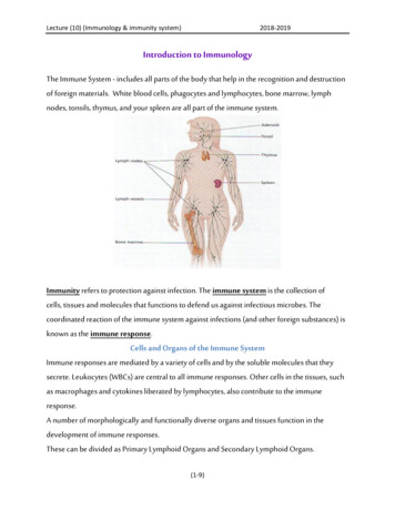
Immune System Speed NotesInnate vs. AdaptiveCells of the Immune SystemINNATE IMMUNE-prevents pathogens fromentering the body-present from birth-non specificADAPTIVE IMMUNE-as an infection continues thebody recognizes the antigenand produces antibodiesagainst it-antigen specific and is moreeffective with increasedexposure to pathogenBoth systems fight pathogens thatthe body comes in contact with.Cells of the Immune fenses75.jpgStem Cell: Cell that can differentiate into other cells.Lymphoid Stem Cell: Stem cell that specifically differentiates into lymphatic cells.Lymphocytes: Make up 15% of blood leukocytes.Natural Killer Cell: Attack and lyse a virus, infected or cancerous cell of the body; makeup 5-10% of lymphocytes in the blood.T-Cell progenitor: May differentiate into a Tc cell or Th cell.Tc cell: Recognize and kill virus infected cells and other altered body cells.Th cell: Assist cellular and humoral immune systems by secreting cytokines.B-Cell progenitor: Differentiates into plasma or memory cells.Memory cell: Provides immunity to future infections from familiar pathogen.Plasma cell: Secretes antibodies.Myeloid Progenitor: Differentiates into granulocytes and dendritic cells.Granulocytes: Cells containing granules made of mediators.Neutrophil: Responds rapidly to inflammation. The cells move from blood stream toinflamed tissue where they phagocytize debris and pathogens.Eosinophil: Migrate from bone marrow to other tissues and kill antibody coated parasites.11
Basophil: Plays a role in inflammation and the allergic response. Found in the bloodstream.Mast Cell: Releases histamine and a number of other inflammatory cytokines andmediators. Plays a role in inflammation and the allergic response. Found in tissue.Monocyte: Circulate in the blood stream and migrate to other tissues. Can differentiateinto a macrophage or dendritic cell.Macrophage: Phagocytic cell that engulfs and digests microorganisms. Activates Tcells by releasing cytokines.Dendritic Cell: Antigen presenting cells that present antigens to T-cells.All of the above are white blood cells (leukocytes).12
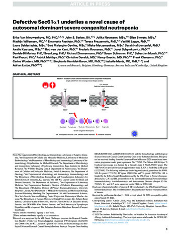
Days 3 and 4:Micro pipetting mini-lab (Optional)This lab was necessary for my students as many had never used amicropipette before. There are an abundance of instructional labs online. Ifound a few that directly addressed the needs of my g.pdfDays 5-7:Case Study #1 HIVStudents will read about a person diagnosed with HIV and complete amock ELISA.Book: Case Studies in Immunology, a Clinical Companion, Fifth edition byRaif Geha and Fred Rosen. Case #31 Acquired Immune DeficiencySyndrome (AIDS), pages 187-191Videos: HIV/AIDS 101 by the Center for Disease Control. Great videoabout the transmission ofHIV. https://www.youtube.com/watch?v I o wkr7N8AHIV life cycle: video by Howard Hughes Medical Institute about the lifecycle of HIV. https://www.youtube.com/watch?v odRyv7V8LAE Follow the PowerPoint (Case studies slide show) while guiding thestudents through the reading. Using the purchased ELISA (mock) kit from Carolina Biological.Follow the instructions provided for HIV. The kit comes withdiagrams, instructions and questions to further the studentsunderstanding of how an ELISA works. Note: Follow all safety procedures. Consult school, district, and stateregulations before beginning. Wear goggles, gloves and storechemicals properly. Read statement concerning proper handling anddisposal in the Materials section of this publication.13
14
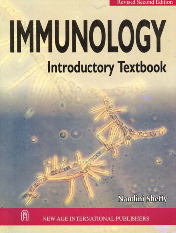
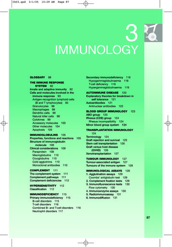
Days 8-10:Case Study #2 MRSAStudents will read about a person with skin rashes and eczema whilecompleting the antibiotic disc lab.Optional: Observe slides of gram-positive and gram negative bacteria foridentification under a microscope.Book: Case Studies in Immunology, a Clinical Companion, Fifth edition byRaif Geha and Fred Rosen. Case #34 Atopic Dermatitis, pages 207-212.Video: Atopic Eczema by Eczema Society ofCanada; https://www.youtube.com/watch?v DVRVnvlvUPAVideo: How do antibiotics work? By eBug. https://www.youtube.com/watch?v X1GT2bKgci8 Before reading: Have students prepare agarose plates according topurchased agarose instructions, swab with bacteria and placeantibiotic discs on plate. Watch video that shows streaking technique and placement ofantibiotic discs. https://www.youtube.com/watch?v O5NwOGazOAA Watch video that shows how to measure the zone ofinhibition https://www.youtube.com/watch?v LzmEwpL2 zI Tape the entire perimeter of the petri dishes to enclose them, afterstreaking bacteria and placing antibiotic discs on plate. I also placethe petri dishes in a Ziploc baggy as an additional barrier and tapethe opening of the Ziploc baggy. The students are not allowed totake the petri dish out of the Ziploc baggy. Place the petri dishes in the incubator. ***Ensure the petri dishes arenot incubating at a temperature close to human temperature (98.6F);this is also a precaution to hopefully prevent bacteria from growingthat could potentially harm humans. Follow the Safety Precautions and Storage procedures above fordisposal of the petri dishes when done with the lab. Note: Follow all safety procedures. Consult school, district, and stateregulations before beginning. Wear goggles, gloves and storechemicals properly. Read statement concerning proper handling anddisposal in the Materials section of this publication. Follow the PowerPoint (Case studies slide show) while guiding thestudents through the reading.15
After the reading, measure the zones of inhibition for the antibioticdiscs.16
17
18
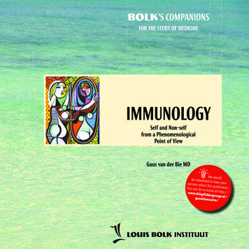
Days 11-13:Case Study #3 Hypersensitivity and AllergyStudents will read about people diagnosed with hypersensitivity to peanutsand allergic asthma, while completing the immunodiffusion lab.Book: Case Studies in Immunology, a Clinical Companion, Fifth edition byRaif Geha and Fred Rosen. Cases #32 Acute Systemic Anaphylaxis and#33 Allergic Asthma, pages 193-205.Video: How our bodies develop allergies by SuperScienced; https://www.youtube.com/watch?v I16cXams0 4Video: Understanding Asthma by NHSchoices; https://www.youtube.com/watch?v 7EDo9pUYvPEPreparation: Have students prepare agarose plates. Punch holes withstraws and fill with antigens/antibodies before beginning to read so thatreaction has time to take place and ready to observe after the reading. Follow the PowerPoint (Case study slide show) while guiding thestudents through the reading. After the reading, have students observe precipitant lines on platesusing light box. Tape the entire perimeter of the petri dishes after micro-pipetting theantigens and antibodies in the wells. Place the petri dishes in the incubator at approximately 72F/22C.Allow to incubate 12 hours minimum. **Be careful incubatingovernight, I unplug my incubator before leaving school for the eveningaccording to safety standards at my school. Worksheet: Immunodiffusion Worksheethttps://www.dshs.state.tx.us/lab/serology id.shtm (website helps withreading precipitant lines for Ouchterlony worksheet)https://www.youtube.com/watch?v Fnx5CkGRBEM (Video procedure forOuchterlony, used to complete Ouchterlony worksheet)Note: Follow all safety procedures. Consult school, district, and stateregulations before beginning. Wear goggles, gloves and store chemicalsproperly. Read statement concerning proper handling and disposal in theMaterials section of this publication.19
20
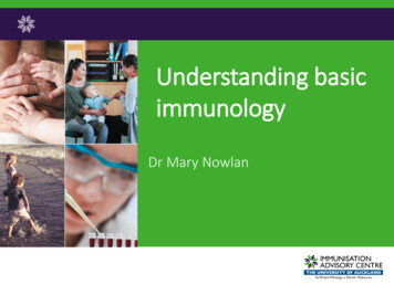
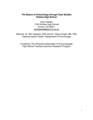
Immunodiffusion (Ouchterlony Assay)Briefly describe in your own words what the assay indicates for:An antigen and antibody diffuse towards each other in agarose, where the two meet a precipitantline forms. This line indicates sensitivity to the antigen. If a person is not producing theantibodies then their serum will not form a line of precipitate with the antigen.Write down the procedure for the Ouchterlony Assay:1.Prepare agarose. (Ensure this is the non-nutrient type)2.Pour agarose into petri-dishes. Let cool.3.Use a drinking straw to punch wells into agarose in star design. *see below diagram.Use a sharpie to label the bottom of the petri dish according to instructions.4.Fill middle well with 10 microliters of antigen.5.Fill outer wells with patient antibody and positive and negative controls.6.Place lid on petri dish and wrap perimeter with parafilm. (DO not remove parafilm toobserve results as bacteria may grow.)7.Incubate for 24 hours at approximately 37C.8.Observe precipitant lines by placing sealed petri dish on light box. (DO not removeparafilm as bacteria might have grown.)9.To safely dispose of petri dishes place them in a sink of bleach water (one capful ofbleach). Underneath the water, remove the parafilm to allow the bleach water to infiltratethe petri dish. Let petri dishes sit in bleach bath for several hours. Make sure to washhands thoroughly. The petri dishes may be thrown in the garbage after sitting in thebleach bath for several opetridishesat https://www.tk.de/rochelexikon/pics/a28264.000-1 big.gif-figure I: Since the lines of precipitate cross each other, the reactions are not fully conclusive.This shows that the reactions between A and B affected the reaction of A to AK and B to AK.-figure II: Since the lines of precipitate do not cross each other, the reactions are conclusive. Theantigen AK has reactions with antibodies in outer wells, without the antibodies and controlsreacting with each other.21
Summative Assessment:Days 14-15:Hinkley Wellness ClinicFaculty and staff at your school will come to your classroom displayingsymptoms of common conditions discussed during this unit. Students willbe required to prescribe assays and a possible diagnosis based onsymptoms. Students will be required to write a paper describing theprescribed assay and diagnosis for their “patient” citing evidence in supportof their diagnosis. Provide patient symptom sheets for faculty and staff. Provide patient questionnaire and patient pain assessment sheets forstudents to complete while interviewing/diagnosing faculty and staff. After students have interviewed/diagnosed a patient, each studentshould complete the following constructed response based on theirinterview;1. Based on the evidence you recorded, what is your diagnosis foryour patient?2. Cite ALL evidence that lead you to that diagnosis.3. What assay would you recommend the lab conduct to helpdiagnose your patient?4. Write a 3-5 sentence summary of the condition you diagnosedyour patient with. Answers: A student should get the correct condition and symptomsthat are listed on the patient symptom sheet. The “patient” should notsimply give the student the information on the sheet. A studentshould obtain all the symptoms and conclude the condition based ontheir questioning and the information they gather from interviewingthe “patient”.22
Patient Symptom sheet 1Male or FemaleBackground: You are a bank manager in downtown Denver. You are slightly overweight buthave recently lost 20 pounds without much effort. You equate your weight loss to stress as thebank is currently merging with another bank, so there have been many late night meetings.You have 2 children. The oldest (5 years old) just recently adopted a kitten. While playing withthe kitten, it scratched you. Usually a small scratch is no big deal, but the next morning thelocation of the scratch (hand/arm) was red, swollen, and hot to the touch. The swell on yourhand/arm is the size of a baseball. The location of the scratch seems to be infected. This is thereason for your visit to the clinic. You assume that the kitten had some bacteria underneath itsclaws that gave you an infection when it scratched you. You are at the clinic to get someantibiotics.Back in college (8 years ago) you engaged in some crazy behavior as college students sometimesdo. You faintly remember shooting up (needle) some illicit drugs at a party one night while youwere rather drunk. You are not sure if the needle was used previously. This only happened once.You are not on any prescription medications.You are healthy other than being slightly overweight.There is no family history of allergies, anaphylaxis, or angioedema.Surgeries: appendectomy 15 years ago****If the doctor asks you a question that has not been addressed on this symptom sheet feel freeto make up an answer that would be consistent with this patient’s scenario.Do not share with student the following information:Suggested Assay: ELISA for HIV and Kirby-Bauer Antibiotic disc assay for the possiblebacterial infection due to the cat scratch.23
Patient Symptom Sheet 2FemaleBackground: You are a teenager at a local high school and heard that Hinkley was having a freeclinic today. You recently had unprotected sex with a guy at a party. He was super cute and saidhe didn’t have a condom, so you figured it would be OK just this once. Now, your menstrualcycle is 8 days late, and you are afraid you’re pregnant. You are here to get a pregnancy test.Absolutely NO ONE knows what you did at the party, (except the guy of course), so you beg thedoctor to
in their lab, I observed many of the lab technicians conducting ELISA's, immunodiffusion assays, Kirby-Bauer disc susceptibility assays, and flow cytometry. While at NJMC, I studied the science behind each assay and developed ways to make the assays and the science accessible to high school students.