Transcription
CMEJOURNAL OF MAGNETIC RESONANCE IMAGING 37:501–530 (2013)Special CommunicationACR Guidance Document on MR Safe Practices: 2013Expert Panel on MR Safety: Emanuel Kanal, MD,1* A. James Barkovich, MD,2Charlotte Bell, MD,3 James P. Borgstede, MD,4 William G. Bradley Jr, MD, PhD,5Jerry W. Froelich, MD,6 J. Rod Gimbel, MD,7 John W. Gosbee, MD,8Ellisa Kuhni-Kaminski, RT,1 Paul A. Larson, MD,9 James W. Lester Jr, MD,10John Nyenhuis, PhD,11 Daniel Joe Schaefer, PhD,12 Elizabeth A. Sebek, RN, BSN,1Jeffrey Weinreb, MD,13 Bruce L. Wilkoff, MD,14 Terry O. Woods, PhD,15Leonard Lucey, JD,16 and Dina Hernandez, BSRT16Because there are many potential risks in the MR environment and reports of adverse incidents involvingpatients, equipment and personnel, the need for a guidance document on MR safe practices emerged. Initiallypublished in 2002, the ACR MR Safe Practices Guidelinesestablished de facto industry standards for safe andresponsible practices in clinical and research MR environments. As the MR industry changes the document isreviewed, modified and updated. The most recent versionwill reflect these changes.Key Words: MR safety; MR; MR safe practicesJ. Magn. Reson. Imaging 2013;37:501–530.C 2013 Wiley Periodicals, Inc.V1Department of Radiology, University of Pittsburgh Medical Center,Pittsburgh, Pennsylvania, USA.2Department of Radiology and Biomedical Imaging, University ofCalifornia, San Francisco, California, USA.3Milford Anesthesia Associates, Milford, Connecticut, USA.4University of Colorado, Denver, Colorado, USA.5Department of Radiology, University of California San Diego MedicalCenter, San Diego, California, USA.6Department of Radiology, University of Minnesota, Minneapolis,Minnesota, USA.7Cardiology Associates of E. Tennessee, Knoxville, Tennessee, USA.8University of Michigan Health System and Red Forest Consulting LLC,Ann Arbor, Michigan, USA.9Radiology Associates of the Fox Valley, Neenah, Wisconsin, USA.10Durham Radiology Associates, Raleigh, North Carolina, USA.11Department of Electrical and Computer Engineering, PurdueUniversity, West Lafayette, Indiana, USA.12MR Systems Engineering, GE Healthcare, Waukesha, Wisconsin, USA.13Yale School of Medicine, New Haven, Connecticut, USA.14Cleveland Clinic, Cleveland, Ohio, USA.15FDA Center for Devices & Radiological Health, Silver Spring,Maryland, USA.16American College of Radiology, Reston, Virginia, USA.Reprint requests to: Department of Quality & Safety, American College of Radiology, 1891 Preston White Drive, Reston, VA 20191-4397.*Address reprint requests to: E.K., University of Pittsburgh MedicalCenter, Presbyterian University Hospital\Presbyterian South Tower,Room 4776, Pittsburgh, PA 15213. E-mail: ekanal@pitt.eduReceived October 3, 2012; Accepted December 4, 2012.DOI 10.1002/jmri.24011View this article online at wileyonlinelibrary.com.C 2013 Wiley Periodicals, Inc.VTHERE ARE POTENTIAL risks in the MR environment, not only for the patient (1,2) but also for theaccompanying family members, attending health careprofessionals, and others who find themselves onlyoccasionally or rarely in the magnetic fields of MRscanners, such as security or housekeeping personnel, firefighters, police, etc. (3–6). There have beenreports in the medical literature and print-mediadetailing Magnetic Resonance Imaging (MRI) adverseincidents involving patients, equipment and personnelthat spotlighted the need for a safety review by anexpert panel. To this end, the American College ofRadiology originally formed the Blue Ribbon Panel onMR Safety. First constituted in 2001, the panel wascharged with reviewing existing MR safe practices andguidelines (5–8) and issuing new ones as appropriatefor MR examinations. Published initially in 2002 (4),the ACR MR Safe Practice Guidelines established defacto industry standards for safe and responsiblepractices in clinical and research MR environments.These were subsequently reviewed and updated inMay of 2004 (3). After reviewing substantial feedbackfrom the field and installed base, as well as changesthat had transpired throughout the MR industry sincethe publication of the 2004 version of this document,the panel extensively reviewed, modified, and updatedthe entire document in 2006–2007.The present panel consists of the followingmembers: A. James Barkovich, MD, Charlotte Bell,MD, (American Society of Anesthesiologists), James P.Borgstede, MD, FACR, William G. Bradley, MD, PhD,FACR, Jerry W. Froelich, MD, FACR, J. Rod Gimbel,MD, FACC, Cardiologist, John Gosbee, MD, MS, EllisaKuhni-Kaminski, RT (R)(MR), Emanuel Kanal, MD,FACR, FISMRM (chair), James W. Lester Jr., MD,John Nyenhuis, PhD, Daniel Joe Schaefer, PhD Engineer, Elizabeth A. Sebek, RN, BSN, CRN, JeffreyWeinreb, MD, Terry Woods, PhD, FDA, Pamela Wilcox,RN, MBA (ACR Staff), Leonard Lucey, JD, LLM (ACRStaff), and Dina Hernandez, RT (R) (CT) (QM) (ACRStaff). The following represents the most recently501
502modified and updated version of the combined priorthree reports (3,4,9) issued by the American College ofRadiology Blue Ribbon Panel on MR Safety, chaired byEmanuel Kanal, MD, FACR. It is important to note thatnothing that appears herein is the result of a ‘‘majorityvote’’ of the member of this panel. As with each priorpublication of these ACR MR Safe Practice Guidelines,the entire document, from introduction to the markedly expanded appendices, represents the unanimousconsensus of each and every member of this SafetyCommittee and the various areas of expertise that theyrepresent. This includes representation from fields andbackgrounds as diverse as MR physicists, research/academic radiologists, private practice radiologists, MRsafety experts, patient safety experts/researchers, MRtechnologists, MR nursing, National Electrical Manufacturers Association, the Food and Drug Administration, the American Society of Anesthesiologists, legalcounsel, and others. Lay personnel, physicians,Ph.D.s, department chairs and house-staff/residents,government employees and private practitioners,doctors, nurses, technologists, radiologists, anesthesiologists, cardiologists, attorneys—these are allrepresented on this Committee. It was believed thatachieving unanimity for these Guidelines was criticalto demonstrate to all that these Guidelines are notonly appropriate from a scientific point of view, butreasonably applicable in the real world in which we allmust live, with all its patient care, financial, andthroughput pressures and considerations. The viewsexpressed in this study are solely those of the authorsand in no way suggest a policy or position of any of theorganizations represented by the authors.The following MR safe practice guidelines documentis intended to be used as a template for MR facilities tofollow in the development of an MR safety program.These guidelines were developed to help guide MR practitioners regarding these issues and to provide a basisfor them to develop and implement their own MR policies and practices. It is intended that these MR safepractice guidelines (and the policies and procedures towhich they give rise) be reviewed and updated on a regular basis as the field of MR safety continues to evolve.The principles behind these MR Safe PracticeGuidelines are specifically intended to apply not onlyto diagnostic settings but also to patient, researchsubject, and health care personnel safety for all MRIsettings, including those designed for clinical diagnostic imaging, research, interventional, and intraoperative MR applications.With the increasing advent and use of 3.0-Tesla andhigher strength magnets, users need to recognize thatone should never assume MR compatibility or safetyinformation about a device if it is not clearly documented in writing. Decisions based on published MRsafety and compatibility claims should recognize thatall such claims apply only to specifically tested conditions, such as static magnetic field strengths, staticgradient magnetic field strengths and spatial distributions, and the strengths and rates of change of gradient and radiofrequency (RF) magnetic fields.Finally, there are many issues that impact MRsafety which should be considered during siteKanal et al.planning for a given MR installation. We include inthis manuscript, as separate appendices, sectionsthat address such issues as well, including cryogenemergency vent locations and pathways, 5-Gaussline, siting considerations, patient access pathways,etc. Yet despite their appearance herein, these issues,and many others, should be reviewed with those experienced with MR site planning and familiar with thepatient safety and patient flow considerations beforecommitting construction to a specific site design. Inthis regard, enlisting the assistance of an architectural firm experienced in this area, and doing so earlyin the design stages of the planning process, mayprove most valuable.It remains the intent of the ACR that these MR SafePractice Guidelines will prove helpful as the field ofMRI continues to evolve and mature, providing MRservices that are among the most powerful, yet safest,of all diagnostic procedures to be developed in thehistory of modern medicine.ACR GUIDANCE DOCUMENT ON MR SAFEPRACTICES: 2013A. Establish, Implement, and Maintain CurrentMR Safety Policies and Procedures1. All clinical and research MR sites, irrespective ofmagnet format or field strength, including installations for diagnostic, research, interventional,and/or surgical applications, should maintainMR safety policies.2. These policies and procedures should also bereviewed concurrently with the introduction ofany significant changes in safety parameters ofthe MR environment of the site (e.g., addingfaster or stronger gradient capabilities or higherRF duty cycle studies) and updated as needed. Inthis review process, national and internationalstandards and recommendations should betaken into consideration before establishing localguidelines, policies, and procedures3. Each site will name a MR medical director whoseresponsibilities will include ensuring that MRsafe practice guidelines are established andmaintained as current and appropriate for thesite. It is the responsibility of the site’s administration to ensure that the policies and proceduresthat result from these MR safe practice guidelines are implemented and adhered to at all timesby all of the site’s personnel.4. Procedures should be in place to ensure that anyand all adverse events, MR safety incidents, or‘‘near incidents’’ that occur in the MR site are tobe reported to the medical director in a timelymanner (e.g., within 24 hours or 1 business dayof their occurrence) and used in continuous quality improvement efforts. It should be stressedthat the Food and Drug Administration statesthat it is incumbent upon the sites to also reportadverse events and incidents to them by meansof their Medwatch program. The ACR supportsthis requirement and believes that it is in the
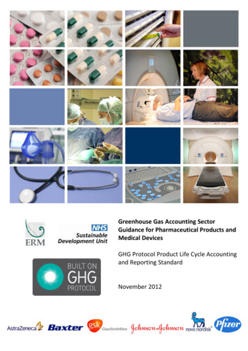
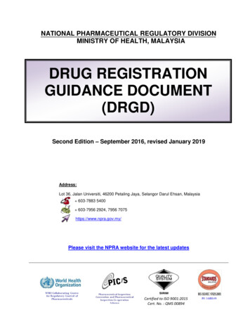
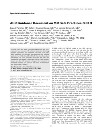
ACR Guidance on MR Safe Practices503Figure 1. Idealized sample floorplan illustrates site access restriction considerations. Other MRpotential safety issues, such asmagnet site planning related tofringe magnetic field considerations,are not meant to be include herein.See Appendix 1 for personnel andzone definitions. Note—In any zoneof the facility, there should be compliance with Health Insurance PortabilityandAccountabilityAct(HIPAA) regulations in regard to privacy of patient information. However, in Zone III, there should be aprivacy barrier so that unauthorizedpersons cannot view control panels.Note: In any zone of the facility,there should be compliance withHIPAA regulations in regard to privacy of patient information. However, in Zone III, there should be aprivacy barrier so that unauthorizedpersons cannot view the controlpanels. Please note that this diagram is an example intended foreducational, illustration purposesonly. The MR Functional Diagramwas obtained from and modifiedwith the permission of the ‘‘Department of Veterans Affairs Office ofConstruction & Facilities Management, Strategic Management Office’’.ultimate best interest of all MR practitioners tocreate and maintain this consolidated databaseof such events to help us all learn about themand how to better avoid them in the future (10).B. Static Magnetic Field Issues: Site AccessRestriction1. ZoningThe MR site is conceptually divided into four Zones[see Fig. 1 and Appendices 1 and 3]:a. Zone I: This region includes all areas that arefreely accessible to the general public. This areais typically outside the MR environment itselfand is the area through which patients, healthcare personnel, and other employees of the MRsite access the MR environment.b. Zone II: This area is the interface between thepublicly accessible, uncontrolled. Zone I and thestrictly controlled Zones III and IV. Typically,patients are greeted in Zone II and are not free tomove throughout Zone II at will, but are rather
504under the supervision of MR personnel (seesection B.2.b, below). It is in Zone II that theanswers to MR screening questions, patienthistories, medical insurance questions, etc. aretypically obtained.c. Zone III: This area is the region in which freeaccess by unscreened non-MR personnel orferromagnetic objects or equipment can result inserious injury or death as a result of interactionsbetween the individuals or equipment and theMR scanner’s particular environment. Theseinteractions include, but are not limited to, thoseinvolving the MR scanner’s static and time-varying magnetic fields. All access to Zone III is to bestrictly restricted, with access to regions within it(including Zone IV see below) controlled by, andentirely under the supervision of, MR personnel(see Section B.2.b, below). Specifically identifiedMR personnel (typically, but not necessarily only,the MR technologists) are to be charged withensuring that this MR safe practice guideline isstrictly adhered to for the safety of the patientsand other non-MR personnel, the health care personnel, and the equipment itself. This function ofthe MR personnel is directly under the authorityand responsibility of the MR medical director orthe level 2-designated (see section B.2.b, below)physician of the day for the MR site.Zone III regions should be physically restrictedfrom general public access by, for example, keylocks, passkey locking systems, or any other reliable, physically restricting method that can differentiate between MR personnel and non-MRpersonnel. The use of combination locks is discouraged as combinations often become morewidely distributed than initially intended, resulting in site restriction violations being more likelywith these devices. Only MR personnel shall beprovided free access, such as the access keys orpasskeys, to Zone III.There should be no exceptions to this guideline.Specifically, this includes hospital or site administration, physician, security, and other non-MR personnel (see section B.2.c, below). Non-MRpersonnel are not to be provided with independentZone III access until such time as they undergo theproper education and training to become MR personnel themselves. Zone III, or at the very least thearea within it wherein the static magnetic field’sstrength exceeds 5-Gauss should be demarcatedand clearly marked as being potentially hazardous.Because magnetic fields are three-dimensionalvolumes, Zone III controlled access areas mayproject through floors and ceilings of MRI suites,imposing magnetic field hazards on persons onfloors other than that of the MR scanner. Zonesof magnetic field hazard should be clearlydelineated, even in typically nonoccupied areassuch as rooftops or storage rooms, and access tothese Zone III areas should be similarly restrictedfrom non-MR personnel as they would be insideany other Zone III region associated with the MRIsuite. For this reason, magnetic field strengthKanal et al.plots for all MRI systems should be analyzed invertical section as well as in horizontal plan,identifying areas above or below, in addition toareas on the same level, where persons may beat risk of interactions with the magnetic field.d. Zone IV: This area is synonymous with the MRscanner magnet room itself, i.e., the physicalconfines of the room within which the MRscanner is located (see Appendix 3). Zone IV, bydefinition, will always be located within Zone III,as it is the MR magnet and its associated magnetic field that generates the existence of ZoneIII. Zone IV should also be demarcated andclearly marked as being potentially hazardousdue to the presence of very strong magneticfields. As part of the Zone IV site restriction, allMR installations should provide for direct visualobservation by level 2 personnel to access pathways into Zone IV. By means of illustration only,the MR technologists would be able to directlyobserve and control, by means of line of site orby means of video monitors, the entrances oraccess corridors to Zone IV from their normalpositions when stationed at their desks in thescan control room.Zone IV should be clearly marked with a red lightand lighted sign stating, ‘‘The Magnet is On’’.Ideally, signage should inform the public that themagnetic field is active even when power to thefacility is deactivated. Except for resistive systems, this light and sign should be illuminated atall times and should be provided with a batterybackup energy source to continue to remainilluminated in the event of a loss of power tothe site.In case of cardiac or respiratory arrest or othermedical emergency within Zone IV for whichemergent medical intervention or resuscitation isrequired, appropriately trained and certified MRpersonnel should immediately initiate basic lifesupport or CPR as required by the situationwhile the patient is being emergently removedfrom Zone IV to a predetermined, magneticallysafe location. All priorities should be focused onstabilizing (e.g., basic life support with cardiaccompressions and manual ventilation) and thenevacuating the patient as rapidly and safely aspossible from the magnetic environment thatmight restrict safe resuscitative efforts.Furthermore, for logistical safety reasons, thepatient should always be moved from Zone IV to theprospectively identified location where full resuscitative efforts are to continue (see Appendix 3).Quenching the magnet (for superconducting systems only) is not routinely advised for cardiac orrespiratory arrest or other medical emergency,because quenching the magnet and having themagnetic field dissipate could easily take morethan a minute. Furthermore, as quenching amagnet can theoretically be hazardous, ideallyone should evacuate the magnet room, whenpossible, for an intentional quench. One shouldrather use that time wisely to initiate life support
ACR Guidance on MR Safe Practicesmeasures while removing the patient from ZoneIV to a location where the strength of the magnetic field is insufficient to be a medical concern.Zones III and IV site access restriction must bemaintained during resuscitation and other emergent situations for the protection of all involved.2. MR Personnel and non-MR personnela. All individuals working within at least Zone III of theMR environment should be documented as havingsuccessfully completed at least one of the MR safetylive lectures or prerecorded presentations approvedby the MR medical director. Attendance should berepeated at least annually, and appropriate documentation should be provided to confirm theseongoing educational efforts. These individuals shallbe referred to henceforth as MR personnel.b. There are two levels of MR personnel:1. Level 1 MR personnel: Those who have passedminimal safety educational efforts to ensure theirown safety as they work within Zone III will bereferred to henceforth as level 1 MR personnel.2. Level 2 MR personnel: Those who have beenmore extensively trained and educated in thebroader aspects of MR safety issues, including,for example, issues related to the potential forthermal loading or burns and direct neuromuscular excitation from rapidly changing gradients,will be referred to henceforth as level 2 MR personnel. It is the responsibility of the MR medicaldirector not only to identify the necessary training, but also to identify those individuals whoqualify as level 2 MR personnel. It is understoodthat the medical director will have the necessaryeducation and experience in MR safety to qualifyas level 2 MR personnel. (See Appendix 1.)c. All those not having successfully complied withthese MR safety instruction guidelines shall bereferred to henceforth as non-MR personnel. Specifically, non-MR personnel will be the terminologyused to refer to any individual or group who has notwithin the previous 12 months undergone the designated formal training in MR safety issues defined bythe MR safety director of that installation.3. Patient and non-MR personnel screeninga. All non-MR personnel wishing to enter Zone IIImust first pass an MR safety screening process.Only MR personnel are authorized to perform anMR safety screen before permitting non-MR personnel into Zone III.b. The screening process and screening forms forpatients, non-MR personnel, and MR personnelshould be essentially identical. Specifically, oneshould assume that screened non-MR personnel,health care practitioners, or MR personnel mayenter the bore of the MR imager during the MRimaging process.Examples of this might include if a pediatricpatient cries for his mother, who then leans into505the bore, or if the anesthetist leans into the boreto manually ventilate a patient in the event of aproblem.c. Metal detectorsThe usage in MR environments of conventionalmetal detectors which do not differentiate betweenferrous and nonferromagnetic materials is not recommended. Reasons for this recommendationagainst conventional metal detector usage include,among others:1. They have varied—and variable—sensitivitysettings.2. The skills of the operators can vary.3. Today’s conventional metal detectors cannotdetect, for example, a 2 3 mm, potentiallydangerous ferromagnetic metal fragment in theorbit or near the spinal cord or heart.4. Today’s conventional metal detectors do not differentiate between ferromagnetic and nonferromagnetic metallic objects, implants, or foreignbodies.5. Metal detectors should not be necessary for thedetection of large metallic objects, such as oxygen tanks on the gurney with the patients.These objects are fully expected to be detected –and physically excluded – during the routinepatient screening process.However, ferromagnetic detection systems are currently available that are simple to operate, capableof detecting even very small ferromagnetic objectsexternal to the patient, and differentiating betweenferromagnetic and non-ferromagnetic materials.While the use of conventional metal detectors isnot recommended, the use of ferromagnetic detection systems is recommended as an adjunct tothorough and conscientious screening of personsand devices approaching Zone IV. It should be reiterated that their use is in no way meant to replacea thorough screening practice, which rathershould be supplemented by their usage.d. Non-MR personnel should be accompanied by, orunder the immediate supervision of and in visualor verbal contact with, one specifically identifiedlevel 2 MR person for the entirety of their durationwithin Zone III or IV restricted regions. However, itis acceptable to have them in a changing room orrestroom in Zone III without visual contact as longas the personnel and the patient can communicateverbally with each other.Level 1 MR personnel are permitted unaccompanied access throughout Zones III and IV. Level 1MR personnel are also explicitly permitted to beresponsible for accompanying non-MR personnelinto and throughout Zone III, excluding Zone IV.However, level 1 MR personnel are not permittedto directly admit, or be designated responsible for,non-MR personnel in Zone IV.In the event of a shift change, lunch break, etc.,no level 2 MR personnel shall relinquish theirresponsibility to supervise non-MR personnel stillwithin Zone III or IV until such supervision hasbeen formally transferred to another of the site’slevel 2 MR personnel.
506e. Nonemergent patients should be MR safety screenedon site by a minimum of 2 separate individuals. Atleast one of these individuals should be level 2 MRpersonnel. At least one of these 2 screens should beperformed verbally or interactively.Emergent patients and their accompanying nonMR personnel may be screened only once, providing the screening individual is level 2 MR personnel. There should be no exceptions to this.f. Any individual undergoing an MR procedure mustremove all readily removable metallic personalbelongings and devices on or in them (e.g.,watches, jewelry, pagers, cell phones, body piercings (if removable), contraceptive diaphragms, metallic drug delivery patches (see Section I, below),cosmetics containing metallic particles (such aseye make-up), and clothing items which maycontain metallic fasteners, hooks, zippers, loosemetallic components or metallic threads). It istherefore advisable to require that the patients orresearch subjects wear a site-supplied gown withno metal fasteners when feasible.g. All patients and non-MR personnel with a historyof potential ferromagnetic foreign object penetration must undergo further investigation beforebeing permitted entrance to Zone III. Examples ofacceptable methods of screening include patienthistory, plain X-ray films, prior CT or MR studiesof the questioned anatomic area, or access to written documentation as to the type of implant or foreign object that might be present. Once positiveidentification has been made as to the type ofimplant or foreign object that is within a patient,best effort assessments should be made to identifythe MR compatibility or MR safety of the implantor object. Efforts at identification might includewritten records of the results of formal testing ofthe implant before implantation (preferred), product labeling regarding the implant or object, andpeer-reviewed publications regarding MR compatibility and MR safety testing of the specific make,model, and type of the object. MR safety testingwould be of value only if the object or device hadnot been altered since such testing had beenpublished and only if it can be confirmed that thetesting was performed on an object of precisely thesame make, model, and type.All patients who have a history of orbit trauma bya potential ferromagnetic foreign body for whichthey sought medical attention are to have theirorbits cleared by either plain X-ray orbit films(2 views) (11,12) or by a radiologist’s review andassessment of contiguous cut prior CT or MRimages (obtained since the suspected traumaticevent) if available.h. Conscious, nonemergent patients and researchand volunteer subjects are to complete written MRsafety screening questionnaires before their introduction to Zone III. Family or guardians of nonresponsive patients or of patients who cannot reliablyprovide their own medical histories are to completea written MR safety screening questionnaire beforetheir introduction to Zone III. These completedKanal et al.questionnaires are then to be reviewed orally withthe patient, guardian, or research subject in theirentirety before permitting the patient or researchsubject to be cleared into Zone III.The patient, guardian, or research subject as wellas the screening MR staff member must both signthe completed form. This form should then becomepart of the patient’s medical record. No emptyresponses will be accepted—each question must beanswered with a ‘‘yes’’ or ‘‘no’’ or specific further information must be provided as requested. A sample pre-MR screening form is provided (seeAppendix 2). This is the minimum information tobe obtained; more may be added if the site sodesires.i. Screening of the patient or non-MR personnelwith, or suspected of having, an intracranial aneurysm clip should be performed as per the separateMR safe practice guideline addressing this particular topic (see section M, below).j. Screening of patients for whom an MR examination is deemed clinically indicated or necessary,but who are unconscious or unresponsive, whocannot provide their own reliable histories regarding prior possible exposures to surgery, trauma, ormetallic foreign objects, and for whom such histories cannot be reliably obtained from others:1. If no reliable patient metal exposure history canbe obtained, and if the requested MR examination cannot reasonably wait until a reliable history might be obtained, it is recommended thatsuch patients be physically examined by level 2MR personnel. All areas of scars or deformitiesthat might be anatomically indicative of animplant, such as on the chest or spine region,and whose origins are unknown and which mayhave been caused by ferromagnetic foreignbodies, implants, etc., should be subject toplain-film radiography (if recently obtainedplain films or CT or MR studies of such areasare not already available). The investigationdescribed above should be made to ensurethere are no potentially harmful embedded orimplanted metallic foreign objects or devices. Allsuch patients should also undergo plain filmimaging of the skull or orbits and chest toexclude metallic foreign objects (if recentlyobtained such radiographic or MR informationnot already available).2. Monitoring of patients in the MR scanner issometimes necessary. However, monitoringmethods should be chosen carefully due to therisk of thermal injury associated with monitoring equipment in the MR environment. Sedated,anesthetized, or unconscious patients may notbe able to express symptoms of such injury.This potential for injury is greater on especiallyhigher field whole body scanners (e.g., 1 Teslaand above), but exists at least theoretically atall MR imaging field strengths. MR ConditionalEKG electrodes should be used and leadsshould be kept from touching the patients during the scan. Patients who require EKG page
Special Communication ACR Guidance Document on MR Safe Practices: 2013 Expert Panel on MR Safety: Emanuel Kanal, MD,1* A. James Barkovich, MD,2 Charlotte Bell, MD,3 .