Transcription
Mechanisms of allergic diseasesSeries editors: Joshua A. Boyce, MD, Fred Finkelman, MD, William T. Shearer, MD, PhD, and Donata Vercelli, MDPrimary immunodeficiency diseases associated withincreased susceptibility to viral infections and malignanciesNima Rezaei, MD, PhD,a,b,c Mona Hedayat, MD,a Asghar Aghamohammadi, MD, PhD,a and Kim E. Nichols, MDdTehran, Iran, Sheffield, United Kingdom, and Philadelphia, PaINFORMATION FOR CATEGORY 1 CME CREDITCredit can now be obtained, free for a limited time, by reading the reviewarticles in this issue. Please note the following instructions.Method of Physician Participation in Learning Process: The core material for these activities can be read in this issue of the Journal or online atthe JACI Web site: www.jacionline.org. The accompanying tests may onlybe submitted online at www.jacionline.org. Fax or other copies will not beaccepted.Date of Original Release: June 2011. Credit may be obtained for thesecourses until May 31, 2013.Copyright Statement: Copyright Ó 2011-2013. All rights reserved.Overall Purpose/Goal: To provide excellent reviews on key aspects ofallergic disease to those who research, treat, or manage allergic disease.Target Audience: Physicians and researchers within the field of allergicdisease.Accreditation/Provider Statements and Credit Designation: TheAmerican Academy of Allergy, Asthma & Immunology (AAAAI) is accredited by the Accreditation Council for Continuing Medical Education(ACCME) to provide continuing medical education for physicians. ThePrimary immunodeficiencies (PIDs) are commonlycharacterized by an increased susceptibility to specific infectionsand, in certain instances, a higher than usual incidence ofmalignancies. Although improved diagnosis and early treatmentof PIDs have reduced early morbidity and mortality frominfection, the development of cancer remains a significant causeof premature death. The emergence of cancer in patients withPIDs often results from impairments in the immune responsethat lead to weakened surveillance against oncogenic viruses,From athe Research Center for Immunodeficiencies, Pediatrics Center of Excellence,Children’s Medical Center, Tehran University of Medical Sciences; bthe MolecularImmunology Research Center and the Department of Immunology, School of Medicine, Tehran University of Medical Sciences; cthe Department of Infection and Immunity, School of Medicine and Biomedical Sciences, University of Sheffield, Sheffield;and dthe Division of Oncology, Children’s Hospital of Philadelphia.Supported in part by a grant from Tehran University of Medical Sciences (to N.R.), theXLP Research Trust (to K.E.N.), and the Grundy Vision of Life Fund (to K.E.N.).Received for publication December 8, 2010; revised February 24, 2011; accepted forpublication February 24, 2011.Available online April 22, 2011.Reprint requests: Nima Rezaei, MD, PhD, Children’s Medical Center Hospital, Dr QaribSt, Keshavarz Blvd, Tehran, 14194, Iran. E-mail: rezaei nima@tums.ac.ir OR Kim E.Nichols, MD, Children’s Hospital of Philadelphia, Colket Translational ResearchBuilding, Rm 3012, 3501 Civic Center Blvd, Philadelphia, PA 19104. E-mail:nicholsk@email.chop.edu.0091-6749/ 36.00Ó 2011 American Academy of Allergy, Asthma & Immunologydoi:10.1016/j.jaci.2011.02.047Terms in boldface and italics are defined in the glossary on page 1330.AAAAI designates these educational activities for a maximum of 1 AMAPRA Category 1 Creditä. Physicians should only claim credit commensurate with the extent of their participation in the activity.List of Design Committee Members: Nima Rezaei, MD, PhD,Mona Hedayat, MD, Asghar Aghamohammadi, MD, PhD, and Kim E.Nichols, MDActivity Objectives1. To identify cutaneous viral infections associated with specific primary immunodeficiency diseases.2. To identify how molecular defects associated with specific primaryimmunodeficiency diseases predispose affected subjects to an increased risk of virus-induced malignancy.Recognition of Commercial Support: This CME activity has notreceived external commercial support.Disclosure of Significant Relationships with Relevant CommercialCompanies/Organizations: The authors have declared that they have noconflict of interest.premalignant or malignant cells, or both. Here we review theclinical and biologic features of several PIDs associated withenhanced susceptibility to viral infections and cancer, includingX-linked lymphoproliferative disease; IL-2–inducible T-cellkinase deficiency; epidermodysplasia verruciformis; warts,hypogammaglobulinemia, infections, and myelokathexissyndrome; autosomal recessive hyper-IgE syndrome; X-linkedagammaglobulinemia; and common variable immunodeficiency.It is of importance that we gain in-depth insights into thefundamental molecular nature of these unique PIDs to betterunderstand the pathogenesis of virus-associated malignanciesand to develop innovative therapeutic strategies. (J Allergy ClinImmunol 2011;127:1329-41.)Key words: Immunodeficiency, viral infection, malignancies,X-linked lymphoproliferative syndromePrimary immunodeficiencies (PIDs) comprise a rare group ofgenetic disorders associated with an enhanced susceptibility tospecific infections and, in certain cases, an increased incidence ofmalignancy.1 The susceptibility to develop tumors depends onseveral factors, including a defective DNA damage response(DDR) and a dysregulated immune response. The DDR pathwayis responsible for sensing and repairing damaged DNA2 and thuscomprises the most powerful tumor surveillance mechanism.3Given the importance of specific DNA-altering mechanisms1329
1330 REZAEI ET ALduring T- and B-lymphocyte development, it is not surprising thatPIDs associated with DDR defects are characterized by compromised immune responses, as well as enhanced formation of lymphoid malignancies.4 Alternatively, immune dysregulation leadsto reduced clearance of viruses, such as EBV, hepatitis B virus,hepatitis C virus, human papilloma virus (HPV), human T-celllymphotropic virus, and Kaposi sarcoma–associated virus, whichcontribute to cellular immortalization and transformation and collectively account for 10% to 15% of cancers worldwide.5 Inabilityto eliminate viral pathogens also creates a hostile inflammatoryenvironment that promotes cell survival and proliferation.6 As aresult, there is an increased risk that rapidly dividing cells willsustain oncogenic mutations.The emergence of malignancies in a heterogeneous group ofpatients with PIDs associated with cellular and/or humoralimmune dysfunction (including X-linked lymphoproliferativedisease [XLP]; IL-2–inducible T-cell kinase [ITK] deficiency;epidermodysplasia verruciformis [EV]; warts, hypogammaglobulinemia, infections, and myelokathexis [WHIM] syndrome;autosomal recessive hyper-IgE syndrome [AR-HIES]; X-linkedagammaglobulinemia [XLA]; and common variable immunodeficiency [CVID]) results from the interplay between the underlying genetic defect or defects, immune dysregulation, andincreased susceptibility to specific viruses. Through the study ofthese rare diseases, we will gain critical insights into themechanisms controlling host antiviral and antitumor immunity,J ALLERGY CLIN IMMUNOLJUNE 2011which will facilitate the development of new treatments forpatients with these and related disorders of the immune system.XLPXLP, also known as Duncan disease in recognition of a wellstudied kindred,7 is a rare immunodeficiency characterized by theclinical triad of fulminant infectious mononucleosis (FIM), dysgammaglobulinemia, and lymphoma (see Table E1 in this article’s Online Repository at www.jacionline.org).8,9 Affectedpatients commonly present with FIM, an inappropriate immuneresponse to EBV infection that is characterized by the uncontrolled expansion of EBV-infected B cells, as well as reactiveCD81 T cells and macrophages, often with evidence of hemophagocytic lymphohistiocytosis (HLH).8,10 Abnormal humoral immune responses, ranging from increased IgA or IgM levels,selective IgG or IgG subclass deficiencies, or both11 to hypogammaglobulinemia, and lymphoproliferative disorders, typically ofB-cell origin, are the 2 other common manifestations of XLP.8,9Affected persons can also rarely present with aplastic anemia,lymphoid vasculitis, pulmonary lymphoid granulomatosis, andautoimmune features.8On a molecular basis, XLP is caused by mutations in the Srchomology 2 domain–containing gene 1A (SH2D1A),12-14 whichencodes the signaling lymphocytic activation molecule(SLAM)–associated protein (SAP). A second XLP-like disorderGLOSSARYACTIVATION-INDUCED CYTIDINE DEAMINASE (AID): A key enzymeinvolved in antibody class-switching and affinity maturation. Mutationsin AID are associated with autosomal recessive hyper-IgM syndrome.CD45RA1 T CELLS: Naive T cells.EBV: EBV, also known as human herpes virus 4, has a double-strandedDNA genome. EBV plays a role in infectious mononucleosis, XLP, oralhairy leukoplakia, hemophagocytic lymphohistiocytosis, and certainmalignant diseases. EBV binds to CD21, also known as the C3d receptoror CR2.ETOPOSIDE: A chemotherapeutic agent used to treat a variety ofcancers. It is in the category of topoisomerase inhibitors and works bylysing cells entering mitosis.FRAMESHIFT MUTATION: A mutation involving the insertion or deletion of a number of nucleotides not divisible by 3, causing incorrectreading of triplet codons.IMMUNOLOGIC SYNAPSE: The region of physical contact between theT cell and the antigen-presenting cell, also known as the supramolecularactivation cluster (SMAC). T-cell specific signaling molecules are rapidlymobilized to the center of the synapse, including the TCR, CD3, and zchains; CD4 or CD8; and receptors for costimulators, signaling, andadapter proteins.LAMELLIPODIA: Motile cytoplasmic extensions characteristic of somemigrating cells.MISSENSE MUTATION: A genetic mutation that can lead to theexchange of 1 amino acid for a different amino acid.NATURAL KILLER T (NKT) CELLS: A small population of T cells that alsoexpress markers found on NK cells. All NKT cells recognize lipids boundto CD1, an MHC-like molecule. They are capable of rapidly secretingcytokines after stimulation.GENODERMATOSIS: A congenital disease of the skin with a geneticcause.NON-HODGKIN B-CELL LYMPHOMA: Broad categories of lymphomaare Hodgkin and non-Hodgkin lymphoma. The hallmark of Hodgkindisease is the presence of the Reed-Sternberg cell.GENOME-WIDE LINKAGE STUDIES: Studies investigating DNAmarkers from affected and unaffected patients to examine whether themarkers cosegregate with phenotypes of interest.NONSENSE MUTATION: Genetic information that does not code for anyamino acid and usually causes termination of the molecular chain inprotein synthesis.HEMOPHAGOCYTIC LYMPHOHISTIOCYTOSIS: A proliferative disorderthat is the result of uncontrolled hemophagocytosis and uncontrolledactivation of inflammatory cytokines similar to the macrophage activation syndrome. Tissue lesions are characterized by infiltration of theinvolved organ with activated phagocytic macrophages and lymphocytes. Diagnostic criteria include having 5 of the following characteristics: fever; splenomegaly; cytopenia in at least 2 cell lines; triglyceridelevels of 265 mg/dL or greater, fibrinogen levels of 150 mg/dL or less, orboth; serum ferritin levels of 500 mg/L or greater plus soluble CD25 levelsof 2400 U/mL or greater; low or absent NK cell activity; hemophagocytosis in the bone marrow, spleen, or lymph node; and no evidence ofmalignancy.ONCOGENIC MUTATIONS: Gain-of-function mutations in alleles resulting in promotion of tumorigenesis.PLECKSTRIN HOMOLOGY DOMAIN: Phospholipid-binding domainslocated on many different signaling molecules, such as BTK.SUBARACHNOID: The space between the arachnoid (a thin membrane)and the pia mater through which the cerebrospinal fluid circulates.WESTERN BLOTTING: An assay that allows for the identification ofspecific proteins in complex mixtures by means of charge- or size-basedseparation or a combination of both.XIAP GENE: X-linked inhibitor of apoptosis geneThe Editors wish to acknowledge Daniel A. Searing, MD, for preparing this glossary.
J ALLERGY CLIN IMMUNOLVOLUME 127, NUMBER 6Abbreviations usedAR-HIES: Autosomal recessive hyper-IgE syndromeCVID: Common variable immunodeficiencyDDR: DNA damage responseDOCK8: Dedicator of cytokinesis 8EV: Epidermodysplasia verruciformisFIM: Fulminant infectious mononucleosisGPCR: G protein–coupled receptorHIES: Hyper-IgE syndromeHL: Hodgkin lymphomaHLH: Hemophagocytic lymphohistiocytosisHPV: Human papilloma virusHSCT: Hematopoietic stem cell transplantationiNKT: Invariant natural killer TITK: IL-2–inducible T-cell kinaseIVIG: Intravenous immunoglobulinNK: Natural killerPID: Primary immunodeficiencyPLC-g: Phospholipase C-gSAP: SLAM-associated proteinSDF-1: Stromal cell–derived factor 1SH2D1A: Src homology 2 domain–containing gene 1ASLAM: Signaling lymphocytic activation moleculeSTAT3: Signal transducer and activator of transcription 3TYK2: Tyrosine kinase 2WHIM: Warts, hypogammaglobulinemia, infections, andmyelokathexisXLA: X-linked agammaglobulinemiaXLP: X-linked lymphoproliferative diseasecaused by mutations in the X-linked inhibitor of apoptosis gene(XIAP gene) was recently described; however, this latter condition is predominantly associated with recurrent HLH,15,16 andthus far no cases of lymphoma have been reported.15-17 Thereforethis condition will not be discussed further here. Most affectedpatients harbor inactivating SH2D1A alterations, including missense mutations, nonsense mutations, and deletion mutations.18Mutations affecting regulatory regions of the gene might exist inthe remaining patients in whom no coding region or intronic abnormalities are identified. Although DNA sequencing remains thegold standard for the molecular diagnosis of XLP, Western blotting19 and flow cytometric analysis20,21 to detect the deficiencyof SAP protein expression are of great value in facilitating amore timely diagnosis and initiation of treatment for thiscondition.SH2D1A encodes the 128-amino-acid Src homology 2 domain–containing protein SAP, which interacts with a conservedtyrosine-based motif within the cytoplasmic domain of theSLAM family of immunomodulatory receptors, includingSLAM (CD150, SLAMF1), LY9 (CD229, SLAMF3), 2B4(CD244, SLAMF4), CD84 (SLAMF5), NTB-A (SLAMF6), andperhaps CRACC (CD319, CS1, SLAMF7).22 In human subjectsSAP is expressed predominantly in thymocytes, T cells, naturalkiller (NK) cells, natural killer T (NKT) cells, and a minor population of B cells.8,20,23,24 Given that SAP binds to and propagatessignals initiated by the SLAM family receptors, the phenotypes ofdisease are likely the result of altered SLAM receptor–associatedimmunomodulatory functions within T, NK, NKT, and possibly Bcells (Fig 1).There is accumulating evidence pointing to the important rolesfor SAP and specific SLAM receptors in the control of primaryREZAEI ET AL 1331EBV infection. Particularly crucial is the receptor 2B4 in NKcell– and CD81 T cell–mediated cytotoxicity against EBVinfected B cells.25,26 CD48, the natural ligand for 2B4, is upregulated on EBV-infected B cells,27 thus directing NK and CD81T-cell cytotoxic activity toward these targets. Accordingly, 2B4associated defects in NK and CD81 T-cell function are likelycentral to the enhanced susceptibility to EBV infection in SAPdeficient patients (Fig 1).28,29 The SLAM receptor NTB-A mightalso facilitate clearance of EBV-infected B cells. Interestingly,engagement of this receptor on SAP-sufficient effectors promotescytolysis, whereas engagement on cells from patients with XLPinhibits target cell killing.30,31 Thus the absence of SAP impairsboth 2B4 and NTB-A function, which likely contributes to a defective anti-EBV immune response. More recently, a proapoptoticfunction has been attributed to SAP during T-cell receptor (TCR)restimulation, a process that also depends on NTB-A.32 Impairedcell death of SAP-deficient T lymphocytes might promote the expansion of activated CD81 T cells that occurs in patients withFIM. Patients with XLP also lack iNKT cells, a lineage ofinnate-like T lymphocytes with regulatory properties, and it hasbeen speculated that the marked reduction in these cells impairshost immunity to EBV (Fig 1).24,33,34 Nonetheless, the exactrole of iNKT cells in mediating antiviral responses, particularlyto EBV, remains to be elucidated.SAP-deficient patients with XLP are at increased risk oflymphomas and other lymphoproliferative diseases, which aretypically high-grade non-Hodgkin B-cell lymphomas.9,35 It ispossible that altered 2B4 signaling and subsequent defects inNK and CD81 T-cell cytotoxicity could facilitate the survivaland persistence of EBV-transformed B cells (Fig 1). Interestingly,not all B-cell lymphomas in patients with XLP express the EBVgenome, and there is no evidence of prior EBV exposure in a proportion of patients with this manifestation.18,36,37 Thus disruptionof lymphocyte function and signaling might extend beyond EBVinfection, and the genetic defect per se might predispose to lymphomagenesis. Similar to its proapoptotic role in T cells, SAPfunctions under certain conditions to promote the death of Bcells.38 The loss of this property could hinder the elimination ofDNA-damaged premalignant B cells independent of their infection by EBV. Furthermore, given the potential importance ofiNKT cells in antitumor immune surveillance,39,40 their absencemight also contribute to the enhanced development of lymphomasin SAP-deficient patients with XLP (Fig 1).Symptomatic, preventive, and curative therapies are thecornerstones of XLP management. Patients with HLH generallyrespond to early initiation of therapy, consisting of etoposide,glucocorticoids, and cyclosporine A and often accompaniedby rituximab (anti-CD20 antibodies) and intravenous immunoglobulin (IVIG) to control acute EBV infection.41,42 Patientswith lymphoma can be successfully treated with standard chemotherapy appropriate to the tumor diagnosis; however, theymust be monitored closely for the development of infectioncaused by associated defects in humoral and cellular immune responses.8 Hematopoietic stem cell transplantation (HSCT), theonly curative therapy for XLP, should be strongly consideredfor patients with HLH who are stabilized with chemoimmunotherapy,43-45 as well as asymptomatic patients who have unaffected HLA-matched siblings. Patients with a suspected orconfirmed diagnosis of XLP might benefit from rituximab incombination with IVIG to prevent primary EBV infection beforeHSCT.8
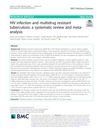
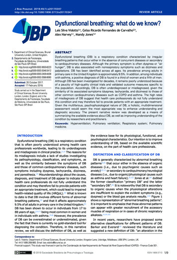
1332 REZAEI ET ALJ ALLERGY CLIN IMMUNOLJUNE 2011FIG 1. SAP deficiency is associated with defective cellular and humoral immune responses in patients withXLP. A-C, SAP deficiency profoundly affects NK (Fig 1, A) and CD81 T-cell (Fig 1, B) cytotoxicity and cytokineproduction and NKT cell development (Fig 1, C). Consequently, increased survival of EBV-infected B cells,together with impaired restimulation-induced cell death (RICD) of T cells, contributes to the development ofFIM, malignant lymphoma, or both. D, SAP deficiency reduces T cell–dependent B-cell activation and immunoglobulin production.ITK DEFICIENCYITK deficiency is a novel PID characterized by severe EBVassociated immune dysregulation that clinically resembles XLP(see Table E1). It was originally identified in 2 female siblings froma consanguineous Turkish family.46 Both ITK-deficient patientshad uncontrolled EBV infection and exhibited clinical and laboratory features consistent with HLH. EBV-induced B-cell proliferation progressed to Hodgkin lymphoma (HL) in both affectedpatients. More recently, 3 cases from a family of Arab origin,who presented with EBV-positive HL, were subsequently foundto harbor a homozygous nonsense mutation in the ITK gene.47All 3 patients responded favorably to initial chemotherapy; however, the clinical phenotype varied considerably among the affected members. One patient had relapsed HL and HLH, which,despite reinitiation of therapy, proved to be fatal. Conversely,complete remission with no evidence of EBV infection, with andwithout HSCT, was observed in the other 2 patients. Lastly, 7additional ITK-deficient patients were identified in a broaderscreen of patients with autoimmune lymphoproliferative syndromeor suspicion of congenital forms of HLH.48,49 Among these newercases, 4 also received diagnoses of HL.48 Together the resultsof these studies suggest that sequence analysis of the ITK geneshould be considered in all patients with EBV-associated lymphoproliferative diseases, particularly HLH and HL.47,48DNA sequencing of affected patients has revealed missenseor nonsense germline ITK mutations that reduce or eliminateITK expression. ITK is a cytoplasmic nonreceptor tyrosinekinase that is expressed in thymocytes and mature T cells, aswell as NK cells, iNKT cells, and mast cells.50,51 Its Pleckstrinhomology domain preferentially binds to phosphatidylinositol(3,4,5)-triphosphate, leading to the recruitment of ITK to theplasma membrane.50 In T cells ITK is activated in response toantigen receptor engagement and is required for full TCRinduced phospholipase C-g (PLC-g) activation and Ca21 mobilization (Fig 2). Indeed, PLC-g activation is reduced rather thaneliminated in Itk2/2 T cells,52,53 thus emphasizing a modulatoryrather than an indispensible role for this kinase in the TCRsignaling cascade.
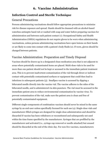
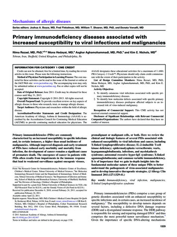
REZAEI ET AL 1333J ALLERGY CLIN IMMUNOLVOLUME 127, NUMBER 6FIG 2. Contribution of the TEC family kinase ITK to TCR-mediated signaling pathways. ITK is required for fullTCR-induced PLC-g activation and Ca21 mobilization, as well as Vav1 localization, signifying its role in TCRinduced signaling complex formation and stability. ITK deficiency alters actin cytoskeletal rearrangement,which probably results in reduced cellular cytotoxicity, as well as gene transcription (eg, decreased Fas ligand expression), which contribute to impaired activation-induced cell death. DAG, Diacylglycerol; ERK, extracellular signal-regulated kinase; IP3, inositol trisphosphate; LAT, linker for activation of T cells; MEK,mitogen-activated protein kinase kinase; NFAT, nuclear factor of activated T cells; RICD, restimulationinduced cell death; SOS, son of sevenless; ZAP-70, zeta-chain-associated protein kinase 70.The strength of the TCR signal plays a critical role in T-celldevelopment and lineage determination.54,55 Consistent with thisnotion, the positive and negative selection of CD41 and CD81 Tcells are defective in the absence of ITK in mice.53,56 In addition,ITK plays an important role in the regulation of conventional versus innate-type CD81 T-cell development.54,57 As opposed toconventional CD81 T cells, innate-type T cells display immediateeffector functions on stimulation and are characterized by expression of activation markers, such as CD44, CD122, and NK1.1. InITK-deficient mice, the majority of CD81 T cells have an innatetype phenotype.58,59 Similarly, ITK-deficient patients havedramatically reduced numbers of naive CD45RA1 T cells.46-48Despite their activated phenotype, Itk2/2 CD81 T cells fail tomount an effective primary or memory immune response to a variety of viral infections.54,60,61 The defective antiviral immune response has been attributed to impaired cytotoxic CD81 T-cellactivity, reduced CD81 T-cell proliferation, and diminishedIFN-g and TNF-a production.60,61 Although not yet formallytested, similar defects might underlie the increased susceptibilityof ITK-deficient patients to EBV infection.ITK is also crucial for iNKT cell development in mice, as wellas efficient iNKT cell cytokine production and survival in theperiphery.62 A characteristic reduction of iNKT cells has beendemonstrated in ITK-deficient patients,46-48 which is consistentwith the experimental data from mutant mice. Hence, as withXLP, the severely reduced number of iNKT cells might also contribute to the increased susceptibility of ITK-deficient patients toprimary EBV infection.46-48The increased development of HL in ITK-deficient patientsmight reflect their perturbation of innate and adaptive antitumorimmune responses, including lack of iNKT cells, which play anindispensable role in antitumor immune surveillance. It is also
1334 REZAEI ET ALpossible that abnormal induction of Fas ligand on TCR stimulation, which results in reduced activation-induced cell death ofstimulated Itk2/2 T cells,63 might contribute to tumorigenesis.Because ITK regulates distinct NK cell–activating receptorsand subsequently NK cell–mediated cytotoxicity,64 NK cell–mediated clearance of premalignant or malignant cells might be impaired in the absence of ITK protein.Given the limited number of patients reported, the efficacy ofvarious treatments has not yet been evaluated systematically.Rituximab, antiviral agents, and steroids can result in a markedreduction in EBV load and temporary improvement of symptoms;however, the disease follows a progressive course in manycases.46,47 EBV-associated HL has been treated successfullywith chemotherapy.46-48 Additionally, successful HSCT hasbeen performed in 2 patients.46-48EVEV is a rare genodermatosis that is characterized by increasedsusceptibility to cutaneous infection with specific HPV genotypes.65,66 EV begins during infancy or childhood and is manifested by disseminated highly polymorphic lesions, includingpityriasis versicolor–like macules and flat wart-like papules.The cutaneous lesions carry a considerable risk of malignanttransformation to squamous cell carcinoma in situ or invasivesquamous cell carcinoma and occur mainly on sun-exposed areas(see Table E1).65-67EV is thought to be an autosomal recessive disease68; however,both X-linked recessive69 and autosomal dominant70 modes of inheritance have been reported. Genome-wide linkage studies haveidentified 2 susceptibility loci,71 including EV1 and EV2 on chromosomal regions 17q25 and 2p21-2p24, respectively. EV1 containsthe EVER1 and EVER2 genes,72 in which 10 truncating or loss-offunction mutations have thus far been reported.65 Homozygous mutations in EVER1 or EVER2 have been identified in approximately75% of patients given a diagnosis of EV,65 leaving a considerableproportion of patients with an unexplained genetic cause.EVER1 and EVER2 belong to an evolutionarily conservedtransmembrane channel–like gene family,73 which constitutes anovel group of modifiers of channels and ion transporters. TheEVER genes are predicted to encode transmembrane proteins located in the endoplasmic reticulum of human keratinocytes.72 TheEVER proteins interact with ZnT-1,74 a zinc transporter that regulates cellular zinc homeostasis. In transfected human keratinocyte cell lines, EVER and ZnT-1 negatively regulate the activityof the metal transcription factor 1 and the cytokine-stimulatedtranscription factors c-Jun and Elk-1.75 Because activator protein1 is a key transcription factor in the HPV lifecycle, it is proposedthat a loss of function of either EVER gene would disrupt zinc homeostasis and consequently enhance the expression of viralgenes, particularly the pro-oncogenic E6 and E7 genes, and contribute to HPV-mediated carcinogenesis.74The EVER proteins are expressed in T and B lymphocytes, NKcells, endothelial cells, bone marrow myeloid cells, and dendriticcells,76 thus pointing to their potential role in immunity againstHPV. Indeed, impaired nonspecific cell-mediated immunity, including decreased total T-lymphocyte and TH lymphocyte counts,reduced T-cell responsiveness to mitogens, and cutaneous anergyto common skin antigens,77 and defective cell-mediated immunity toward EV-specific HPV (EV-HPV) types or infectedJ ALLERGY CLIN IMMUNOLJUNE 2011keratinocytes78,79 have been demonstrated, which could compromise viral clearance and lead to malignant transformation.65,74Despite research on the causative role of HPV infection incervical and anogenital cancers, the role of HPV in cutaneousoncogenesis has yet to be elucidated.66,80 In favor of a protumorigenic role, the E6 oncoprotein of EV-HPV types inhibits UVinduced apoptosis through proteolytic degradation of theproapoptotic protein Bcl-2 (B-cell lymphoma 2) homologousantagonist killer.81 Failure to repair UV-induced thyminedimers has also been demonstrated in cells expressing the E6 protein of HPV-5.82 Moreover, the E6 protein of HPV-8 directlybinds the XRCC1 protein required for DNA single-strand breakrepair,83 thus compromising genetic stability. On the contrary,what is not in favor of the causality hypothesis is the dramatic decrease in the positivity of HPV DNA in malignant skin lesionsdevoid of the upper dermis,84 which might represent a mere contamination from the surrounding skin. However, EV-HPV typescould be involved in the early stages of cutaneous oncogenesisbut not in the maintenance of the malignant state.85Clinical vigilance and stringent sun protection are mandatedfor early diagnosis and surgical treatment of malignant orpremalignant lesions. However, in the case of multiple cutaneousmalignancies or persistent widespread nonmalignant lesions,surgical intervention is less desirable. Several nonsurgicalmodalities, including topical 5-fluorouracil,86 5% imiquimod,87tacalcitol,88 systemic retinoids combined with IFN-a,89 cimetidine,90 and topical 5-aminolevulinic acid photodynamic therapy,91 have been used, with inconsistent results. Hence theirlong-term efficacy merits further evaluation in larger randomizedtrials.WHIM SYNDROMEWHIM syndrome is a rare immunodeficiency syndrome characterized by warts, hypogammaglobulinemia, infection, andmyelokathexis (ie, retention of mature neutrophils in the bonemarrow).92-94 It typically manifests as recurrent bacterial infections from infancy or early childhood, most commonly gastrointestinal, pulmonary, and cutaneous infections, which, despitemarked immunologic abnormalities, often follow a relativelymild clinical course (see Table E1).93,94 The only common laboratory feature is neutropenia, whi
American Academy of Allergy, Asthma & Immunology (AAAAI) is ac-credited by the Accreditation Council for Continuing Medical Education (ACCME) to provide continuing medical education for physicians. The AAAAI designates these educational activities for a maximum of 1 AMA PRA Category 1 Credit . Physicians should only claim credit commensu-