Transcription
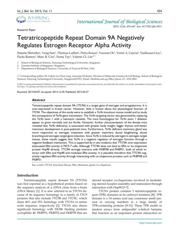
Estrogen receptor α mediates the nongenomic activationof endothelial nitric oxide synthase by estrogenZhong Chen,1 Ivan S. Yuhanna,1 Zoya Galcheva-Gargova,2 Richard H. Karas,2Michael E. Mendelsohn,2 and Philip W. Shaul11Departmentof Pediatrics, University of Texas Southwestern Medical Center, Dallas, Texas 75235, USACardiology Research Institute, New England Medical Center and Tufts University School of Medicine,Boston, Massachusetts 02111, USA2MolecularAddress correspondence to: Philip W. Shaul, Department of Pediatrics, University of Texas Southwestern Medical Center,5323 Harry Hines Boulevard, Dallas, Texas 75235-9063, USA. Phone: (214) 648-2015; Fax: (214) 648-2481;E-mail: pshaul@mednet.swmed.eduReceived for publication September 28, 1998, and accepted in revised form December 15, 1998.Estrogen is an important vasoprotective molecule that causes the rapid dilation of blood vessels by activating endothelial nitric oxide synthase (eNOS) through an unknown mechanism. In studies of intact ovineendothelial cells, 17β-estradiol (E2) caused acute (five-minute) activation of eNOS that was unaffected byactinomycin D but was fully inhibited by concomitant acute treatment with specific estrogen receptor (ER)antagonists. Overexpression of the known transcription factor ERα led to marked enhancement of the acuteresponse to E2, and this was blocked by ER antagonists, was specific to E2, and required the ERα hormonebinding domain. In addition, the acute response of eNOS to E2 was reconstituted in COS-7 cells cotransfected with wild-type ERα and eNOS, but not by transfection with eNOS alone. Furthermore, the inhibition of tyrosine kinases or mitogen-activated protein (MAP) kinase kinase prevented the activation of eNOSby E2, and E2 caused rapid ER-dependent activation of MAP kinase. These findings demonstrate that theshort-term effects of estrogen central to cardiovascular physiology are mediated by ERα functioning in anovel, nongenomic manner to activate eNOS via MAP kinase–dependent mechanisms.J. Clin. Invest. 103:401–406 (1999).IntroductionThe hormone estrogen classically exerts its effects by modifying gene expression (1–3). However, there are also important rapid, presumably nongenomic, effects of estrogen andother related steroid hormones in a variety of tissues including the vasculature, brain, and bone (4–9). Recent evidencesuggests that the vascular effects of estrogen play a criticalrole in the atheroprotective properties of the hormone (10,11). Premenopausal women have very little coronary arterydisease compared to men, the incidence of the disease risesmarkedly after menopause, and hormone replacement therapy reduces the risks to premenopausal levels (12–15). Inaddition, acute estrogen administration rapidly restores theendothelium-dependent dilation of atherosclerotic arteriesin primate models, and it acutely improves endotheliumdependent responses in healthy postmenopausal women(16–19). The rapid vasodilatory effect of estrogen is at leastpartially related to its ability to enhance the bioavailabilityof nitric oxide (NO) (10, 11, 16–19), which is a potent regulator of blood pressure, platelet aggregation, leukocyteadhesion, and vascular smooth muscle mitogenesis (20).Endothelial NO is produced by the endothelial isoform ofNO synthase (eNOS) upon the conversion of the substrateL-arginine to L-citrulline (20). To better understand themechanism(s) by which estrogen acutely increases endothelial NO production, the present experiments were performed in cultured endothelial cells to determine the potential role of estrogen receptor (ER) α in this process. We andothers have recently shown that estrogen rapidly stimulateseNOS activity in endothelial cells and that ERα is expressedThe Journal of Clinical Investigation in endothelium (21–24). ERα and the second, more recently discovered ER isoform, ERβ, are known to function astranscription factors mediating estrogen-induced geneexpression in both reproductive and nonreproductive tissues (10, 11, 25). However, ER may also be involved in acute,nongenomic physiologic responses because the rapideffects of estrogen in certain cell types are inhibited by ERantagonists (4, 5, 7, 8).MethodsCell culture. Pulmonary artery endothelial cells (PAEC) wereobtained from the intrapulmonary arteries of fetal lambs at125–135 days gestation (term 144 days) by collagenase digestionand were propagated as described previously (26). These cells havebeen used previously to evaluate the acute effects of varying oxygenation on eNOS activation (26). Animal care and euthanasiaprocedures were approved by the Institutional Review Board forAnimal Research. Near-confluent PAEC were studied at passage4–6. COS-7 cells (American Type Culture Collection, Rockville,Maryland, USA) were grown in DMEM (Life Technologies Inc.,Grand Island, New York, USA) supplemented with 10% heat-inactivated FBS plus 200 U/ml penicillin and 200 µg/ml streptomycin.eNOS activation in whole cells. eNOS activation was assessed inwhole cells by measuring [3H]L-arginine conversion to [3H]L-citrulline during acute incubations, using methods reported previously (21). This procedure provides a direct evaluation of the acuteactivation of existing eNOS, keeping signal transduction mechanisms intact (27). Adherent cells grown in 24-well plates wereplaced in L-arginine–deficient, serum-free endothelial-SFMGrowth Media (Life Technologies Inc.) for 6 h and then preincubated in PBS (pH 7.4) containing 120 mM NaCl, 4.2 mM KCl, 2.5February 1999 Volume 103 Number 3401
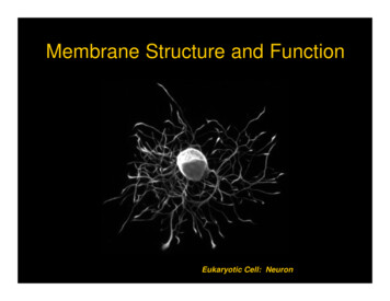
Figure 1Rapid activation of eNOS in endothelial cells. (a) Effect of E2 on eNOSactivity in intact PAEC. [3H]L-arginine conversion to [3H]L-citrulline wasmeasured over 5–15 min in the presence of 10–8 M E2. (b) Effect of actinomycin D (Act D) on the rapid activation of eNOS. After 120 min preincubation in the absence or presence of 25 µg/ml Act D, 15 min incubations were done with or without continued Act D and either 10–8 M E2 orthe calcium ionophore A23187 (10–5 M). (c) Effect of tamoxifen on E2stimulated eNOS activity. Fifteen-minute incubations were performed inthe absence or presence of 10–8 M E2, with or without 10–6 M tamoxifen(Tam) added simultaneously. Partial inhibition (50%–70%) was also notedwith 10–8 M Tam (13). (d) Effect of ICI 182,780 on E2-stimulated eNOSactivity. Fifteen-minute incubations were performed in the absence or presence of 10–8 M E2, with or without 10–5 M ICI 182,780 added simultaneously. Full inhibition was also observed with 10–6 M ICI 182,780 (13). Values are mean SEM; n 4–6. *P 0.05 vs. basal. E2, estradiol-17β; eNOS,endothelial nitric oxide; PAEC, pulmonary artery endothelial cells.mM CaCl2, 1.3 mM MgSO4, 7.5 mM glucose, 10 mM HEPES, 1.2mM Na2HPO4, and 0.37 mM KH2PO4 for 15 min at 37 C. Theincubation for eNOS activity was initiated by replacing the preincubate solution with PBS containing 1.5 µCi/ml [3H]L-arginine.After 5–15 min, the reaction was stopped by adding 1 N TCA, thecells were freeze-fractured in liquid nitrogen and scraped with arubber spatula, the contents of each well were ether extracted, andthe [3H]L-citrulline generated was isolated using Dowex AG50WX8 columns (Sigma Chemical Co., St. Louis, Missouri, USA) andquantified by liquid scintillation spectroscopy. In individual experiments, four to six wells were used for each treatment group. Allfindings were confirmed in at least three independent studies.Basal [3H]L-citrulline generation over 15 min ranged from 4 to 7fmol/100,000 cells on different experimental days. Results areexpressed as the percent of basal eNOS activity determined in thesame 24-well plate. To study the acute effects of estrogen on eNOSin PAEC, [3H]L-arginine conversion to [3H]L-citrulline was measured in whole cells either under basal conditions or in the presenceof 10–8 M estradiol-17β (E2). In previous experiments, the maximaleffect of E2 was obtained at 10–8 M, and the threshold concentration was 10–10 M (21). The activation of eNOS was also evaluated402The Journal of Clinical Investigation in the presence of estradiol-17α (10–12 to 10–6 M) or in the presenceof the known eNOS agonist acetylcholine (10–6 M) or the calciumionophore A23187 (10–5 M) (21). Acetylcholine has been used previously in this cell culture model to mimic mechanisms in intactarteries (21, 28), and it has also been used by others in studies ofnitric oxide (NO)-release processes in a variety of endothelial celltypes (29–31). Both basal and stimulated eNOS activity were fullyinhibited by 2.0 mM nitro-L-arginine methyl ester. To determine ifthe effect of E2 on PAEC eNOS is mediated at the level of gene transcription, cells were treated with 25 µg/ml actinomycin D for 120min, and eNOS activation was then determined in the continuedpresence of actinomycin D for 15 min. Studies of cyclooxygenasetype 1 and malate dehydrogenase gene expression have revealedthat such treatment fully inhibits gene transcription in the PAEC(32). The role of ER in the rapid response to E2 was determinedduring 15-min incubations done in the absence or presence of 10–8M E2, with or without 10–6 M tamoxifen or 10–5 M ICI 182,780added simultaneously (33).Cell transfection studies. To determine the specific role of ERα inthe acute endothelial cell response to E2, the abundance of functional ERα was augmented by transiently transfecting the expression plasmid pCMV3-ERα, or pCMV3 alone, into PAEC usingLipofectamine (Life Technologies Inc.) (34). pCMV3-ERα was constructed by cloning full-length human ERα cDNA (35) as an EcoRIfragment into the CMV-driven plasmid pCDNA 3.1 (InvitrogenCorp., San Diego, California, USA). The construct was confirmedby sequencing in both directions. Cotransfection with a plasmidcontaining SV40-driven β-galactosidase was performed to normalize for transfection efficiency (34). Transfected cells were placedin phenol red–free, estrogen-free media. Seventy-two hours aftertransfection, acute E2-stimulated eNOS activation was examined.Enhanced ER-mRNA expression was documented in reverse transcription-PCR assays (21), and greater ERα protein abundance wasconfirmed by both immunocytochemistry and immunoblotanalysis with the mouse monoclonal antibody AER 320 directedagainst amino acids 495–595 of human ERα (Neomarkers Inc.,Fremont, California, USA), using methods described previously(34). Immunocytochemistry for β-galactosidase revealed successful expression of the protein in 15%–20% of cells, and immunostaining for ERα confirmed enhanced expression in 20%–40% ofcells transfected with ERα cDNA. To determine the effects of ERαoverexpression on estrogen-induced transcriptional transactivation, cotransfection was performed with a luciferase reporter plasmid that contains three copies of the Xenopus vitellogenin estrogen-response element (ERE), ERE-Luc,7 or the control plasmidTK-Luc (34). After transfection, cells were placed in either phenolred–free, estrogen-free media, or phenol red–free media containing 10–8 M E2 for 48 h, and reporter activity was measured (34).Additional experiments were performed to delineate the role ofthe hormone-binding domain of ERα. Acute eNOS activation wasassessed in PAEC transfected with pCMV3, pCMV3-ERα, orpCMV3-ERα-271-NTF. The latter expression plasmid was constructed by first cloning full-length human ERα cDNA into thepCDNA3.1 plasmid, followed by insertion of the coding sequencefor the 8–amino acid “Flag” epitope (NTF), recognized by M2monoclonal antibody (Kodak, Rochester, New York, USA), immediately after the initiation codon at the 5′ end of the ERα codingsequence to yield pCMV3-ERα-NTF. Then pCMV3-ERα-NTF wasdigested with Xcm1 and Xho1 to remove all coding sequence distalto amino acid 271, which excludes the hormone-binding domain(HBD), followed by blunting of the construct ends and religationto yield pCMV3-ERα-271-NTF. Expression from these plasmids ofboth full-length and truncated epitope-tagged proteins of the predicted size was demonstrated by transient transfection of COS-1cells followed by immunoprecipitation and immunoblot analysis.To generalize the findings in the native endothelial cells, furtherstudies were done to reconstitute the ERα-mediated activation ofeNOS in a cell type that does not constitutively express either ERFebruary 1999 Volume 103 Number 3
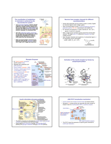
Figure 2Effect of ERα overexpression on transcriptional transactivation and on basal and E2-stimulated eNOS activity. (a) Effect of ERα overexpression onERE-mediated gene transcription in PAEC. Transient transfections were performed with either the estrogen-responsive reporter plasmid ERE-Luc orthe control plasmid TK-Luc, in combination with either sham plasmid or ERα cDNA. Reporter activity was then determined in control cells (upperpanel) and cells exposed to 10–8 M E2 for 48 h (lower panel). Reporter activity is expressed as luciferase activity/β galactosidase activity (LUC/β-gal). *P 0.05 vs. TK-Luc, †P 0.05 vs. control cells, ΨP 0.05 vs. sham. Similar findings were obtained in three independent experiments. (b) Effect of ERαoverexpression on basal eNOS activity in PAEC. Cells were transfected with sham plasmid or ERα cDNA, and 72 h later [3H]L-arginine conversion to[3H]L-citrulline was measured over 15 min in nonstimulated, intact cells. (c). Effect of ERα overexpression on acute eNOS activation by E2. PAECwere transiently transfected with sham plasmid or ERα cDNA, and 72 h later [3H]L-arginine conversion to [3H]L-citrulline was measured in intactcells over 15 min in the absence or presence of 10–8 M E2, with or without 10–5 M ICI 182,780 added simultaneously. Values are mean SEM; n 4–6. *P 0.05 vs. basal, †P 0.05 vs. sham. ERα, estrogen receptor α; ERE, estrogen response element.or eNOS and that is not estrogen responsive. COS-7 cells werecotransfected with human eNOS cDNA (36) and either ERαcDNA or sham plasmid, and the acute effects of E2 were assessed.Tyrosine kinase–MAP kinase inhibition and measurement of MAPkinase activity. Additional studies were performed to begin to elucidate the signal transduction mechanisms involved in acute E2stimulation of eNOS. In nonendothelial cell types, E2 can causethe rapid activation of several signaling pathways, includingthose involving c-src–related tyrosine kinases and mitogen-activated protein (MAP) kinases (7, 8). Therefore, the effects of specific inhibitors of these signaling pathways on eNOS activationwere assessed in intact PAEC. Cells were treated with either thetyrosine kinase inhibitors genistein (50 µM) or herbimycin A (10µM) for 20 h, or with the MAP kinase kinase (MEK) inhibitorPD98059 (50 µM) for 45 min (37, 38). Acute E2-induced eNOSactivation was then evaluated in the continued presence of genistein, herbimycin A, or PD98059 over a 15-min period.The effect of E2 on MAP kinase activity was also assessed (8).PAEC were treated for 5 min with 10–8 M E2 in the absence or presence of 10–5 M tamoxifen or 10–5 M ICI 182,780, or with serum toserve as a positive control. The cells were solubilized with 20 mMTris buffer (pH 7.5) containing 10% glycerol, 1% Triton X-100, 137mM NaCl, 25 mM β-glycerophosphate, 2 mM EDTA, 0.5 mMdithiothreitol, 1 mM sodium orthovanadate, 2 mM sodiumpyrophosphate, 10 µg/ml leupeptin, and 1 mM phenylmethylsulfonyl fluoride. The extracts were centrifuged at 15,000 g for 20min at 4 C, and the endogenous kinase was immunoprecipitated from the supernatant by incubation for 2 h at 4 C with polyclonal anti-Erk2 antibody (Santa Cruz Biotechnology Inc., SantaCruz, California, USA) bound to protein A–Sepharose. Theimmunoprecipitates were washed twice with the 20-mM Trisbuffer and twice with buffer containing 25 mM HEPES (pH 7.4),25 mM β-glycerophosphate, 25 mM MgCl2, 0.5 mM dithiothreitol, and 0.1 mM sodium orthovanadate. Protein kinase assayswere then performed using 1 µg of myelin basic protein and 50µM [γ-32P]ATP (10 Ci/mmol) in a final volume of 20 µl of the 25mM HEPES buffer. The phosphorylation reaction was linear withtime for at least 60 min. The reaction was terminated after 30 minat 30 C by the addition of Laemmli sample buffer. Phosphorylation of the substrate protein was examined after SDS-PAGE byautoradiography and PhosphorImager ( Molecular Dynamics,Sunnyvale, California, USA) analysis.The Journal of Clinical Investigation ResultseNOS activation by E2. The time period over which physiologic concentrations of E2 cause changes in eNOS activityin PAEC is shown in Fig. 1a. E2 (10–8 M) stimulated anincrease in eNOS activity, which reached maximal levelswithin 5 min of exposure to the hormone. The responsewas specific to E2, because 17α-estradiol (10–12 to 10–6 M)had no effect (data not shown). The responses to both E2and the calcium ionophore A23187 were not altered by theinhibition of gene transcription with actinomycin D (Fig.1b). To determine if this acute process involves rapid ERactivation, the effect of concomitant treatment with the ERantagonist tamoxifen was determined (Fig. 1c). Tamoxifencaused no change in basal eNOS activity but fully inhibited the acute response to E2. In addition, simultaneoustreatment with the pure ER antagonist ICI 182,780 alsocompletely negated the rapid, E2-stimulated increase ineNOS activity but did not alter basal activity (Fig. 1d).Effect of ERα overexpression. The potential role of ERα inacute eNOS activation was evaluated in overexpressionstudies. To first confirm the overexpression of functional ERα, estrogen-induced transcriptional transactivationwas assessed by cotransfection of the estrogen-responsive reporter plasmid ERE-Luc or the control plasmidTK-Luc into PAEC with either ERα cDNA or the shamplasmid. In the absence of estrogen, reporter activity wasnot detectable in any paradigm (Fig. 2a, upper). In contrast, after prolonged treatment with 10–8 M E2, ERE-Lucreporter activity was eightfold greater than TK-Lucreporter activity in cells having a normal complement ofER (Fig. 2a, lower). Overexpression of ERα caused a further threefold augmentation of estrogen-induced transcriptional transactivation.The effect of ERα overexpression on basal eNOS activityin intact cells is depicted in Fig. 2b. Basal eNOS activity wasunchanged by ERα overexpression. In addition,immunoblot analysis revealed that eNOS protein abundance was not affected by ERα overexpression (data notFebruary 1999 Volume 103 Number 3403
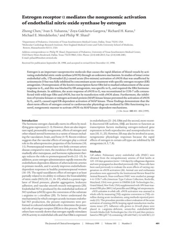
Figure 3Specificity of the augmentation of eNOS activation by ERα overexpression.(a and b) Comparison of the effects of ERα overexpression on acute eNOSactivation by acetylcholine (ACh) and E2. PAEC were transiently transfected with sham plasmid or with ERα cDNA. After 72 h, [3H]L-arginine conversion to [3H]L-citrulline was measured in intact cells over 15 min in theabsence or presence of 10–6 M ACh (a), or in the absence or presence of10–8 M E2 (b). (c) Role of HBD in acute eNOS activation by E2. PAEC weretransfected with either sham plasmid, wild-type ERα cDNA, or an ERαmutant lacking coding sequence distal to amino acid 271 (labeled 271),which excludes the HBD. The eNOS activity in the absence or presence of10–8 M E2 (15 min) was evaluated after 72 h. (d) Reconstitution of acute E2response in COS-7 cells. Transfections were performed with human eNOScDNA and either sham plasmid or ERα cDNA, and eNOS activation wasmeasured 72 h later over 15 min in the absence or presence of 10–8 M E2.Additional studies were done in ERα-transfected cells with 10–5 M ICI182,780 added simultaneously. Values are mean SEM; n 4–6. *P 0.05vs. basal, †P 0.05 vs. sham. HBD, hormone-binding domain.shown). Experiments were then performed to determine theeffect of enhanced ERα expression on the acute response toE2 (Fig. 2c). It was found that the rapid response was augmented four- to fivefold in cells transfected with ERα compared to sham-transfected cells (Fig. 2c). Furthermore, theenhanced response was inhibited completely by concomitant acute treatment with ICI 182,780.To determine the specificity of the augmentation ineNOS activity in response to E2, experiments were donecomparing the rapid effects of acetylcholine and E2 inPAEC overexpressing ERα. With acetylcholine, there werecomparable increases in eNOS activity in sham and ERαtransfected cells (Fig. 3a). This contrasted with the augmented response to E2 after ERα transfection (Fig. 3b). Todetermine the role of the ERα HBD, additional studieswere performed in PAEC transfected with a truncationmutant of ERα lacking coding sequence distal to amino404The Journal of Clinical Investigation acid 271 (Fig. 3c). In contrast to the enhanced E2-mediatedresponse observed after wild-type ERα overexpression, E2mediated eNOS activity was not augmented in cellsexpressing the truncation mutant lacking the HBD. Tofurther define the specific role of ERα, studies were performed to reconstitute the phenomenon in COS-7 cellsthat do not constitutively express either ER or eNOS andthat are not estrogen responsive. In COS-7 cells transfected with eNOS alone, E2 had no effect on eNOS activity(Fig. 3d). However, in cells transfected with both eNOS andERα, there was a more than threefold increase in eNOSactivity upon acute stimulation with E2. In contrast, ERαexpression had no effect on basal eNOS activity (102 33%of activity in sham-transfected cells, or ERα–). In addition,activity stimulated by A23187 was unchanged, being 376 105% and 357 73% of basal levels in ERα– and ERα cells, respectively. The rapid stimulation of eNOS by E2 inthe COS-7 cells transfected with eNOS and ERα was completely inhibited by concomitant ICI 182,780 treatment.Role of tyrosine kinase–MAP kinase pathway. The role of thetyrosine kinase–MAP kinase signaling pathway in theendothelial cell response to E2 was assessed by both pharmacologic intervention and measurements of MAP kinaseactivity. The tyrosine kinase inhibitor genistein did not alterbasal eNOS activity in the PAEC but completely inhibitedthe response to E2 (Fig. 4a). Similar results were obtainedwith herbimycin A (data not shown). In addition, basaleNOS activity was not modified by the specific MEKinhibitor PD98059, but the stimulatory effect of E2 wasfully negated by PD98059 (Fig. 4b). Furthermore, a fiveminute exposure to 10–8 M E2 caused an increase in MAPkinase activity (Fig. 4c), and this effect was completely inhibited by the ER antagonists ICI 182,780 and tamoxifen.DiscussionIn this study we have demonstrated that E2 causes acuteactivation of eNOS in cultured endothelial cells (withinfive minutes). The response is specific to E2 because 17αestradiol had no effect. When the rapidity of the responseis considered along with the observation that it was notaltered by the inhibition of gene transcription with actinomycin D, this suggests that the process does not requirethe classical nuclear effects of the hormone. However, theacute response was fully inhibited by concomitant acutetreatment with the ER antagonists tamoxifen and ICI182,780, suggesting that this occurs via rapid ER activation. The specific level of E2 that was studied is readilyachieved during pregnancy (39, 40), and we have previously shown that the response observed in this model isalso evident at concentrations that are well below thosefound in normal cycling women (21, 41, 42). These cumulative observations are consistent with a novel, nongenomic physiologic role for ER in endothelial cells.To specifically define the role of ERα in the rapidresponse to E2, overexpression studies were performed.Overexpression of ERα resulted in augmented estrogeninduced transcriptional transactivation as expected, andit had no effect on the level of basal eNOS activity oreNOS protein abundance in the intact cells. However,the acute response to E2 was augmented four- to fivefoldin cells transfected with ERα compared with sham-transfected cells, and the enhanced rapid response was inhib-February 1999 Volume 103 Number 3
Figure 4Role of tyrosine kinase–MAP kinase signaling pathway. (a) Role of tyrosine kinase in acute eNOS activation by E2. [3H]L-arginine conversion to [3H]L-citrulline was measured over 15 min in intact PAEC in the absence or presence of 10–8 M E2, with or without treatment with the tyrosine kinase inhibitor (TKI)genistein (50 µM). (b) Role of MEK in acute eNOS activation by E2. eNOS activity was measured in the absence or presence of 10–8 M E2, with or withouttreatment with the MEK inhibitor (MEKI) PD98059 (50 µM). Values are mean SEM; n 4–6. *P 0.05 vs. basal. (c) Effect of E2 on MAP kinase activityin PAEC. Cells were treated for 5 min with 10–8 M E2 in the absence or presence of 10–5 M tamoxifen (TAM) or 10–5 M ICI 182,780, or with serum to serveas a positive control. Endogenous kinase was immunoprecipitated with anti-Erk2 antibody, and protein kinase activity was measured by evaluating thecapacity to phosphorylate myelin basic protein. Quantification by PhosphorImager yielded values of 1, 2.6, 1, 1, 1, and 7.5, respectively, relative to untreated cells. Results shown are representative of five independent experiments. MAP, mitogen-activated protein; MEK, mitogen-activated protein kinase.ited completely by concomitant acute treatment with ICI182,780. These findings suggest that ERα is capable ofmediating the acute response.To determine the specificity of the augmentation ineNOS activity in response to E2, further experiments weredone comparing the rapid effects of acetylcholine and E2in PAEC-overexpressing ERα. With acetylcholine, therewere comparable increases in eNOS activity in sham- andERα-transfected cells, which contrasted with the augmented response to E2 after ERα transfection. As such, theenhanced acute response to E2 in the presence of increasedERα is specific to the hormone, and it is not due to changesin the expression or function of other components of theeNOS signaling cascade. In contrast to the findings withwild-type ERα overexpression, E2-mediated eNOS activitywas not augmented in cells expressing a truncation mutantof ERα lacking the HBD. This is consistent with the inhibition of the acute eNOS response to E2 in native endothelial cells by tamoxifen and ICI 182,780, both of which causeER antagonism via interaction with the ER HBD (10), supporting the conclusion that the HBD of ERα is requiredfor the acute activation of eNOS by E2. Moreover, we haverecently found that physiologic concentrations of E2 causeactivation of eNOS in isolated plasma membranes fromPAEC that is fully inhibited by ICI 182,780 and that ERαprotein is readily detectable in the plasma membrane byimmunoblot analysis (43). These observations suggest thata specific subpopulation of cell-surface receptors may beinvolved in the rapid, nongenomic response.To generalize the findings in the native endothelialcells, experiments were performed to reconstitute theERα-mediated nongenomic activation of eNOS in a celltype that does not constitutively express either ER oreNOS and that is not estrogen responsive. In COS-7 cellstransfected with both eNOS and ERα, there was a morethan threefold increase in eNOS activity upon acute stimulation with E2, whereas there was no response in cellstransfected with eNOS alone. As in native endothelialThe Journal of Clinical Investigation cells, the rapid activation of eNOS by E2 in the COS-7cells transfected with eNOS and ERα was completelyinhibited by concomitant ICI 182,780 treatment, indicating that the response is mediated by acute ERα activation. Thus, the cotransfection of ERα into cells expressing only eNOS confers the ability of E2 to rapidly activateeNOS, confirming the role of ERα in this process.Because E2 can cause the rapid activation of signalingpathways involving c-src–related tyrosine kinases andMAP kinases in nonendothelial cells (7, 8), the potential role of tyrosine kinase–MAP kinase signaling inacute ERα-mediated eNOS activation was evaluated.The tyrosine kinase inhibitors genistein and herbimycin A completely inhibited the response to E2. Inaddition, the specific MEK inhibitor PD98059 alsofully negated the stimulatory effect of E2. Furthermore,E2 caused a rapid increase in MAP kinase activity in thePAEC, and this effect was prevented by both tamoxifenand ICI 182,780. These data indicate that the acutestimulation of eNOS by E2 and ERα entails the activation of tyrosine kinase/MAP kinase. It has been shownthat calcium signaling in endothelial cells in responseto agonists such as bradykinin involves tyrosinekinase/MAP kinase activation (44). We therefore postulate that the rapid stimulation of eNOS by E2 is dueto an increase in intracellular calcium that is mediatedby tyrosine kinase/MAP kinase activation.There is strong evidence from both human and animalstudies that estrogen is protective against vascular injuryand atherosclerosis. This occurs both indirectly by an effecton lipoprotein metabolism and directly through effects onthe vessel wall, including alterations in vascular cell geneexpression, mediated at least in part by ERα acting as a ligand-activated transcription factor. However, the beneficialeffects of estrogen also have an important rapid component in the vasculature, which includes the acute activationof endothelial NO production (10, 11, 16–19). The presentobservations indicate that ERα may mediate the latterFebruary 1999 Volume 103 Number 3405
process, revealing for the first time that this protein regulates physiologic responses in a nongenomic fashion independent of its known ability to control transcription. Assuch, our current conceptualization of steroid hormonereceptor function may be too narrow.Along with the implications regarding estrogen andvascular endothelial function, the present findings areimportant to the mechanisms underlying the rapideffects of estrogen in a variety of other cell types. Forexample, the effects of opioids on hypothalamic neuronsare acutely blunted by E2, and this response is attenuated by ER antagonism (5). In addition, E2 rapidly inhibitsacid production and alters cell shape in osteoclasts in areceptor-depen
eNOS activation in whole cells. eNOS activation was assessed in whole cells by measuring [3H] L-arginine conversion to [ H] -cit-rulline during acute incubations, using methods reported previ-ously (21). This procedure provides a direct evaluation of the acute activation of existing eNOS, keeping signal transduction mecha-nisms intact (27).